Block 5 Immuno
 Call Kai
 Learn
 Practice Test
 Spaced Repetition
 Match
 Flashcards
 Knowt Play
1/78

There's no tags or description
Looks like no tags are added yet.
79 Terms
What are the main differences between innate and adaptive immunity?
Innate (natural, native)
no memory
quick response
low specificity
Adaptive (acquired)
memory (enhanced by vaccine)
slow response
specific
B and T lymphocytes
Humoral vs cell-mediated immunity components
Humoral = soluble factors in serum and fluids
most effective against extracellular bacteria
antibodies
complement and CRP
Cell-Mediated Immunity = cells that do the work themselves
effective against intra and extracellular pathogen, etc
effector cells: phagocytes, granulocytes, and cytotoxic cells
THEY WORK TOGETHER = OPSONIZATION
What is clonal selection?
before infection: each T and B cell has its own receptor shape
antigen selection: antigen fits into receptor and activates it
proliferation: chosen cell copies itself a lot (clones) → each with same receptor as original
Differentiation: each clone takes on 1 of 2 jobs (plasma cell or memory cell)
plasma cell = effector cell (pump out antibody to fight infection NOW; very short lived)
Memory cells = stick around for long time. (ready for next time same antigen attacks so response is faster and stronger; think vaccines)

What is opsonization and what contributes to it?
coating a pathogen with molecules so phagocytes can grab and eat it more easily
opsonins = antibody, complement, CRP





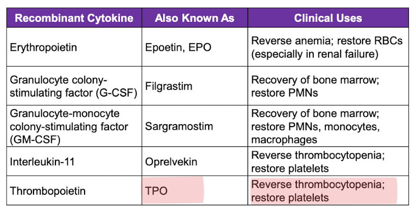
What are the cell markers for B cells?
CD19,20,21 (antibody secretion - humoral immunity)
What are the cell markers for T cells? (all, Helpers, Regulatory, Cytotoxic)
all = CD3
helper = & CD4 (cytokine secretion)
Regulatory = & CD4 & CD25 (prevent autoimmunity)
Cytotoxic = & CD8 (kill infected cells, tumors, mismatch grafts)
What are the cell markers for Natural Killer cells?
CD16,56 (kill infected cells, tumors, mismatched graft)
Adaptive immune system overall function and the major cell player
function = build a targeted attack against one specific pathogen and remembers it
major cell players = B & T cells (plasma cells and antibodies)
Granulocytic cell types
neutrophils, basophils, eosinophils, mast cells
phagocytic cell types
neutrophils, monocytes/macrophages, eosinophils
5 resident tissue macrophages
Kupffer cell
microglia
osteoclasts
langerhans cells
synovial A cells
Kupffer cells
help maintain the liver by removal of debris, fighting infection and orchestrating hepatic immune tolerance
microglia
resident macrophage of the CNS
respond to infection, injury, and provide maintenance
osteoclasts
breakdown and resorb old bone tissue
langerhans cells
cells with macrophage and dendritic cell-like characteristics. present antigens to lymphocytes in the skin
synovial A cells
clear debris and help maintain the synovial fluid in joints such as the knee
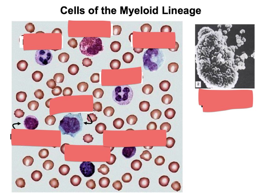

differential white blood cell counts
leukocytosis = increase WBC (above normal)
leukopenia = decreased WBC
lymphocytosis
high lymphocytes = acute viral infection
neutrophilia
high neutrophils = acute bacterial infection
left shift
neutrophilia due to increased bands/stabs (immature neutrophils)
eosinophilia
high eosinophils = acute worm infection or allergy
Primary vs secondary T-Cell development
primary (central) = thymus
T cells mature and first express their TCR (before they meet their antigen)
Secondary (peripheral) = lymph nodes, spleen, MALT
where mature T cells meet and respond to antigen → clonal selection and proliferation
How do T-cells mature in thymus?
precursors in subscapular region are double negative (no CD 4 or 8)
thymocytes become double positive in the cortex (both CD4 and CD8 present)
thymocytes become single positive in the medulla (express one or other but not both CD’s)
**any T cells specific for self are ELIMINATED
What does atrophy of thymus look like and when does it occur in humans?
largest at puberty and then atrophies with age

DiGeorge Syndrome
Thymus (congenital thymic aplasia)
chromosome 22q11.2 microdeletion
little to no thymus = few to no T cells → heighten susceptibility to viruses and cancer
deformed ears, facial anomalies, heart and kidney defects

secondary lymphoid organ: lymph node structure
cortex = B cells
primary follicles = naive B cells
secondary = germinal centers with dividing B cells
paracortex = T cells
medulla
corders = lymphocytes and plasma cells
sinuses = macrophages, drain into efferent lymphatics
what are germinal centers and what does it means if they aren’t present?
germinal centers are found in the cortex of lymph node
secondary follicles = germinal centers contain dividing B cells

what does asplenia mean?
immunocompromised = more susceptible to encapsulated bacteria
**bonus: spleen is major site of antibody synthesis
splenic white pulp
PALS = T cells surround a central artery
B cell-rich follicles = primary and secondary germinal centers

MALT = Mucosa-associated lymphoid tissue
lymphoid tissue sitting under mucosal surfaces (gut-G, airways-B, urogenital tract-GU) that responds to antigens coming in through those surfaces
B cells produces secretory IgA
Peyer’s patches of small intestine
what antibodies may be secreted by structures like Peyer’s patch?
IgA is secreted by B cells (or sIgA) - an antibody that can be secreted onto the surface and survive there
M cells = epithelial cells that mediate transcytosis of antigens from lumen to subepithelial T cells
what happens after an antigen enters the gut?
an antigen enters the gut → M cells carry it to the Peyer’s patch → B cells there make IgA → IgA is secreted into the lumen → it blocks pathogens before they get in
Basic antibody structure
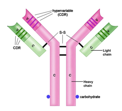
2 heavy chains and 2 light chains (held together by disulfide bonds)
each chain has variable (out toward antigen) and constant (opp side) region
Fab end = antigen binding end (grabs the antigen w/ both light and heavy chain V ends)
Fc end = constant (only heavy chains)
recognized by phagocytes, NK cells and granulocytes and Fc receptors)
binds complement C1q and CRP
how is the isotype or class determined based on antibody structure?
Which antibody crosses the placenta?
IgG = provides the fetus and newborn with passive protection
(dissipates by age 5-6 months)
IgG functions & how many subclasses?
4 subclasses (1,2,3,4)
toxin and virus neutralization
complement activation
opsonization
agglutination
ADCC

what do elevated serum IgG indicate for a person over 6 months?
past infection or the convalescence phase of a current infection
prior vaccination
chronic infection
Primary vs secondary infection immune response. When does IgG dominate?
primary infection = clonal expansion; IgM early & IgG later
secondary infection = memory B cells immediately differentiate into IgG secreting plasma cells
IgM
major class of the primary immune response
made of 5 IgG structure with J chain (like a star)
great agglutinator - 10 binding sites
elevated levels = acute phase of infection
only class produced by the fetus
pentameric, valence of 10

What is the only class of antibody produced by the fetus?
IgM
what does if mean if fetus or newborn has elevated IgM to particular pathogen?
baby is infected by that pathogen
IgA
1 & 2
in seromucous secretions (saliva, colostrum, breast milk, sweat, mucus, etc)
dimeric secretory IgA contains a J chain
valance of 4
secretory component protects sIgA from proteases
oral and intranasal vaccines induce IgA synthesis

What induces IgA synthesis?
oral and intranasal vaccines induce IgA synthesis
IgE
similar structure to IgG (monomeric with valance of 2)
binds FceR on basophils, mast cells, and eosinophils
cross linking causes degranulation
interacts with worms and allergens
immediate hypersensitivity reactions

SCID
persons who lack functional Rag genes can not make antibodies or TCR molecules
(RAG components generate loops in the DNA which bring together ends of randomly selected V)
How does VDJ recombination lead to genetic recombination?
A randomly selected V, D, or J segment is discarded as two segments come together. Since each B cell does this independently and the combinations multiply there is a lot of variability.
differentiate light chains and heavy chains in VDJ recombination
light = NO D → forming antigen binding site
one step
types: kappa and lambda
Fab only
heavy = VDJ → decision maker
two steps (D-J then V joins)
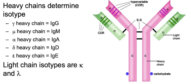
μ, δ, γ, α, ε
determines antibody class
Fab + all of Fc
What is AID enzymes main role?
AID enzyme is required for class-switching to IgG, IgA or IgE (noticing when regions aren’t being used)
reason that once switched from IgM you can’t go back bc H chain was cleaved
activated when B cell receives T cell help (CD40 on B interacts with CD40L on Th cell)
Th cell makes cytokines that determine antibody class synthesized by B cell

What happens when a person is deficient in CD40, CD40L or AID?
they suffer from hyper-IgM syndrome
What is AID’s role in hypermutation and affinity maturation?
AID enzyme → point mutation in V exons of H and L chains
Point mutations occur at a high rate → somatic hypermutation
somatic hypermutation leads to → affinity maturation
which coreceptor binds to each class of MHC for MHC-TCR interactions?
CD4 binds MHC class II on APC and is found on all TH and Treg cells
CD8 binds MHC class I and is found on all Tc cells
What HLA genes correspond to each MHC class?
Major histocompatibility complex (MHC)
class I = HLA-A, B, C
expressed by all nucleated cells and platelets → NO RBC
α chain
class II = HLA-DP, DQ, DR
expression on professional APCs: dendritic cells, macrophages, monocytes, B cells
α chain + β chain

what is a haplotype?
a group of genes inherited en bloc (as a unit) from a single parent
humans have 2 (one from mom and one from dad)
components of MHC class I
α chain (biggest portion)+ β2-microglobulin
presents to CD8
HLA-A,B,C
endogenous antigens come from inside the APC (intracellular viruses, parasites, bacteria)

components of MHC class II
α chain + β chain
presented to CD4
HLA-DP,DQ,DR
exogenous antigens come from outside the APC (phagocytosed or endocytosed pathogens)

CD3 coreceptor domain and its helps with downstream signaling
always attached to the TCR on every T cell (marker that tags all T cells)
made of 6 protein chains
recognizes the antigen, but can’t send the signal into the cell by itself
when binds antigen on MHC
tail of CD3 gets phosphorylated → down stream signaling to nucleus
results in T cell activation, proliferation, and response to infection
Antigen processing pathway: how antigens are placed on MHC complexes
endogenous antigens come from inside the APC (intracellular viruses, parasites, bacteria)
MHC class I → CD8
exogenous antigens come from outside the APC (phagocytosed or endocytosed pathogens)
MHC class II → CD4

TCR, CD complex all put together
TCR recognizes the peptide antigen.
CD4 or CD8 grips the MHC class (II or I) and stabilizes the binding.
CD3 sends the “activate” signal into the cell.
big picture: Antigen processing pathway: how antigens are placed on MHC complexes

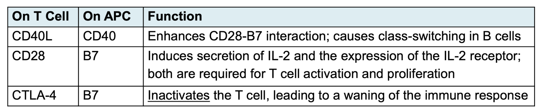
T cells require 2 signals
Signal 1: interaction between the TCR and the peptide/MHC complex on the APC
CD4 or CD8 acts as a co-receptor molecule (this is the right antigen)
Signal 2 is provided by multiple co-stimulatory interactions between cell surface molecules on the T cell and the APC (Yes, really activate)

examples of T cell signal 2 (3 of them)
signal transduction: what are Lck and Zap70?
Lck and Zap70 exist and activate the transcription factors: AP-1, NF-AT, and NF-KB

CD4 T cell functions overview (KNOW THIS WHOLE IMAGE)

X-linked immunodysregulation, Polyendocrinopathy, and enteropathy (IPEX)
mutation in FOXP3 gene = NO Treg cells
autoimmune disease, allergy, inflammatory bowel disease
classic triad
enteropathy (severe diarrhea)
endocrinopathy (type I diabetees mellitus, thyroiditis)
dermatitis
Eosinophilia
severe atopy with elevated IgE
increased infections
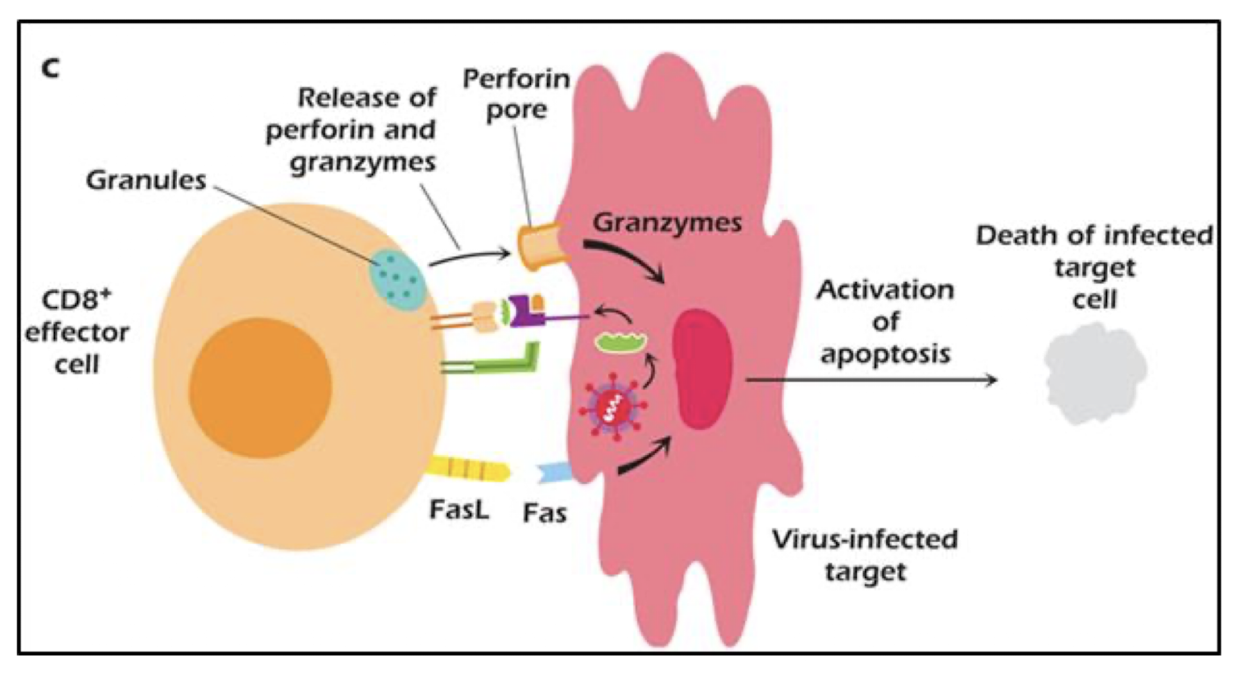
CD8 T cells overall function & what do they secrete and express?
kill: infected host cells, tumors, grafts
secrete:
perforin = punches holes in target cell membrane
granzymes = activates caspases, induces apoptosis
granulysin = kills intracellular bacteria listeria, mycobacterium
express:
FasL = kills target cells expressing Fas by apoptosis (apop does not cause inflam)
What are superantigens?
induce massive cytokine release by T cells and APCs (e.g. macrophages)
no processing needed
clamps the outside of Class II MHC to the B chain of the TCR
induces 20% T cell activation
superantigens (3)
TSST-1 : toxic shock syndrome toxin-1 from S. aureus
Staphylococcal enterotoxin causing food poisoning
pyrogenic toxins from strep. pyogenes
cytokines associated with superantigens and what do they induce?
TNF and IL-1 → induce:
low blood pressure
fever
DIC (disseminated intravascular coagulation: platelet deposition)
cardiovascular shock
cachexia (wasting of muscle and fat)
pleotropism
a cytokine has more than one function
redundancy
multiple cytokines have the same funciton
antagonism
one cytokine blocks the function of another cytokine
synergism
2 or more cytokines enhance each other’s activities
JAK-STAT signaling: know it is associated with cytokine signaling
JAK-STAT is how cytokines signal into a cell. Cytokine → receptor → JAK → STAT → nucleus → genes turn on.
Too much JAK-STAT signaling leads to autoimmune disease and cancer, so JAK-STAT inhibitors treat them (e.g., rheumatoid arthritis, psoriasis, ulcerative colitis).
X-linked SCID: the common gamma chain is broken, so cytokine receptors can’t signal → severe immunodeficiency.