Acute inflammation
 Call Kai
 Learn
Learn Practice Test
 Spaced Repetition
 Match
Match Flashcards
 Knowt Play
1/34

There's no tags or description
Looks like no tags are added yet.
Name | Mastery | Learn | Test | Matching | Spaced | Call with Kai | Chat |
|---|
No analytics yet
Send a link to your students to track their progress
35 Terms
what is inflammation
its aim is to
Get rid of damaged or necrotic tissue
Remove microorganisms or other foreign material
Sets the scene for regeneration or repair of tissues
inflammation can be harmful when you have
Hypersensitivities
Autoimmune reactions
Prolonged inflammation (covered more in the lecture on chronic inflammation)
what is inflammation? the stages/cardinal signs
Cardinal signs of inflammation
1-Rubor (redness)
2-Tumor (swelling) Celsus,
3- Calor (heat)
4- Dolor (pain)
5- Functio laesa (loss of function)

acute inflammation
Rapid response to stimuli
3 components:
• Increased blood flow
• Increased vascular permeability
• Leukocyte emigration

stimuli for acute inflammation
Microorganisms
Parasites, fungi, bacteria and viruses
Microbial toxins
Receptors e.g. Toll-like receptors (TLRs) and cytoplasmic receptors
Necrosis
Ischaemia, trauma, physical and chemical injury
Molecules released from necrotic cells
E.g. uric acid, ATP, HMGB-1 (DNA-binding protein), DNA
Hypoxia
Hypoxia-induced factor 1α (HIF-1α)
Induces vascular endothelial growth factor (VEGF) – increases vascular
permeability
Foreign bodies
Cause trauma or carry microbes
Hypersensitivity reactions / autoimmune disease
Self antigens
Excessive reactions to environmental or microbial antigens
Reactions whereby the immune system damages the animal’s own tissues
reaction of blood vessels to inflammation-vasodilation
When the insult to the tissue occurs there may be a few seconds of vasoconstriction
This is followed by vasodilation
First the arterioles dilate opening new capillary beds in the region
• Allows increased blood flow to the tissue
• Causes erythema (heat and redness) at the site
Vasodilation is induced by histamine and nitric oxide (NO) acting on the smooth muscle of the vessels

reaction of blood vessels to inflammation- increased vascular permeability
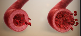
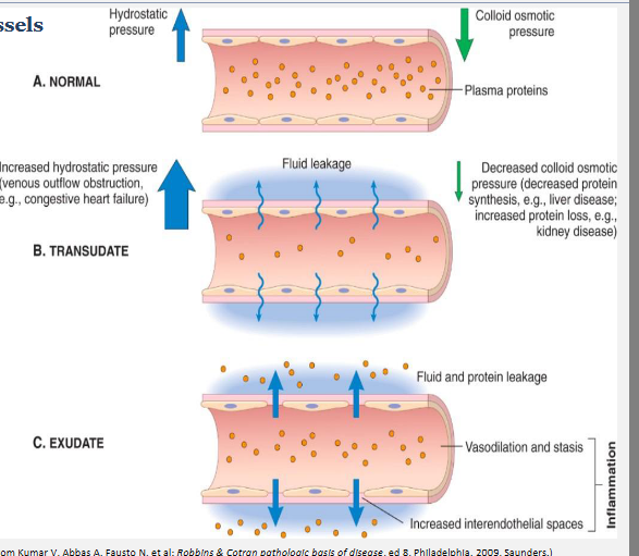
Increased vascular permeability allows escape of protein-rich exudate into the tissue (oedema)
Three mechanisms promote the increase in
vascular permeability:
1. Contraction of endothelial cells (increasing interendothelial spaces)
2. Endothelial injury (necrosis and detachment)
3. Increased transport through endothelial cells (transcytosis)
These mechanisms are not independent and may all be occurring at the same time with some stimuli

reactions of blood vessels
Blood vessels change in order to allow plasma proteins and cells out of the circulation into the site of the stimulus
• Release of excess fluid into tissue or body cavities is called oedema
• Release of fluid and cells from the circulation is called exudation (rich in leukocytes)
— An exudate is an extracellular fluid rich in protein and containing cells
— Has a high specific gravity
— Pus (purulent exudate) is rich in leukocytes (mainly neutrophils), debris of dead cells +/- microbes
• Ultrafiltrates of plasma (i.e. no increased vascular permeability) are caused by loss of osmotic pressure or high hydrostatic pressure
— Creates a transudate with low protein content and little or no cellular material
— Has a low specific gravity
fibrin
Plasma leaving the vessel acts to dilute the stimulus of acute inflammation
• Fibrinogen is one of the proteins which leaves vessels in exudates
• Fibrinogen polymerises to form fibrin
— Fibrin stops the stimulus spreading into nearby tissue
— Allows the leukocytes to target the inciting cause of inflammation
— Assists in blood clotting
— Acts as a scaffold for endothelial migration during wound healing

stasis -a state of balance, inactivity, or a stoppage of normal movement and flow
Fluid loss + increased vessel diameter —> slower blood flow
Increased concentration of red cells —> increased viscosity of blood
Slower blood flow + increased viscosity stasis —> stasis
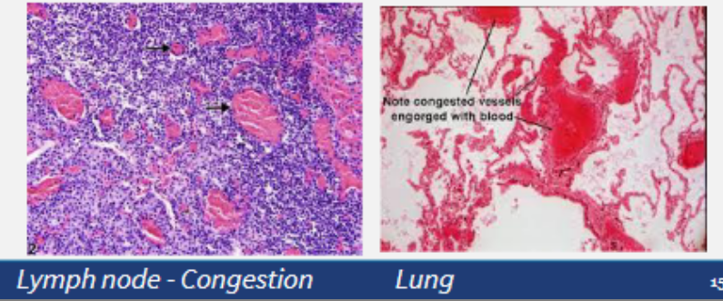
Stasis is observed as vascular congestion
• Allows leukocytes to accumulate on the vascular endothelium
• Endothelial cells are activated by mediators of inflammation, so express increased levels of adhesion molecules
• Leukocytes can then adhere to the endothelium and then migrate through the vessel wall
reactions of lymphatic vessels
Lymphatics normally drain the extravascular fluid (lymph) and take it to lymph nodes
Lymph flow is increased in inflammation, draining the oedema fluid, Leukocytes and cell debris also enter the lymph
Both lymphatic vessels and blood vessels proliferate during inflammation
Secondary inflammation of the lymphatic vessels can occur = lymphangitis
The draining lymph nodes may also become inflamed = lymphadenitis
The lymph nodes will often increase in size too, due to hyperplasia of the lymphoid follicles = reactive/inflammatory lymphadenitis
recruitment of leukocytes
1. Adhesion to the endothelium
2. Migration across the vessel wall
3. Migration to the stimulus
adhesion to the endothelium
When blood is flowing normally through vessels, the red blood cells travel centrally and the leukocytes travel peripherally
When blood flow slows and stasis occurs, due to inflammation, the leukocytes start to make contact with the endothelial surface = margination
These cells start rolling along the endothelium before coming to rest and adhering with the endothelium
Adhesion between the leukocyte and endothelial cell is by complementary adhesion molecules
Cytokines, secreted by cells as part of the inflammatory response, enhance the expression of these adhesion molecules

adhesion to the endothelium
Selectins are proteins which mediate rolling
L-selectin is expressed on leukocytes
E-selectin is expressed on endothelial cells
P-selectin is expressed on platelets and endothelial cells
The selectins bind to ligands
They are sialylated oligosaccharides bound to mucin-like glycoprotein backbones
Both the selectins and their ligands are expressed in response to cytokines
— Within a couple of hours of endothelial cells begin to express E-selectin and the ligands — for L-selectin
— P-selectin is redistributed from its intracellular stores (Weibel-Palade bodies) to the cell surface
— L-selectin is expressed on the tips of the microvilli of leukocytes, along with
ligands for E- and P-selectin
The interactions between selectins and their ligands is low-affinity, so that the bound leukocytes and bind, detach and re-attach quickly, allowing rolling along the endothelium

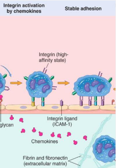
Adhesion to the endothelium
Rolling slows down the leukocytes, allowing stronger adhesions to form
These are mediated by integrins
ICAM-1 on endothelial cells binds to β2 integrins on neutrophils, monocytes and lymphocytes
VCAM-1 on endothelial cells binds to β1 integrins on eosinophils, monocytes and lymphocytes
This stops the rolling, reorganizes the cytoskeleton so that the leukocyte spreads out on the endothelial surface


Migration across the vessel wall
Once the leukocytes have adhered to the endothelial cell they need to move through
the endothelium = diapedesis (or transmigration)
Predominantly in post-capillary venules
Migration occurs down a chemokine concentration gradient
PECAM-1 (platelet endothelial cell adhesion molecule or CD31), JAM A, and JAM C
(Junctional Adhesion Molecule) on endothelial cells mediate transmigration
Once passing the endothelial cells the leukocyte needs to get through the basement
membrane to get into the extravascular tissue
Thought to be due to secreting collagenases
The leukocytes adhere to the extracellular matrix in the connective tissue using integrins and CD44 to bind to matrix proteins – keeping them where they are needed
Leukocyte adhesion deficiencies are characterised by recurrent bacterial infections due to the inability of the leukocytes to perform the steps described so far

leukocyte adhesion deficiencies
Numerous molecular
defects in animals
and humans
• Include
Canine and
Bovine
Leukocyte
adhesion
deficiency
(Canine LAD
and BLAD)
• Affect adhesion of
leukocytes and
therefore the ability of
leukocytes to reach
the site of
inflammation
only needto know canine and bovone Leukocyte adhesion deficiency - missing a adhesoin molecule but cant adhere to blood vessel - inflammation would occur but wont be abke to pass through blood vessel to infecrion - so animals have more infections

migration to the stimulus
Chemotaxis = migration of cells along a chemical gradient
Used by leukocytes migrating towards sites of injury
Exogenous:
Bacterial products, such as lipids and peptides (see your microbiology lectures
for details)
Endogenous:
Cytokines
Complement system
Arachidonic acid metabolites
All these chemotactic agents bind to receptors on the surface of leukocytes.
Downstream signalling alters the cytoskeleton of leukocytes, causing filopodia to
move the cell in the direction of the stimulus, along the chemical gradient
chemokines attract inflmmatory cells to the area
cytokine is type of chemokine

receptors - recogntion of forein organisms and dead tissue
opsonin makes the bacteria more visible to attack on
G protein–coupled receptors
Found on most leukocytes
Recognise short bacterial peptides containing N-formylmethionyl residues
Toll-like receptors (TLRs)
Recognise components of different types of microbes e.g. bacterial
lipopolysaccharide
Cytokine receptors
Leukocytes express receptors for cytokines that are produced in response to
microbes.
Opsonin receptors
Proteins can be used to coat organisms of particles for phagocytosis e.g.
antibodies, complement proteins and lectins
These proteins are called opsonins and the act of coating is called opsonisation
Receptors for opsonins are present on leukocytes

phagocytosis
Removal of material
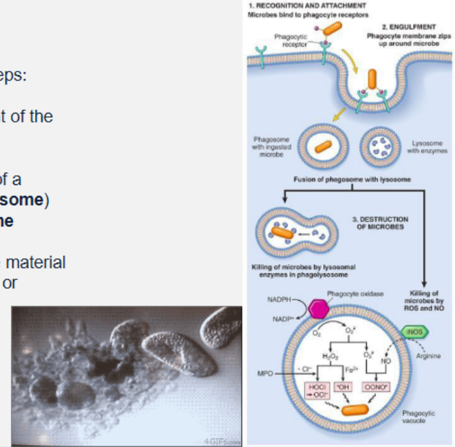
Phagocytosis involves three steps:
1. Recognition and attachment of the
particle by the leukocyte
2. Engulfment and formation of a
phagocytic vacuole (phagosome) within cytosoplasm
which fuses with a lysosome
3. Killing or degradation of the material
by reactive oxygen species or
lysosomal enzymes within
phagolysosome
some microbial agents can avoid step 2
actions of mediators of acute inflammation
Mediators are either cell derived or plasma protein derived
Active mediators are produced in response to various stimuli
One mediator can stimulate the release of other mediators
Mediators vary in their range of cellular targets
There is redundancy
Once activated and released from the cell, most of these mediators are short- lived - because once the issue is fixed why keep it going? inflammation is to heal tissue right - gotta stop inflammation after tissue is healed
Forms the basis of many therapeutic interventions of inflammation - e.g. reduce inflammation, fever, the magnitude of natural responses in the body to fight infection/injury to body
An understanding of inflammation will aid your understanding of rational choice of therapy
vasoactive amines (histamine and seratonine)
Histamine and Serotonin
• Called vasoactive amines because their main action is on vessels
• Stored preformed in cells – so some of the first mediators on the scene
Histamine
• In mast cell granules (also in basophils and platelets)
• Released by degranulation in response to:
• Trauma, cold, heat, binding antibodies to mast cells, anaphylatoxins
(complement C3a and C5a), leukocyte histamine-releasing proteins,
neuropeptides and cytokines
• Binds to H1 receptors on microvascular endothelial cells
• Causes dilation of arterioles and increases the permeability of venules
Serotonin (5-hydroxytryptamine)
• Acts similarly to histamine
• In platelets, certain neuroendocrine cells and in mast cells in some species
• Release of serotonin (and histamine) from platelets is stimulated when platelets
aggregate
— After contact with collagen, thrombin, adenosine diphosphate, and antigen-
antibody complexes
• The platelet release reaction, which is a key component of coagulation, also results
in increased vascular permeability - link between clotting and inflammation
image- arachidonic acid - pro inflammatory molecule - dont need to memorise all steps, just to know the generl concepts and role it plays in mediating inflammation - notice what it leads to - prostaglandn to enhance inflammatory resposne like vasodilation, and chemotaxis, increased vascular permbeability

platelet-activating factor (PAF)
• Phospholipid-derived mediator
— From platelets, basophils, mast cells, neutrophils, macrophages and
endothelial cells
• PAF can elicit most of the vascular and cellular reactions of inflammation:
—Causes platelet aggregation, vasoconstriction and bronchoconstriction
— At extremely low concentrations it induces vasodilation and increased
venular permeability
• Causes increased leukocyte adhesion to endothelium, chemotaxis,
degranulation and the oxidative burst
• Increases the synthesis of other mediators by leukocytes and other cells
reactive oxygen species (ROS)
Oxygen-derived free radicals
• Released extracellularly from leukocytes after exposure to microbes,
chemokines, and immune complexes, or following a phagocytic challenge
• Their production is dependent on the activation of the NADPH oxidase
system.
Superoxide anion, hydrogen peroxide, and hydroxyl radical are the major
species produced within cells
— Can combine with NO to form reactive nitrogen species
• Extracellular release can increase the expression of chemokines, cytokines, and
endothelial leukocyte adhesion molecules, amplifying the inflammatory response
• ROS in leukocytes destroy phagocytosed microbes, so their release can be
damaging to the host:
— Endothelial cell damage, with resultant increased vascular permeability
— Injury to other cell types (parenchymal cells, red blood cells).
— Inactivation of antiproteases, such as α1-antitrypsin. This leads to
unopposed protease activity, with increased destruction of extracellular
matrix
• Serum, tissue fluids, and host cells possess antioxidant mechanisms that protect
against these potentially harmful oxygen-derived radicals
— Superoxide dismutase, catalase, glutathione peroxidase, ceruloplasmin
and transferrin
nitric oxide (NO)
• Produced by endothelial cells and macrophages
• Relaxes vascular smooth muscle
— Promoting vasodilation
• Inhibitor of the cellular component of the inflammatory response
• Microbicidal derivatives of NO (NO-derived free radicals)
cytokines and chemokines (cell derived mediators)
Cytokines
• Tumour necrosis factor (TNF) and Interleukin 1 (IL1)
Produced by activated macrophages
— Local effects
• Increased expression of leukocyte adhesion molecules (inflammation)
• Increased procoagulant activity (inflammation)
• Activation of leukocytes (inflammation)
• Production of further cytokines (inflammation)
• Proliferation of fibroblasts (repair)
• Increased collagen synthesis (repair)
— Systemic effects
• Fever
• Leukocytosis
• Increased acute phase proteins
• Decreased appetite
• Lethargy
Chemokines
• Chemoattractants for different leukocytes
neuropeptides
Secreted by sensory nerves and leukocytes
• Include substance P and neurokinin A
Substance P
• Transmission of pain signals
• Regulation of blood pressure
• Stimulation of secretion of endocrine cells
• Increasing vascular permeability
Sensory neurons can also produce other pro-inflammatory molecules
• Thought to link painful stimuli to protective host responses
Plasma Protein Derived Mediator- complement system
Group of plasma proteins triggered by antibody fixation or microbial antigens
Cause inflammation, phagocytosis, and cell lysis


kinin
Vasoactive peptides
• Derived from kininogens by the action of kallikreins (specific
proteases)
• Prekallikrein is converted to kallikrein
• Kallikrein cleaves high molecular weight kininogen (HMWK)
to produce bradykinin
Bradykinin:
• Increases vascular permeability
• Contraction of smooth muscle
• Dilation of blood vessels
• Pain
in the top pic - dont memorise - just know it plays a role and is related to clotting, fibronylytic system

prevention of excessive damage to the host - termination of acute inflammation
Half life of mediators is short and mediators are rapidly degraded once released - short lived so they can degrade rapidly
Neutrophils have short half lives and die by apoptosis within hours after their release from the circulation
Switch from pro- to anti-inflammatory mediators:
Anti-inflammatory lipoxins (from arachidonic acid)
Macrophages produce anti-inflammatory cytokines e.g. TGF-β and IL-10
Resolvins and protectins (from polyunsaturated fatty acids)
Neural inhibition (cholinergic discharge) that inhibit TNF production
serous inflammation
• The nonhaired skin of the nose is covered by a crust resulting from
dehydration of the serous exudate (fluid from plasma or mesothelial linings)
released from injured blood vessels following a short exposure to the sun
(photosensitivity)
• Microscopically, there is moderate expansion of the superficial dermis with
oedema secondary to vascular leakage (serous inflammation)

catarrhal inflammation
A, Abomasum, cow. The mucosal epithelium is moderately thickened,
covered by a glistening layer of clear mucus, and has a subtle nodular
appearance caused by accumulation of mucinous secretory products
(catarrhal exudate) in the gastric pits
B, Colon, cow. Microscopically, there is a catarrhal colitis with hyperplasia of
mucosal epithelial cells and increased accumulation of mucus on the mucosal
surface

fibrinous inflammation
As vascular permeability increases, fibrinogen can pass the vascular barrier
and fibrin is formed and deposited in the extravascular space
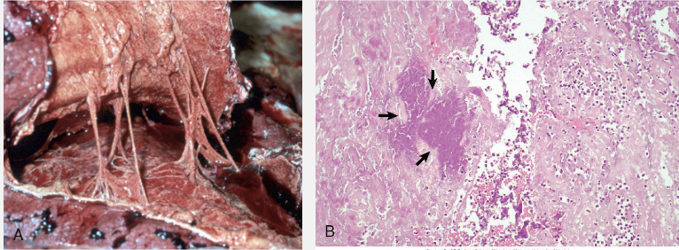
• The pleural surfaces are covered by a yellow-grey, thick, friable exudate
consisting of fibrin mixed with other plasma proteins. This exudate can be
easily pulled apart and should not be confused with fibrous adhesions.
• Microscopically, there are layers of homogeneous eosinophilic material
(fibrin) that contain occasional neutrophils and a focus of bacteria (arrows).
suppurative inflammation
A, The cranioventral areas of the lung are firm and beige to brown. This
lesion is caused by neutrophils transmigrating into alveoli in an acute
inflammatory response secondary to bacterial infection of the lung
B, Microscopically, alveoli contain numerous neutrophils (suppurative
exudate) and sloughed pneumocytes.

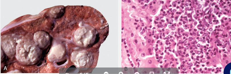
abscess
A, Abscess, lung, cow. A cut section of lung has numerous abscesses. Note
the white-to-grey exudate and how it bulges from the cut surface
B, The exudate in A consists of cell debris and a large number of neutrophils
admixed with lesser numbers of degenerating macrophages and
lymphocytes, and bacteria (the latter not visible with H&E stain).