Patho prac 1: Liver
 Call Kai
 Learn
Learn Practice Test
 Spaced Repetition
 Match
Match Flashcards
 Knowt Play
1/36

There's no tags or description
Looks like no tags are added yet.
Name | Mastery | Learn | Test | Matching | Spaced | Call with Kai | Chat |
|---|
No analytics yet
Send a link to your students to track their progress
37 Terms
Reversible hepatic changes
Hepatocyte swelling by fat, water, bile pigment
Irreversible hepatic changes
Apoptosis, Necrosis
Pattern of liver damage: Zonal cause
Toxin / Hypoxia
Pattern of liver damage: Bridging cause
Viral severe
Pattern of liver damage: Interface cause
Immune
Pattern of liver damage: Apoptotic cause
Viral
Apoptosis histological marker in liver
Councilman body
Focal necrosis microscopic appearance (M/E)
Necrotic foci indicated by aggregates of lymphocytes & macrophages


Zonal necrosis: Central zonal location and causes
Affects zone III due to acute viral hepatitis & C.V.C.
Zonal necrosis: Mid-zonal location and cause
Affects zone II due to yellow fever

Zonal necrosis: Peripheral zonal (Piecemeal necrosis) location and cause
Affects zone I due to chronic hepatitis

Diffuse necrosis definition
Complete (Massive) or near complete (Sub-massive) destruction of hepatocytes affecting almost the whole liver
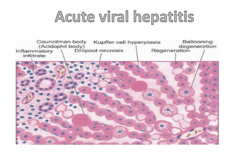
Acute viral hepatitis histological features
Councilman body (Acidophil body), Kupffer cell hyperplasia, Inflammatory infiltrate, Dropout necrosis, Regeneration, Ballooning degeneration
Types of hepatocyte degeneration
Feathery degeneration, Ballooning degeneration

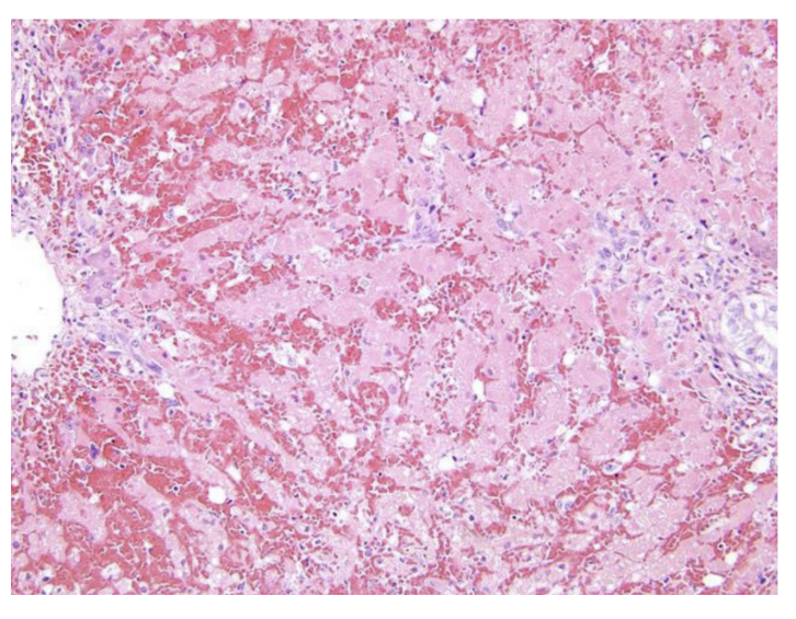
Classic viral hepatitis hepatocyte changes
Councilman bodies, Spotty/Focal necrosis

Acute viral hepatitis gross appearance
Liver gets smaller in size, soft, with wrinkled capsule with yellow patchy foci or large areas

Acute viral hepatitis microscopic appearance (M/E)
Extensive hepatocyte necrosis with few surviving ones with reticulin framework collapse

Chronic hepatitis features (M/E)
Hepatocyte destruction and lymphocytes and focal necrosis

Chronic hepatitis B histological marker
Scattered ground glass hepatocytes

Chronic hepatitis C histological feature
Moderate fatty change

Congenital alpha-1 antitrypsin deficiency histological finding
Presence of PAS +ve globules inside the hepatocytes

Cirrhosis gross features (N/E): Size, Consistency, Surface
Size: Enlarged then become reduced (shrunken).
Consistency: Firm due to fibrosis.
Surface & C/S: Nodular (Macronodular: 3mm to 1cm, may reach up to 6cm, Micronodular: 2 - 3mm, and Mixed.)
The image is wrong, the text under macro and micro are switched

Cirrhosis M/E: Regenerating nodules
Proliferated hepatocytes arranged in thick plates and separated by blood sinusoids. Central vein in abnormal sites (eccentric). Hepatocytes may be small, large, or binucleate.
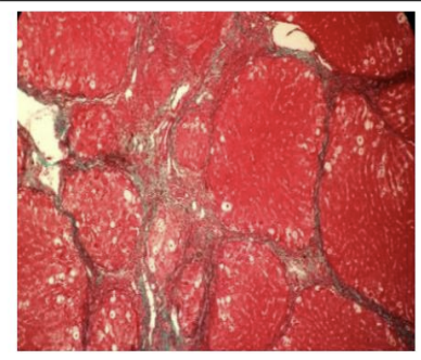
Cirrhosis M/E: Fibrosis patterns
Perivenular, perisinusoidal, pericellular fibrosis

Cirrhosis M/E: Bile ductular proliferation & inflammation
Bile ductular proliferation occurs in fibrous septa with focal cholestasis and feathery degeneration at margins of regenerating nodules.
Inflammation present contains lymphocyte, macrophage, plasma cell infiltrate in fibrous septa.

Etiological classification categories of cirrhosis
Congenital, Acquired
Congenital cirrhosis examples
Primary idiopathic haemochromatosis, and alpha-1-antitrypsin deficiency

Alcoholic cirrhosis progression & gross features
Progresses slowly over few years.
Gross: Liver at first enlarged (fatty change), then normal size, lastly slightly reduced (1.2 kg or more). Micronodular then macronodular then mixed.

Alcoholic cirrhosis M/E features
Hepatocytes: Fatty change that decreases progressively; few show increased intracytoplasmic haemochromatosis.
Fibrous septa: Regular margins with regenerating nodules, moderate lymphocytic infiltrate, slight bile ductular proliferation.
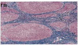
Primary Biliary Cirrhosis (PBC) staged progression
Stage 1: Bile duct inflammation & damage (early portal tracts with lymphocytes and plasma cells destroying ducts).
Stage 2: Granulomatous inflammation surrounding damaged ducts (hallmark).
Stage 3: Cholestatic changes (bile ductular proliferation, periportal Mallory's hyaline, increased copper in periportal hepatocytes).
Stage 4: End stage micronodular cirrhosis with subsided inflammation.
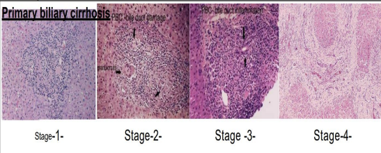
Primary Biliary Cirrhosis (PBC) histological hallmark
Granulomatous inflammation surrounding the damaged and inflamed bile ducts
Liver cell adenoma characteristics
Highly vascularized, liable for rupture with internal hemorrhage. Formed of groups of hepatocytes which may produce pseudocanaliculi and bile.

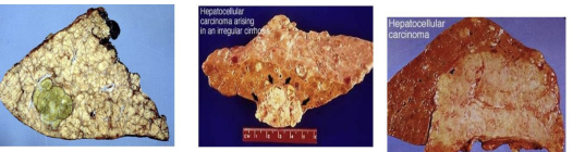
Primary liver carcinoma gross forms and features
Arises in irregular cirrhosis.
Forms:
Single large well-defined mass with hemorrhage & necrosis.
Multiple small nodules scattered all over liver sparing periphery.
Diffuse infiltration. Tissue is yellow-white and may be bile-stained (green).
Hepatocellular carcinoma M/E: Well differentiated
Tumor cells resemble hepatocytes, arranged in trabeculae separated by sinusoids & pseudoglandular structure. Bile may be present.

Hepatocellular carcinoma M/E: Poorly differentiated
Large anaplastic cells, may be bi- or multinucleated giant cells or clear cell type. Stroma is scanty and poorly vascular.
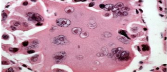
Hepatocellular carcinoma M/E: Fibrolamellar carcinoma
Cords of polygonal cells with acidophilic cytoplasm separated by lamellated fibrous stroma

Secondary liver tumors (Metastases) gross features
Marked hepatomegaly, distorted liver. Surface shows central umbilication (central depression due to necrosis). Green color due to intrahepatic cholestasis. Multiple nodules all over including periphery (rarely 1-2 masses). Color varies by primary (e.g. black in melanoma).
