Lexus Car Models
 Call Kai
 Learn
 Practice Test
 Spaced Repetition
 Match
 Flashcards
 Knowt Play
1/384

There's no tags or description
Looks like no tags are added yet.
385 Terms
Ch. 10 • Q1: The study of the muscular system is known as_________. A) kinesiology B) pathophysiology C) myology D) biology E) neurology
C) myology
Ch. 10 • Q2: Which of the following is not a function of the muscular system? | A) Stability | B) Heat production | C) Control of openings | D) Secretion | E) Breathing
D) Secretion
Ch. 10 • Q3: What term best describes the relationship between the pronator quadratus and supinator? | A) Fixators | B) Antagonists | C) Synergists | D) Prime movers | E) Depressors
B) Antagonists
Ch. 10 • Q4: Muscle fibers are arranged in bundles called_________. | A) compartments | B) fascicles | C) retinacula | D) aponeuroses | E) groups
B) fascicles
Ch. 10 • Q5: Which of the following describes muscular tissue emerging directly from bone rather than being separated from it by an obvious tendon? | A) Direct attachment | B) Indirect attachment | C) Inverted attachment | D) Aponeurosis
A) Direct attachment
Ch. 10 • Q6: The rectus femoris is an example of what muscle shape? | A) Fusiform | B) Parallel | C) Triangular | D) Elongated | E) Pennate
E) Pennate
Ch. 10 • Q7: Groups of muscles are separated by which of the following? | A) Epimysium | B) Fascicles | C) Perimysium | D) Fascia | E) Endomysium
D) Fascia
Ch. 10 • Q8: Which term below best describes the deltoid? | A) Fusiform | B) Intrinsic | C) Involuntary | D) Extrinsic | E) Parallel
B) Intrinsic
Ch. 10 • Q9: Which term best describes the relationship of the deltoid and SITS muscles during shoulder abduction? | A) Prime movers | B) Antagonists | C) Synergists | D) Fixators | E) Adductors
C) Synergists
Ch. 10 • Q10: The semitendinosus is an example of what muscle shape? | A) Triangular | B) Fusiform | C) Sphincter | D) Convergent | E) Pennate
B) Fusiform
Ch. 10 • Q11: Which of the following muscles is the prime mover for inhalation? | A) Internal intercostals | B) External intercostals | C) Diaphragm | D) Stylohyoid | E) Hyoglossus
C) Diaphragm
Ch. 10 • Q12: Which of the following extensors of the head is the most superficial? | A) Trapezius | B) Semispinalis capitis | C) Splenius capitis | D) Sternocleidomastoid | E) Temporalis
A) Trapezius
Ch. 10 • Q13: The occipital and frontal bellies of occipitofrontal are connected via the_________. | A) galea retinaculum | B) galea aponeurotica | C) galea cranium | D) galea orbicularis | E) galea neurotica
B) galea aponeurotica
Ch. 10 • Q14: Which of the following muscles of facial expression is not innervated by the facial nerve? | A) Orbicularis oculi | B) Zygomaticus major | C) Corrugator supercilii | D) Levator palpebrae superioris | E) Buccinator
D) Levator palpebrae superioris
Ch. 10 • Q15: A point of convergence for several muscles of the lower face is the_________. | A) retinaculum | B) aponeurotica | C) modiolus | D) nuchal line | E) galea
C) modiolus
Ch. 10 • Q16: The_________ attaches to the zygomatic arch and also to the angle of the mandible. | A) masseter | B) buccinator | C) temporalis | D) genioglossus | E) stylohyoid
A) masseter
Ch. 10 • Q17: Which one of these muscles helps to open the mouth (depress the mandible)? | A) Zygomaticus major | B) Digastric | C) Sternohyoid | D) Depressor anguli oris | E) Hyoglossus
B) Digastric
Ch. 10 • Q18: The palatoglossus is innervated by which nerve(s)? | A) Hypoglossal | B) Accessory | C) Vagus | D) Hypoglossal and accessory | E) Accessory and vagal
E) Accessory and vagal
Ch. 10 • Q19: Which of the following muscles of mastication would be responsible for mandibular elevation? | A) Temporalis | B) Lateral pterygoid | C) Stylohyoid | D) Zygomaticus major | E) Zygomaticus minor
A) Temporalis
Ch. 10 • Q20: The aponeurosis of the external oblique forms the_________ at its inferior margin. | A) linea alba | B) rectus sheath | C) linea semilunaris | D) medial retinaculum | E) inguinal ligament
E) inguinal ligament
Ch. 10 • Q21: The deepest muscle of the abdominal wall is the_________. | A) transverse abdominal | B) internal oblique | C) rectus abdominis | D) latissimus dorsi | E) pectoralis minor
A) transverse abdominal
Ch. 10 • Q22: While sitting at your desk, you drop your pencil onto the floor. You bend over to pick up the pencil. In order to straighten up and continue your exam you must use which of the following muscles? | A) Tibialis anterior | B) Erector spinae | C) Semimembranosus | D) Tibialis anterior and semimembranosus | E) Extensor hallucis longus
B) Erector spinae
Ch. 10 • Q23: Which of the following muscles is not externally visible on the trunk of the body? | A) Transverse abdominal | B) Latissimus dorsi | C) Trapezius | D) Pectoralis major | E) External abdominal oblique
A) Transverse abdominal
Ch. 10 • Q24: Unilateral contraction of which muscle causes ipsilateral flexion of the lumbar vertebral column? | A) Multifidus | B) Latissimus dorsi | C) External intercostals | D) Serratus anterior | E) Quadratus lumborum
E) Quadratus lumborum
Ch. 10 • Q25: In lifting a heavy weight from the floor, one should use the power of the_________ in order to avoid straining the lower back. | A) biceps brachii and brachialis | B) gastrocnemius and soleus | C) external and internal abdominal obliques | D) knee and hip extensors | E) trapezius and latissimus dorsi
D) knee and hip extensors
Ch. 10 • Q26: In a whiplash injury, the neck undergoes forced hyperextension then hyperflexion, injuring multiple muscles in the neck. Which of the following muscles would be injured during the hyperextension phase of the injury? | A) Trapezius | B) Splenius capitis | C) Sternocleidomastoid | D) Semispinalis capitis | E) Levator scapulae
C) Sternocleidomastoid
Ch. 10 • Q27: Tendinous intersections divide the_________ into segments that are externally visible on the abdomen of a well-muscled person. | A) vastus lateralis | B) serratus anterior | C) rectus abdominis | D) quadriceps femoris | E) transverse abdominal
C) rectus abdominis
Ch. 10 • Q28: The rectus abdominis is a_________ muscle, while the rectus femoris is a_________ muscle. | A) fusiform; parallel | B) parallel; bipennate | C) convergent; unipennate | D) bipennate; parallel | E) multipennate; convergent
B) parallel; bipennate
Ch. 10 • Q29: Which of the following correctly states the proximal and distal attachments of the sternocleidomastoid? | A) Sternal manubrium and lateral one-third of clavicle; mastoid process and lateral half of superior nuchal line | B) Sternal manubrium and medial half of clavicle; mastoid process and medial one-third of superior nuchal line | C) Sternal manubrium and medial one-third of clavicle; mastoid process and lateral half of superior nuchal line | D) Sternal manubrium and lateral half of clavicle; mastoid process and lateral one-third of superior nuchal line | E) Mastoid process and lateral half of superior nuchal line; sternal manubrium, medial one-third of clavicle
C) Sternal manubrium and medial one-third of clavicle; mastoid process and lateral half of superior nuchal line
Ch. 10 • Q30: The muscles that laterally rotate and depress the scapula, as in shrugging and lowering the shoulders, are the_________. | A) trapezius and serratus anterior | B) pectoralis minor and serratus anterior | C) levator scapulae and rhomboideus | D) trapezius and rhomboideus | E) trapezius and latissimus dorsi
A) trapezius and serratus anterior
Ch. 10 • Q31: Which cranial nerve innervates the sternocleidomastoid and trapezius? | A) Abducens | B) Facial | C) Vagus | D) Accessory | E) Trigeminal
D) Accessory
Ch. 10 • Q32: Which muscle(s) of respiration is(are) innervated by the phrenic nerve? | A) Diaphragm | B) External intercostals | C) Internal intercostals | D) Innermost intercostals | E) Pectoralis major
A) Diaphragm
Ch. 10 • Q33: After taking their wedding vows, a bride and groom often ceremonially use which of the following muscles before walking away from the altar? | A) Depressor anguli oris | B) Orbicularis oris | C) Levator labii superioris | D) Platysma | E) Quadriceps femoris
B) Orbicularis oris
Ch. 10 • Q34: Which muscle attaches to the manubrium and the thyroid cartilage, and aids in singing low notes? | A) Omohyoid | B) Thyrohyoid | C) Sternothyroid | D) Stylohyoid | E) Lateral pterygoid
C) Sternothyroid
Ch. 10 • Q35: Some people suffer involuntary urination due to incompetence of what muscle? | A) External urethral sphincter | B) Bulbospongiosus | C) Compressor urethrae | D) Ischiocavernosus | E) Levator ani
A) External urethral sphincter
Ch. 10 • Q36 [Figure: PDF p. 312]: A sacral fracture affecting the S3 and S4 nerve roots would result in a person having difficulty with evacuation of the bowels. A) True B) False
True
Ch. 10 • Q37: A baseball pitcher who injures one of their SITS muscles most commonly sustains tears to the tendon of which muscle? | A) Supraspinatus | B) Infraspinatus | C) Teres minor | D) Subscapularis | E) Deltoid
A) Supraspinatus
Ch. 10 • Q38: Which of the following is true regarding the levator scapulae? | A) It is innervated by the phrenic nerve. | B) It elevates the scapula if the scapula is fixed. | C) It rotates the scapula and depresses the apex of the shoulder. | D) It flexes the neck if the scapula is fixed. | E) It protracts the scapula if the humerus is fixed.
D) It flexes the neck if the scapula is fixed.
Ch. 10 • Q39: Which of the following is an intrinsic muscle of the hand? | A) Extensor carpi radialis brevis | B) Adductor pollicis | C) Extensor pollicis longus | D) Extensor indicis | E) Abductor pollicis longus
B) Adductor pollicis
Ch. 10 • Q40: The serratus anterior is innervated by which nerve? | A) Accessory | B) Dorsal scapular | C) Long thoracic | D) Lateral pectoral | E) Axillary
C) Long thoracic
Ch. 10 • Q41: Which muscle acting on the arm assists in deep inspiration? | A) Pectoralis major | B) Trapezius | C) Teres minor | D) Diaphragm | E) Deltoid
A) Pectoralis major
Ch. 10 • Q42: A butcher who cuts the distal portion of their fingers may cut the_________ tendon. | A) flexor pollicis longus | B) flexor carpi ulnaris | C) palmaris longus | D) adductor pollicis | E) flexor digitorum profundus
E) flexor digitorum profundus
Ch. 10 • Q43: When a nurse draws blood, it is not uncommon for the needle to penetrate which muscle near the cubital region? | A) Pronator quadratus | B) Semimembranosus | C) Pronator teres | D) Buccinator | E) Supinator
C) Pronator teres
Ch. 10 • Q44: The antagonist to the triceps brachii is the_________. | A) deltoid | B) anconeus | C) biceps brachii | D) supinator | E) teres minor
C) biceps brachii
Ch. 10 • Q45: Which of the following best describes the insertion of the extensor digitorum? | A) It indirectly inserts on all digits of the hand. | B) It indirectly inserts on all digits of the hand except the thumb. | C) It directly inserts on all digits of the hand. | D) It directly inserts on all digits of the hand except the thumb. | E) It directly inserts on the index finger only.
B) It indirectly inserts on all digits of the hand except the thumb.
Ch. 10 • Q46: Tennis elbow is a common term describing injury to the attachment of which of the following? | A) Extensor hallucis longus | B) Pronator quadratus | C) Extensor carpi radialis longus | D) Subscapularis | E) Flexor digitorum profundus
C) Extensor carpi radialis longus
Ch. 10 • Q47: An injury that results in complete functional loss of the ulnar nerve will affect which of the following muscles? | A) Abductor digiti minimi | B) Abductor pollicis brevis | C) Flexor carpi radialis | D) Brachioradialis | E) Extensor indicis
A) Abductor digiti minimi
Ch. 10 • Q48: Carpal tunnel pressure is sometimes relieved by surgically excising part or all of which structure? | A) Palmar aponeurosis | B) Extensor retinaculum | C) Flexor retinaculum | D) Extensor aponeurosis | E) Median nerve
C) Flexor retinaculum
Ch. 10 • Q49: What is the longest muscle in the human body? | A) Iliopsoas | B) Sartorius | C) Erector spinae | D) Semitendinosus | E) Semimembranosus
B) Sartorius
Ch. 10 • Q50: Which of the following muscles is in the medial compartment of the thigh? | A) Soleus | B) Adductor longus | C) Piriformis | D) Vastus medialis | E) Semimembranosus
B) Adductor longus
Ch. 10 • Q51: Which of the following muscles attaches on the head of the fibula? | A) Extensor digitorum longus | B) Tibialis anterior | C) Popliteus | D) Biceps femoris | E) Semitendinosus
D) Biceps femoris
Ch. 10 • Q52: Which muscle is innervated by the tibial nerve? | A) Extensor digitorum longus | B) Tibialis anterior | C) Semimembranosus | D) Fibularis longus | E) Vastus lateralis
C) Semimembranosus
Ch. 10 • Q53: Which of the following muscles is responsible for medially rotating the tibia on the femur when the knee is flexed? | A) Piriformis | B) Semitendinosus | C) Adductor brevis | D) Vastus medialis | E) Sartorius
B) Semitendinosus
Ch. 10 • Q54: Which of the following muscles adducts and medially rotates the thigh? | A) Gracilis | B) Piriformis | C) Adductor magnus | D) Rectus femoris | E) Sartorius
C) Adductor magnus
Ch. 10 • Q55: Which of the following muscles is innervated by the obturator nerve? | A) Gracilis | B) Biceps femoris | C) Tibialis anterior | D) Soleus | E) Sartorius
A) Gracilis
Ch. 10 • Q56 [Figure: PDF p. 316]: Injury to the deep fibular nerve would cause inability to extend one's toes. A) True b) False
True
Ch. 10 • Q57 [Figure: PDF p. 316]: The triceps surae consists of three muscles in the lower limb. A) True B) false
False
Ch. 10 • Q58: A clinician induces contraction of the gastrocnemius and soleus and notices that the foot does not plantar flex as expected. Which of the following would be a logical diagnosis? | A) Peroneal nerve injury | B) Ruptured calcaneal tendon | C) Anterior compartment syndrome | D) Shinsplints | E) Lateral plantar nerve injury
B) Ruptured calcaneal tendon
Ch. 10 • Q59: A weightlifter sustains a crushing injury to their foot. After a couple weeks, they begin to notice that they cannot bend the little toe on their right foot. Which of the following would be a logical diagnosis? | A) Atrophy of the flexor hallucis brevis | B) Atrophy of the fibularis brevis | C) Atrophy of the soleus | D) Atrophy of the flexor digiti minimi | E) Atrophy of the adductor hallucis
D) Atrophy of the flexor digiti minimi
Ch. 10 • Q60: An out of shape middle-aged insurance salesperson accepts a friend's invitation to a pickup basketball game. When attempting a jump shot, they fall to the ground in pain, grasping at their calf. There is an enormous bulge in their leg immediately below the popliteal fossa, and they are unable to plantar flex that foot. Most likely they have injured their_________ and the bulge is_________. | A) quadriceps tendon; their rectus femoris | B) calcaneal tendon; their triceps surae | C) patellar ligament; their fibularis longus | D) tibia; a previously undetected bone tumor | E) hamstrings; a large hematoma (blood clot)
B) calcaneal tendon; their triceps surae
Ch. 10 • Q61: An Olympic sprinter readies for a race. After the sound of the starting gun, they propel themselves forward from the starting block and immediately grimace in pain, grabbing for the back of their thigh. Within 48 hours they begin to notice extensive bruising on the back of their thigh extending down behind their knee. They have difficulty standing from a seated position and flexing their knee, and bending at the waist generates more pain. Which muscle is likely injured? | A) Gluteus maximus | B) Popliteus | C) Biceps femoris | D) Gastrocnemius | E) Iliopsoas
C) Biceps femoris
Ch. 10 • Q62: After childbirth, a woman begins experiencing tremendous pain in her groin, making it difficult for her to walk. An X-ray shows a fracture extending from her symphysis pubis to the inferior ramus of the pubis. As a result of the fracture, which group of muscles might be impaired? | A) Hip flexors | B) Thigh abductors | C) Hip extensors | D) Lateral rotators | E) Thigh adductors
E) Thigh adductors
Ch. 10 • Q63: A skydiver's parachute fails to deploy but after some struggle, their reserve chute opens in time to save their life. However, their landing is harder than normal and they feel their knee slightly hyperextend. Later that day they begin experiencing knee pain. Two days later they notice that their knee "pops" when they crouch and soon discover they have dislocated their femur. Which muscle was likely initially injured? | A) Popliteus | B) Tensor fasciae latae | C) Soleus | D) Sartorius | E) Quadriceps femoris
A) Popliteus
Ch. 10 • Q64: A skateboarder falls and lands directly on their buttocks and lower back. They do not feel much pain and continue skating. After a couple of days they begin experiencing right hip pain and notice their right foot and leg are rotated to the right. A doctor diagnoses a muscle spasm. Which of the following muscles is most likely causing the leg and foot rotation? | A) Quadratus lumborum | B) Tibialis posterior | C) Piriformis | D) Adductor longus | E) Sacrospinalis
C) Piriformis
Ch. 10 • Q65: An_________ muscle is contained within a region whereas an_________ muscle acts upon one region but has an attachment elsewhere. | A) intrinsic; extrinsic | B) extrinsic; intrinsic | C) innate; extraneous | D) extraneous; innate
A) intrinsic; extrinsic
Ch. 10 • Q66: You hear a loud noise behind you and look back over your left shoulder. Which of the following is true? | A) This was a contralateral contraction of your right sternocleidomastoid muscle. | B) This was a contralateral contraction of your left sternocleidomastoid muscle. | C) This was an ipsilateral contraction of your right sternocleidomastoid muscle. | D) This was an ipsilateral contraction of your left sternocleidomastoid muscle.
A) This was a contralateral contraction of your right sternocleidomastoid muscle.
Ch. 10 • Q67: Short ribs are a cut of beef often used for barbeque. "Back ribs" are taken from the area near the spine where the meat tends to be thinner. Which muscle forms the meat on this type of rib? | A) Intercostal muscles | B) Diaphragm | C) Rectus abdominus | D) External abdominal oblique | E) Trapezius
A) Intercostal muscles
Ch. 10 • Q68: What is a retinaculum? | A) A band of connective tissue that covers and stabilizes a group of tendons | B) A particularly thick fascia that separates functional muscle groups into compartments | C) A thick, sheet-like tendon | D) A sheath of connective tissue that wraps muscle fibers together to form fascicles | E) A thin sleeve of connective tissue that surrounds each muscle fiber.
A) A band of connective tissue that covers and stabilizes a group of tendons
Ch. 10 • Q69: The skeletal muscles are innervated by_________. | A) spinal nerves and cranial nerves | B) spinal nerves only | C) cranial nerves only | D) spinal nerves and autonomic nerves | E) cranial nerves and autonomic nerves
A) spinal nerves and cranial nerves
Ch. 10 • Q70: Which of these muscles attaches to both the thyroid cartilage of the larynx and to the hyoid bone? | A) Thyrohyoid | B) Sternothyroid | C) Sternohyoid | D) Stylohyoid | E) Sternothyroid
A) Thyrohyoid

Ch. 10 • Q71.1 [Figure: PDF p. 320]: Based on this figure, which two muscles have direct attachments? | A) The brachialis and the lateral head of the triceps brachii | B) The brachialis and the biceps brachii | C) The brachialis and the long head of the triceps brachii | D) The lateral and long heads of the triceps brachii | E) The biceps brachii and the long head of the triceps brachii
A) The brachialis and the lateral head of the triceps brachii

Ch. 10 • Q71.2 [Figure: PDF p. 320]: Based on this figure, which two muscles have indirect attachments? | A) The brachialis and the lateral head of the triceps brachii | B) The brachialis and the biceps brachii | C) The brachialis and the long head of the triceps brachii | D) The lateral and long heads of the triceps brachii | E) The biceps brachii and the long head of the triceps brachii
E) The biceps brachii and the long head of the triceps brachii
Ch. 10 • Q72: The muscles of the head and neck are innervated by the_________. | A) cranial nerves | B) spinal nerves | C) muscle fibers | D) aponeuroses | E) muscle capillaries
A) cranial nerves
Ch. 10 • Q73.1: What is the action of the depressor labii inferioris? | A) Lowers the bottom lip | B) Flexes the smallest finger | C) Aids in urine retention | D) Elevates the scapula | E) Extends the wrist
A) Lowers the bottom lip
Ch. 10 • Q73.2: What is the action of the flexor digiti minimi bevis? | A) Lowers the bottom lip | B) Flexes the smallest finger | C) Aids in urine retention | D) Elevates the scapula | E) Extends the wrist
B) Flexes the smallest finger

Ch. 10 • Q74 [Figure: PDF p. 322]: What type of muscle is this? | A) Fusiform | B) Parallel | C) Triangular | D) Bipennate | E) Multipennate
A) Fusiform

Ch. 10 • Q75 [Figure: PDF p. 323]: What type of muscle is this? | A) Fusiform | B) Parallel | C) Triangular | D) Bipennate | E) Multipennate
B) Parallel

Ch. 10 • Q76 [Figure: PDF p. 324]: What type of muscle is this? | A) Fusiform | B) Parallel | C) Triangular | D) Bipennate | E) Multipennate
C) Triangular
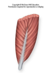
Ch. 10 • Q77 [Figure: PDF p. 326]: Which muscle has this type of fascicle orientation? | A) Rectus femoris | B) Biceps brachii | C) Deltoid | D) Orbicularis oris | E) Pectoralis major
A) Rectus femoris

Ch. 10 • Q78 [Figure: PDF p. 326]: Which muscle has this type of fascicle orientation? | A) Rectus femoris | B) Biceps brachii | C) Deltoid | D) Orbicularis oris | E) Pectoralis major
C) Deltoid

Ch. 10 • Q79.1 [Figure: PDF p. 328]: Name the layer of connective tissue labeled 1. | A) Fascia | B) Epimysium | C) Perimysium | D) Endomysium | E) Retinaculum
A) Fascia

Ch. 10 • Q79.2 [Figure: PDF p. 329]: Name the layer of connective tissue labeled 2. | A) Fascia | B) Epimysium | C) Perimysium | D) Endomysium | E) Retinaculum
B) Epimysium
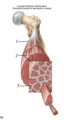
Ch. 10 • Q79.3 [Figure: PDF p. 329]: Name the layer of connective tissue labeled 3. | A) Fascia | B) Epimysium | C) Perimysium | D) Endomysium | E) Retinaculum
C) Perimysium

Ch. 10 • Q80.1 [Figure: PDF p. 330]: Name the arm muscle labeled 1. | A) Long head of triceps brachii | B) Lateral head of triceps brachii | C) Medial head of triceps brachii | D) Brachialis | E) Biceps brachii
A) Long head of triceps brachii

Ch. 10 • Q80.2 [Figure: PDF p. 330]: Name the arm muscle labeled 2. | A) Long head of triceps brachii | B) Lateral head of triceps brachii | C) Medial head of triceps brachii | D) Brachialis | E) Biceps brachii
B) Lateral head of triceps brachii

Ch. 10 • Q80.3 [Figure: PDF p. 330]: Name the arm muscle labeled 3. | A) Long head of triceps brachii | B) Lateral head of triceps brachii | C) Medial head of triceps brachii | D) Brachialis | E) Biceps brachii
E) Biceps brachii

Ch. 10 • Q80.4 [Figure: PDF p. 330]: Name the arm muscle labeled 4. | A) Long head of triceps brachii | B) Lateral head of triceps brachii | C) Medial head of triceps brachii | D) Brachialis | E) Biceps brachii
D) Brachialis

Ch. 10 • Q81.1 [Figure: PDF p. 331]: Name the facial muscle labeled 1. | A) Orbicularis oculi | B) Orbicularis oris | C) Levator palpebrae superioris | D) Occipitalis | E) Corrugator supercilli
A) Orbicularis oculi

Ch. 10 • Q81.2 [Figure: PDF p. 331]: Name the facial muscle labeled 2. | A) Zygomaticus minor | B) Zygomaticus major | C) Levator labii superioris | D) Risorius | E) Buccinator
A) Zygomaticus minor

Ch. 10 • Q81.3 [Figure: PDF p. 331]: Name the facial muscle labeled 3. | A) Zygomaticus minor | B) Zygomaticus major | C) Levator labii superioris | D) Risorius | E) Buccinator
B) Zygomaticus major

Ch. 10 • Q81.4 [Figure: PDF p. 332]: Name the facial muscle labeled 4. | A) Zygomaticus minor | B) Zygomaticus major | C) Masseter | D) Risorius | E) Buccinator
C) Masseter
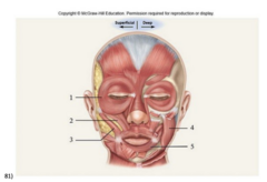
Ch. 10 • Q81.5 [Figure: PDF p. 332]: Name the facial muscle labeled 5. | A) Orbicularis oris | B) Orbicularis oculi | C) Masseter | D) Risorius | E) Buccinator
A) Orbicularis oris
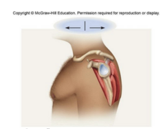
Ch. 10 • Q82 [Figure: PDF p. 332]: Together, the group of muscles shown here is referred to as the _________. | A) Rotator cuff | B) Hamstrings | C) Quadriceps | D) Lateral rotators | E) Iliopsoas
A) Rotator cuff

Ch. 10 • Q83.1 [Figure: PDF p. 334]: Use your three-dimensional understanding of arm anatomy to identify the muscle labeled 1 in this figure. | A) Biceps brachii | B) Brachialis | C) Triceps brachii | D) Brachioradialis | E) Pronator teres
A) Biceps brachii

Ch. 10 • Q83.2 [Figure: PDF p. 334]: Use your three-dimensional understanding of arm anatomy to identify the muscle labeled 2 in this figure. | A) Biceps brachii | B) Brachialis | C) Triceps brachii | D) Brachioradialis | E) Pronator teres
B) Brachialis
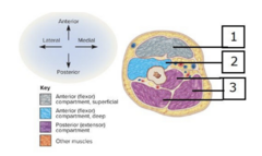
Ch. 10 • Q83.3 [Figure: PDF p. 334]: Use your three-dimensional understanding of arm anatomy to identify the muscle labeled 3 in this figure. | A) Biceps brachii | B) Brachialis | C) Triceps brachii | D) Brachioradialis | E) Pronator teres
C) Triceps brachii

Ch. 10 • Q84 [Figure: PDF p. 336]: The structure labeled 1 is an example of _________. | A) a retinaculum | B) an aponeurosis | C) an interosseous membrane | D) a tendon | E) a ligament
A) a retinaculum

Ch. 10 • Q85 [Figure: PDF p. 336]: Based on their position and attachments, how would you classify the muscles shown here? | A) Adductors | B) Lateral rotators | C) Flexors | D) Extensors | E) Pronators
A) Adductors

Ch. 10 • Q86 [Figure: PDF p. 337]: What is the name of the muscle highlighted in purple? | A) Iliacus | B) Psoas major | C) Pectineus | D) Rectus femoris | E) Gluteus maximus
A) Iliacus
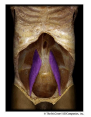
Ch. 10 • Q87 [Figure: PDF p. 338]: What is the name of the muscle highlighted in purple? | A) Iliacus | B) Psoas major | C) Pectineus | D) Rectus femoris | E) Gluteus maximus
B) Psoas major