BIOL155 Midterm Exam Study
 Call Kai
 Learn
 Practice Test
 Spaced Repetition
 Match
 Flashcards
 Knowt Play
1/82

83 Terms
What is homeostasis and how is it preserved?
Homeostasis, stable internal environment of body
Homeostasis is preserved when our physiological mechanisms use energy to keep body conditions steady in spite of disruptions
What is allostasis and what are the two types it is classified in?
Allostasis, refers to the body’s ability to adapt to our set points (behaviours) to meet different conditions
Physiological allostasis, our internal integrators can shift their set points during specific states (ex: blood pressure)
Behavioural allostasis, when we voluntarily perform behaviours that alter a regulated variable away from its current set point (ex: dressing warm for winter)
Both forms are feed-forward, they anticipate a future physiological need
What are trade-offs with respect to anatomy and physiology?
If structure usually determines or predicts function, the exact features of structure may reflect trade-offs (compromises) between multiple competing functions
What is negative feedback and what constitutes a negative feedback loop?
Negative feedback, is when an effector activated by the control center opposes, or negates, the original stimulus
Negative feedback tends to minimize change, keeping variation in key body systems within limits compatible with our long term survival
Negative feedback loops return the controlled variable back to its set point
What is positive feedback and what constitutes a positive feedback loop?
Positive feedback, an initial stimulus produces a response that exaggerates or enhances the change in the original conditions, rather than opposing it
Positive feedback tends to produce extreme responses
An escalating cycle is called a positive feedback loop
Describe an example that illustrates how structure predicts function.
Consider the structure of hand or foot bones, what makes one more suitable for walking and the other for grasping? In feet, large, flat bones help absorb weight vs small short bones in our wrists help with dexterity.
How can failure to regulate the cell cycle lead to tumours and cancers? How can we distinguish between them?
Cancer is an illness characterized by mutations, permanent changes in DNA that disrupt normal control mechanisms that regulate the rate of cell division
As all cells in our bodies are generated by cell divisions, they are normally tightly regulated by gene networks. If the divisions are disregulated, tumors, or even cancer can result.
Tumor, is a mass or swelling produced by abnormal cell growth and division
Benign tumor, cells usually remain within the originating tissue
Malignant tumor, cells divide very rapidly and release chemicals that stimulate the growth of new blood vessels into the area
Metastasis, can produce secondary tumors in tissues remote from the primary tumor site; cancer leads to metastasis due to disregulated cell division
How do molecules and ions move across cell membranes through free diffusion?
Passive transports are free diffusion or facilitated diffusion
Facilitated diffusion, process in which molecules freely move from an area of higher concentration to one of a lower concentration
No ATP is expended in facilitated diffusion
What is osmosis / osmotic flow?
Osmosis, diffusion of water across a selectively permeable membrane
What is the difference between active transport and passive transport?
Active transport, is when ATP provides the energy needed to move ions or molecules across the plasma membrane, the cell can import or export specific substances regardless of their intracellular or extracellular concentrations
Passive transport, is where molecules freely move from an area of higher concentration to one of a lower concentration, no ATP is expended as a result of this facilitated diffusion
What is vesicular transport?
Vesicular transport, is a form of active transport in which materials move into or out of the cell in vesicles
Contrast the similarities and differences between primary and secondary active transport.
Primary active transport, use what all cells contain carrier proteins called ion pumps which actively transport the cations sodium (Na+), potassium (K+), calcium (Ca2+) and magnesium across their plasma membranes
Directly uses ATP
Secondary active transport, transport mechanism does not require ATP but uses a gradient of one molecule (created using ATP) to move another molecule
Cell often needs to expend ATP at a later time to preserve homeostasis
Movement follows an existing concentration gradient for one of two substances transported, typically sodium ions so transport does not require energy
What are examples of molecular transport across cell membranes?
A primary active transport example: the sodium-potassium pump (Na⁺/K⁺-ATPase)
Secondary active transport example:
Glucose molecule with a sodium ion binding with it enters ICF; later it is pumped out with sodium-potassium pump (Na⁺/K⁺-ATPase)
What are the four main types of tissue and give examples of where in the body each is likely to be found.
Epithelial tissue
Lines surfaces and spaces in the body, covers exposed surfaces
Found on superficial layer of skin, lines internal passageways and chambers, secretory glands
Connective tissue
Fills or stiffens internal spaces in body; provides structural support; stores energy
Found in dermis, blood, tendon, cartilage, areolar/adipose tissue, bones
Muscle tissue
Contracts to produce movement or mechanical force; includes skeletal muscle, cardiac muscle and smooth muscle
Found attached to tendons across skeleton, heart and certain organs
Nervous tissue
Send and receive information via electrochemical signals, conducts electrical impulses; carries information
Found across body, mostly in brain and spinal cord
Identify the shared features of epithelial tissue, including glands.
Epithelia, cellular layers of different types that cover exposed surfaces and line internal cavities and passageways
Are avascular, do not have blood vessels and often contain secretory cells, or gland cells scattered among other cell types
Glands, organized groups of cells or organs that contain epithelial-derived cells that synthesize substances for secretion; two types of glands:
Exocrine glands, secrete onto external surfaces or into internal passageways (ducts) that connect to exterior
Endocrine glands, secrete hormones (chemical messengers) or inactive precursors (prohormones) into interstitial fluid that then enter bloodstream for distribution
Shared features include:
Polarity, refers to presence of structural and functional differences between exposed and attached surfaces
Tight junctions, characteristic of epithelial cells lining the intestinal tract, they form a barrier that isolates the basolateral surfaces and deeper tissues from contents of the lumen
‘Tight’ tight junctions do not allow paracellular transport
‘Leaky’ tight junctions allow for paracellular transport of water and solutes
Basement membrane, noncellular structure produced by basal surface of epithelium and underlying connective tissue
Contrast the similarities and differences of key structures and physiological features between simple and stratified epithelial tissue.
Simple epithelia has one layer and all simple epithelial cells are attached to the basement membrane
Stratified epithelia has multiple layers but only the deepest basal layer makes direct contact with the basement membrane
Both simple and stratified epithelium have squamous, cuboidal and columnar arrangements including a pseudo-stratified structure
Squamous are the most flat shaped
Cuboidal are cube shaped
Columnar are tall rectangular shaped
Pseudo-stratified appears stratified but is simple- all epithelial cells are attached to basement membrane
Explain the basic definition of connective tissue and classify whether a tissue is a connective tissue or not based on a description.
Connective tissue is defined by the presence of extracellular protein fibres and cells suspended within a ground substance (matrix), but can fulfill many functions like the following:
Establish a structural framework for the body
Transport fluids and dissolved materials
Protect delicate organs
Support, surround and interconnect other types of tissue
Store energy
Defend body from invading microorganisms
Compare and contrast the structure and general functions of loose and dense connective tissue proper.
Loose connective tissue fibers: areolar tissue, adipose tissue, reticular tissue
Areolar tissue, least specialized and most common form of connective tissue proper in adults; all cell types found in other forms of connective tissue proper can be found in areolar tissue
Adipose tissue is found deep to the skin at the flanks, buttocks and breasts, it forms a layer that provides padding within the orbit of the eyes, abdominopelvic cavity and around the kidneys
Reticular tissue is found in liver, kidney, spleen, lymph nodes, and bone marrow, it forms a tough, flexible network that provides support and resists distortion
Dense connective tissue fibers: dense regular, dense irregular, elastic
Dense regular connective tissue is found in cords (tendons) or sheets connecting skeletal muscles to bone, and in cords (ligaments) that interconnect bones or stabilize the position of internal organs
Dense irregular connective tissue, fibers form an interwoven meshwork in no consistent pattern, strength and support areas subjected to stresses from many directions
Forms a covering or capsule that sheathes visceral organs; a superficial layer covering bones, cartilages, and peripheral nerves; and a thick supporting layer in the skin (dermis)
Elastic tissue, when elastic fibers outnumber collagen fibers, the tissue is springy and resilient allowing it to tolerate cycles of extension and recoil
Elastic tissue is found between vertebrae of the spinal column, in the walls of large blood vessels, in ligaments supporting transitional epithelia and erectile tissue of the penis
Describe the key features of tissue repair in proper connective tissue, and predict consequences for the organism if a component of the repair process is disrupted.
Connective tissue, strategy for repair is mostly increased rates of secretion
Ex: scar tissue forms, when dense web of collagen fibers is secreted at injury site
Consequences if component of inflammation phase is disrupted is infection at the site should harmful bacteria enter the site of injury, or during regeneration, scar tissue not returning to normal after stabilizing injury site
Explain the four major functions of the integument using specific examples of how the properties of the dermis, epidermis or accessory structures contribute to each function.
Vitamin D & Ca2+ Homeostasis
Synthesize Vitamin D3, steroid which subsequently converted to calcitriol, a hormone important to calcium metabolism
Barrier (immune & osmotic homeostasis)
Epidermis is keratinized and cross-linked making skin water resistant
Sebaceous (oil) glands release sebum which moisturizes skin and hair while inhibiting growth of bacteria
Thermal Homeostasis
Sweat glands (apocrine and eccrine) and smooth muscle in the walls of blood vessels supply the skin when homeostasis is disturbed
Sweat glands create sweat which can support evaporative cooling of the body temperature
Sense organ
Detect touch, pressure, pain and temperature stimuli and relay info to nervous system
Touch receptors include free nerve endings, tactile discs, tactile (Meissner) corpuscles, lamellar (Pacinian) corpuscles and bulbous (Ruffini) corpuscles
Explain the general function of melanin and how melanocytes contribute to skin colour at baseline and analyze the physiological response of melanocytes following UV exposure.
Melanin, brown yellow-brown or black pigment produced by melanocytes
Melanin can absorb UV photons and prevent damage to cells particularly DNA while releasing it safely
Melanocytes within the stratum basale produce melanin pigments and transfer them to keratinocytes through melanosomes
In pale skin, transfer occurs in stratum basale and stratum spinosum and cells of more superficial layers lose their pigmentation
In dark skin, melanocytes are more actives, melanosomes are larger and more numerous and transfer may occur in the stratum granulosum as well


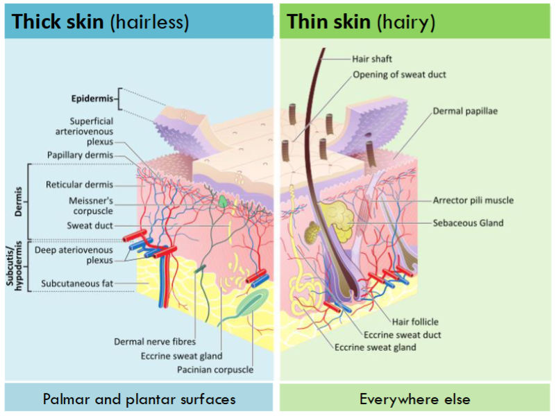
Identify at least three examples of accessory structures within the integument.
Hair shaft - dead keratinized cells forming body of hair
Tactile (Meissner’s) corpuscles - nerve which supports sensation of touch
Sebaceous (oil) gland - produces sebum to protect hair and skin
Arrector pili muscle - flexes hair for “goosebumps”
Sweat gland duct - where sweat is released on surface of skin
Hair follicles - where hair is formed meeting root and shaft
Lamellar (Pacinian) corpuscle - deep sensation nerve
Nerve fibers
Sweat gland (Apocrine or Eccrine) - produces sweat
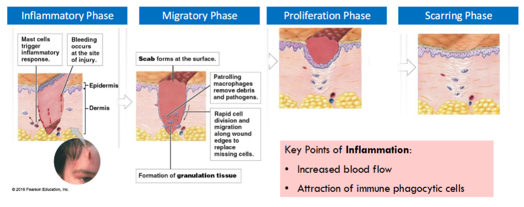
Compare and contrast the processes of tissue repair in the dermis and epidermis, and describe the origin of both granulation tissue, scar tissue, and calluses in the integument.
Granulation tissue is a combination of blood clot, fibroblasts and extensive capillary network
A scab forms on epidermis made from blood cells (platelets) at injury site
Scar tissue forms on dermis, when dense web of collagen fibers is secreted at injury site
A callus is a specific thickening of the stratum corneum, caused by keratinocytes being more strongly cross-linked
Describe the roles of the skin as a key effector organ in thermal homeostasis.
Sweat glands (apocrine and eccrine) and smooth muscle in the walls of blood vessels supply the skin when homeostasis is disturbed
Evaporative cooling, when sweat is produced by the sweat glands and turns to gas through evaporation
Radiant cooling, heat through radiation and convection causes heat loss by blood circulating to surface of the skin to expel heat
Staying warm is the opposite, heat is retained by the body when its cold as blood circulation avoids the surface of skin to retain heat
Explain how skin and UV exposure are linked to Vitamin D and calcium homeostasis, and analyze the trade-offs between melanin and vitamin D synthesis.
Vitamin D through sunlight is necessary to synthesize calcitriol (a hormone) that supports calcium homeostasis in the body, the effect can vary based on an individual's skin tone
In dark skin (more melanin) upper layers of skin absorb more UV light and less reaches the basal layers -> trade-off is less vitamin D but also less cell damage
In light skin (less melanin) upper layers of skin let in more UV light to pass through basal layers -> trade-off is more vitamin D but also more cell damage
Identify key effects of ageing on the integument and discuss how this interacts with UV exposure.
Aging has more of an effect than cosmetic on the integument system, which can be accelerated by exposure to UV as cells can be further damaged
As we age, the epidermis has fewer melanocytes; a drier epidermis (less active sebaceous oil glands); thinning epidermis; decreased perspiration of sweat glands and; fewer active hair follicles
These factor culminate in key effects:
Diminished immune response
Loss of elastin in skin (“stretchiness”)
Reduced blood supply
Altered hair and fat distribution
Slower skin repair
Describe the five main anatomical components of the musculoskeletal system in terms of tissue type and give examples of their roles.
Muscle - muscle tissue which produces movement; provides support; generates heat
Bone - supportive connective tissue which provides support; protects tissues; stores minerals; forms blood cells
Tendons - dense regular connective tissue which anchors bones to muscles
Matrix: mostly collagen and little to no elastin
Ligaments - dense regular connective tissue which anchors bone to other bones
Matrix: collagen and usually some elastin
Cartilage - supportive connective tissue; lines joint surfaces; ex: ears, nose, vertebral discs
Compare and contrast the gross anatomy and histology of bone and cartilage, including the composition of the matrix and the diversity of distinct cell types.
Cartilage and bone are distinct (but related) types of supportive connective tissues that have dense, solid matrix
Bone tissue matrix: strong relatively rigid
Matrix comp: collagen, calcium salts, hydroxyapatite
Cartilage tissue matrix: relatively flexible
Matrix comp: chondroitin sulfates (+ proteins forming proteoglycans)
Cartilage cells (chondrocytes) are the only cells in the avascular cartilage matrix - they occupy small chambers known as lacunae
Bone tissue is highly vascularized and innervated; inside bones are spaces filled with bone marrow
Bone tissue contains multiple cell types:
Osteogenic cells produce other bone cells
Osteoblasts produce bone matrix
Osteocytes maintain bone matrix
Osteoclasts dissolve bone matrix
Compact bone is the weightbearing outer layer that is well organized
Spongy bone is the finer network that lines the internal cavity

What are the types of cartilage and how are they differentiated?
Hyaline cartilage, made of proteoglycans
Found in tips of the ribs, bones of sternum, covering bone surfaces at movable joints, respiratory passageways and forming nasal septum
Elastic cartilage, made of proteoglycans and elastic fibers
Found in external ear and a number of small internal structures
Fibrocartilage, made of proteoglycans and extra collagen
Found within knee joint, pubic bones and intervertebral discs

Compare and contrast the different mechanisms of growth that occur in cartilaginous tissues, including how these processes alter following puberty.
Growth in cartilaginous tissues:
Appositional growth adds new layers of cartilage to the surface
Cells in cellular layer of the perichondrium differentiate into chondroblasts
Chondroblasts secrete new matrix and become separated from each other
Dividing stem cells in the perichondrium continuously produce additional chondroblasts; meanwhile chondroblasts completely surrounded by matrix gradually mature into chondrocytes
Interstitial growth enlarges the cartilage from within
Chondrocyte undergoes division within a lacuna surrounded by cartilage matrix
As daughter cells secrete additional matrix, they move apart and expand the cartilage from within

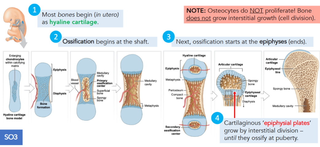
Compare and contrast the different mechanisms of growth that occur in bones, including how these processes alter following puberty.
Appositional bone growth is important in increasing the diameters of existing bones but does NOT form the original bones
Endochondral ossification, cartilages gradually replaced by bone 6 weeks after fertilization
As cartilage enlarges, chondrocytes near center of shaft increase greatly in size
Blood vessels grow around edges of cartilage and cells of the inner layer of the perichondrium differentiate into osteoblasts
Blood vessels penetrate cartilage and invade central region producing spongy bone at primary ossification center
Remodeling occurs as growth continues creating medullary cavity
Capillaries and osteoblasts migrate into epiphyses, creating secondary ossification centers; interstitial growth takes place at each metaphysis
At each metaphysis, epiphyseal cartilage separates the epiphysis from diaphysis
Chondrocytes within epiphyseal cartilage are arranged into epiphyseal side & diaphyseal side
Epiphyseal: chondrocytes continually add new cartilage
Diaphyseal: chondrocytes are degenerating
At puberty, combination of rising levels of sex hormones, growth hormone, and thyroid hormones stimulates bone growth dramatically
Intramembranous ossification begins when mesenchymal cells within embryonic or fibrous connective tissue differentiate into osteoblasts; normally occurs in deeper layers of dermis as dermal bones
Mesenchymal cells first cluster together, differentiate into osteoblasts and start to secrete the organic components of the matrix (ossification center)
Developing bone grows outward from ossification centers in spicules, small struts
Blood vessels begin to grow into area, as spicules meet and fuse together
Continued deposition of bone by osteoblasts located close to blood vessels result in plate of spongy bone with blood vessels weaving throughout
As rate of growth slows, the connective tissues around the bone organizes into fibrous layer of periosteum
Areas of spongy bone are remodeled forming diploe layer between two layers of compact bone
Intramembranous ossification starts approx 8 week of embryonic development
Describe the phases involved in bone repair and compare and contrast the capacity for repair in bone and cartilage.
Steps in fracture repair:
Immediately after fracture, extensive bleeding occurs, over a period of several hours, a large blood clot, or fracture hematoma soon closes off injured vessels and leaves a fibrous meshwork in damaged area
Next cells of the intact endosteum and periosteum undergo rapid cell division and the daughter cells migrate into fracture zone
An internal callus forms as a network of spongy bone unites the inner edges of the fracture; an external callus of cartilage and bone encircles and stabilizes the outer edge of the fracture
As the repair continues, osteoblasts replace the central cartilage of the external callus with spongy bone, which then unites the broken ends
Bone fragments and areas of dead bone closest to the break are removed and replaced, ends of the fracture now held firmly in place and can withstand normal stresses from muscle contraction
A swelling initially marks the location of the fracture; over time, this region will be remodeled by osteoblasts and osteoclasts and little evidence of the fracture will remain
Damage to bones constitutes a fracture, most fractures heal even after severe damage provided that the blood supply and cellular components of the endosteum and periosteum survive
Cartilage does not repair itself well as interstitial growth no longer occurs after puberty
Repairs through appositional growth can occur into adulthood for fibrous and elastic cartilage
Though dense connective tissue proper is not a good substitute for elastic cartilage
Articular cartilage usually cannot go undergo self repair at all as it lacks a perichondrium layer

Explain the pathological processes occurring in osteoarthritis and analyze the potential costs and benefits of treating osteoarthritis with total joint replacement.
Osteoarthritis, is a degenerative disorder caused by damage to articular cartilage
Damage to articular cartilage increases friction at the synovial joint, leading to inflammation, more pressure on the remaining cartilage, and more damage
If severely damaged, only current treatment is artificial joint replacement
Benefits are: less overall pain in the joint and thus less pressure on joint from rest of body
Downside/costs: expensive procedure/implant, temporary as it eventually needs to be replaced again, does not fully provide the original functions of a normal knee joint (ie: can’t run/stress the knee joint too hard)

Explain the role of bone as a mineral reservoir, and predict how the activity of bone cells will respond to changes in body calcium levels.
As the calcium reserve, the skeleton plays the primary role in blood calcium homeostasis
When large numbers of calcium ions are released into blood, the bones become weaker, when calcium salts are deposited, bones become denser and stronger
When blood calcium ion concentration falls below normal, cells of parathyroid glands embedded in thyroid gland of neck release parathyroid hormone (PTH) into bloodstream which has multiple effects that increase blood calcium level
Osteoclasts do not have PTH receptors, the PTH does bind to adjacent osteoblasts which causes them to release a hormone that stimulates immature osteoclasts to differentiate into mature osteoclasts; eroding the bone matrix thereby released stored calcium ions
PTH enhances the calcium absorbing effects of calcitriol on the intestines, as a result the rate of intestinal calcium absorption increases
PTH increases renal production of the hormone calcitriol, under normal circumstances a low level of calcitriol is always present because it is secreted continuously by the kidneys; stimulates calcium reabsorption by the kidneys and calcium absorption in the intestines
When blood calcium ion concentration is above normal, C cells of thyroid gland secrete calcitonin, whose effects are restricted to osteoclasts
Calcitonin decreases osteoclast activity but does not affect osteoblasts which continue to deposit calcium ions within bone matrix
Decreasing PTH or calcitriol level results in a decrease in the rate of calcium absorption by intestines
Increased calcitonin level has an inhibitory effect on kidneys and suppresses calcium ion reabsorption
Define the properties of muscle tissue, including key features shared between its subtypes.
Muscle tissue is able to contract and produce mechanical force
Movement is produced by muscle tissue, which is specialized for contraction
Striated muscle
Skeletal muscle tissue - moves the skeleton
Cardiac muscle tissue - pumps blood through the heart
Smooth muscle
Smooth muscle tissue - blood vessels, digestive tract, and many more places

Describe the organization of a skeletal muscle and the tissue types that it contains, including the relationship of muscle fibres to fascicles, and muscles to fascial compartments.
Skeletal muscles are often organized within layers of fasciae with each individual muscle as an organ
Epimysium, a dense layer of collagen fibers surround the entire muscle
Epimysium separates the muscle from surrounding tissues and organs and is connected to the deep fascia, a dense connective tissue layer
Muscle fascicle is a bundle (or group) of muscle fibers, the fibrous perimysium layer separates the fascicles from each other
Perimysium, is a fibrous layer that divides the skeletal muscle into a series of compartments
Contains collagen and elastic fibers as well as the blood vessels and nerves that supply the muscle fibers within the fascicles
Individual skeletal muscle fibers are sheathed in a delicate endomysium that contains capillaries, myosatellite cells, and axons of the neurons that control the muscle fibers
Endomysium, a thin layer of areolar connective tissue that surrounds each myofiber (muscle fiber), it loosely interconnects nearby muscle fibers, each muscle fiber contains bundles of protein filaments called myofibrils
Tendon, are narrow/rounded and attach the muscle to a specific point on a bone
Where they contact the bone, collagen fibers extend into bone matrix providing a firm attachment, as a result any contraction of muscle pulls on the attached bone or bones
Aponeurosis, are a broad/flat sheet formed from where the ends of a skeletal muscle, collagen fibers of the connective tissue layers merge
Aponeurosis provides attachment over a broad area that may involve more than one bone

Identify the features of a myofibre that make it unusual compared to the features of most cells in the body, and explain how they arise in development and are maintained in adulthood.
The individual cells within skeletal muscles are myofibers, they are produced by fusion of many myoblasts during development, and can be repaired in adulthood by myosatellite cells
Each myofiber is a single cell that contains many nuclei and stretches from muscle origin to insertion (tendon to tendon)
Myosatellite cells are stem cells that function in the repair of damaged muscle tissue

Identify the specialized parts of myofibres (including the neuromuscular junction and the three key organelles in a skeletal muscle triad) by structure and function.
Myofibrils are assemblies of special protein filaments that allow muscle to contract
Sarcolemma (cell membrane)
Sarcoplasm (cytoplasm of myofibril)
Sarcoplasmic reticulum (SR; endoplasmic reticulum of myofibril) - stores & releases calcium
Transverse tubules (T tubules) are narrow tubes that are continuous with the sarcolemma and extend into sarcoplasm at right angles to the cell surface
On either side of T tubule, the tubules of SR enlarge, fuse and form terminal cisternae expanded chambers
Combination of a pair of terminal cisternae + a T tubule is known as a triad
A single myofibre has only one neuromuscular junction (NMJ)
NMJ is made up of axon terminal, synaptic cleft and motor end plate
A single motor neuron can make NMJs with 1-1000 myofibers
This combo of motor neuron and muscle cells forms a motor unit

Explain the difference between excitation and contraction in a skeletal muscle cell.
Contraction = myofibers/myofibrils becoming shorter or producing tension
Excitation = sarcolemma generating electrical signal that precedes contraction
Occurs at NMJ with electrochemical signal
Excitation-contraction coupling refers to mechanism by which excitation triggers the contraction of a muscle fiber

Define the following terms: graded potential, action potential, and, resting membrane potential.
Action potential is an electrical event that involves one location on the excitable membrane, once it develops in one location it spreads along the surface of an axon toward the axon terminals
Graded potential, localized change which decreases with distance away from the stimulus in the resting membrane; last for tens of milliseconds and have different polarity (direction) and size
Amplitude can vary depending on how many ion channels are open
Resting membrane potential all neural activities begin with a change in resting membrane potential
Arises from unequal permeability of ions due to different leak channel abundance
Define the following terms: hyperpolarization, depolarization, and, repolarization.
Hyperpolarization, shift in membrane potential past resting levels, resulting in an increasingly negative membrane potential
Depolarization, any shift from resting membrane potential toward a more positive value
Applies to changes in potential from -70 mV to a smaller negative values (0 mV) as well as to membrane potentials above 0 mV
Repolarization, when the chemical stimulus is removed, membrane returns to its normal resting membrane potential as excess sodium ions are transported out of cytosol
Define the following terms: ligand-gated, voltage-gated, and, mechanically-gated.
All of these gated channels open and close depending on gating factors and ion selectivity:
Chemically-gated or Ligand-gated ion channels open when they bind specific chemicals
Receptors that bind ACh at the NMJ are chemically gated channels
Are the most abundant of the dendrites and cell body of a neuron
Voltage-gated ion channels open or close in response to changes in membrane potential
Sodium channels have two gates that function independently:
Activation gate that opens on stimulation, letting sodium ions into cell
Inactivation gate that closes to stop the entry of sodium ions
Mechanically-gated ion channels, open in response to mechanical stimuli on neuron’s membrane surface
Such channels are important in sensory receptors that respond to stretch, pressure, or vibration and cause the sense of touch and hearing
Explain at least one reason why ion channels have a primary role in creating membrane potential.
Ion channels are diverse and are selectively permeable, categorized by their gating and their ion selectivity through charge separation until equilibrium and the role of the active transport via sodium-potassium pump.
Explain the difference between a gated and non-gated (leak) ion channel, identify which of these classes of channel are involved in establishing the resting membrane potential, and give examples of where channels with different types of gating could be found in skeletal myofibres.
Leak channels are always open
Gated channels open and close depending on gating factors and ion selectivity
RMP is established and maintained primarily by ungated leak channels and by active transport in the sodium-potassium pump (Na⁺/K⁺-ATPase)
Chemically-gated or Ligand-gated ion channels open when they bind specific chemicals
Ex: motor neurons release a neurotransmitter, which causes opening of ion channels which allow sodium into the myofibre
Voltage-gated ion channels open or close in response to changes in membrane potential
Ex: excitation propagates through the myofibre from NMJ due to voltage gated ion channels
Mechanically-gated ion channels, open in response to mechanical stimuli on neuron’s membrane surface
Ex: excitation-contraction coupling of receptors during muscle contraction
Explain the key steps that occur across a neuromuscular junction and within a myofibre during excitation and predict the effects of specific drugs on excitation-contraction coupling of skeletal muscle fibres.
Electrical signal (arriving action potential) reaches myofibre at rest
Chemical signal releases neurotransmitter
Neurotransmitter binding and NMJ excitation
Ex: ACh-receptor membrane channel
Spread of excitation; enzymatic removal of neurotransmitter
Ex: AChE removes ACh
Curare, drug that prevents ACh binding to ACh receptors; lower excitation at NMJ
Novichok, blocks enzyme that breaks down ACh in the synaptic cleft; higher excitation at NMJ
Describe the arrangement of thin filaments in a sarcomere in terms of A & I bands, and Z & M lines, and in terms of the key biochemical features (e.g. protein composition, binding properties) of thin filaments.
At either end of the sarcomere, thin filaments are attached to the Z line
Actinin, protein interconnects the thin filaments at the Z line; composed of 4 main proteins:
F-actin is a twisted strand composed of two rows of 300-400 individual molecules of G-actin (globular actin)
Each G-actin molecule contains an active site to which myosin can bind
Nebulin is a large protein that extends along F-actin strand in the cleft between rows of G-actin molecules, holds the F-actin together
Tropomyosin cover the active sites on G-actin and prevent actin-myosin interaction; tropomyosin molecule is a double-stranded protein that is bound to one molecule of troponin midway along length
Troponin molecule consists of three globular subunits:
One binds to tropomyosin locking them together troponin-tropomyosin complex
Two binds to one G-actin, holding troponin-tropomyosin complex in position
Three has a receptor that binds two calcium ions

Describe the arrangement of thick filaments in a sarcomere in terms of A & I bands, and Z & M lines, and in terms of the key biochemical features (e.g. protein composition, binding properties) of thick filaments.
Thick filaments are composed of myosin protein
All myosin molecules are arranged with their tails pointing towards M line
All myosin heads are arranged in a spiral, each facing one of the surrounding thin filaments
Thick filaments each have a core of an elastic protein called titin, it also extends from each end of the thick filament across the I band to the Z line on that side
Titin is described as a molecular spring as it recoils after stretching
Each myosin tail is bound to other myosin molecules in the thick filament
Connection between head and tail acts as a hinge that lets the head pivot at its base
Free head has two globular protein subunits, during contraction, myosin heads interact with thin filaments

Explain the steps of the contraction cycle in terms of protein and small molecule interactions, including the specific role played by calcium, the interaction between actin and myosin, and the specific role for ATP.
Resting Sarcomere, each myosin head is already “energized” with energy that will be used to power a contraction
Each myosin head points away from the M line, cocked like a spring in a mousetrap, cocking requires energy obtained by breaking down ATP
In this process, myosin head function as ATPase, an enzyme that breaks down ATP
At start of contraction, the breakdown products, ADP and phosphate remain bound to myosin head
Contraction cycle is a series of interrelated steps that begins with the arrival of calcium ions within the zones of overlap in a sarcomere
Calcium ions bind to troponin, weaken the bond between actin and troponin-tropomyosin complex
Troponin molecule then changes position, rolling the tropomyosin molecule away from the active sites on actin and allowing interaction with energized myosin heads
Once active sites are exposed, the energized myosin heads bind to active sites forming cross-bridges
After the cross-bridges form, the energy that was stored in the resting state is released as myosin heads pivot toward the M line
Action called the power stroke, when it occurs the bound ADP and phosphate groups are released; filaments slide against one another
When another ATP binds to the myosin head, link between the myosin head and active site on the actin molecule is broken; active site is now exposed and able to form another bridge
Myosin reactivates when the free myosin head splits ATP into ADP and P; energy released is used to recock the myosin head
*Myosin can change the orientation of myosin head and neck
Entire cycle repeats several times each second, as long as Ca2+ concentration remains elevated and ATP reserves are sufficient
ATP hydrolysis by free myosin heads resets the neck
ATP is required for more than just contraction cycle - also required for myofiber excitation
Explain how the sliding filament model produces muscle shortening and/or tension production.
Interaction of sliding filaments causes the sarcomere to shorten, creating tension in the tendons
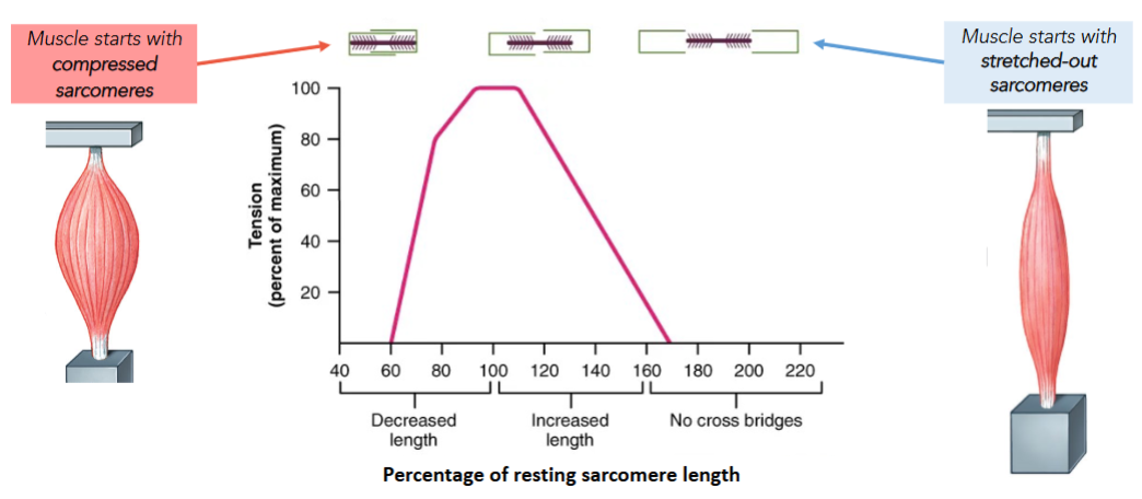
Tension can only be produced by myosin heads that overlap with (thus can bind) actin
Tension can only be when active sites are available, tension depends on Ca2+ levels
Sliding Filament Theory
When a skeletal muscle fiber contracts, thin filaments (actin) slide past the thick filaments (myosin) in a repeating cycle
H bands and I bands get smaller
Zones of overlap get larger
Z lines move closer together
Width of the A band remains constant
Analyze how the sliding filament model of sarcomere predicts different amounts of muscle tension depending on muscle length.
Muscles produce maximum tension when they are excited while at an intermediate sarcomere length
Explain how physiological mechanisms controlling calcium relate to tension production by skeletal muscle during a single muscle twitch, and why overlapping twitch stimuli can summate to produce tetanic contraction.
In a single muscle twitch contraction there are three phases:
Latent period (muscle excitation)
Contraction phase (Ca2+ build up)
Relaxation phase (Ca2+ removal)
Repeatedly stimulating a single muscle fibre in an overlapping twitch can cause summation of tension due to residual Ca2+ build-up
Summation occurs because more Ca2+ enters the cytosol before all the original Ca2+ is removed, revealing more active sites
A muscle producing persistent maximum tension (with little to no relaxation phase) is in tetanus (which occurs when the stimulation (excitation) frequency of the myofibres reaches a maximum)
Explain how muscle shortening can occur at different speeds or reverse depending on the load on the muscle.
Concentric (shortening) contractions occur when the external load is less than internally generated tension
Concentric contractions produce movement of a joint
An external load on a muscle will slow the rate of contraction (shortening) during activation; heavier loads = slower contraction rates
Isometric contractions occur when the muscle produces just enough tension to balance an external load
Isometric contractions are used to stabilize a joint
When the degree of muscle activation (internal tension produced) exactly matches the external force (the weight), there is contraction (tension production) without shortening
Eccentric (lengthening) contractions occur when the external load is higher than the internally generated tension
Eccentric contractions are used to slow down movement at a joint
Eccentric contraction can be used like a ‘brake’, slowing down movement at a joint produced by external forces; although muscle is activated gets longer, not shorter
Describe how tension generation by a muscle can be affected by physiological differences between the types of myofibres that make up a muscle.
Different muscles – and different individuals – exhibit different ‘optima’ in the power vs fatigue trade-off
Ex: a world-class powerlifter after a few reps vs a world-class marathon runner
Powerlifter would have more developed type II (fast twitch) fibers
High forces and rapid contractions but fatigue easily and recover slowly
Runner with more developed type I (slow twitch) fibers
Fatigue resistant and fast to recover
Compare and contrast the different mechanisms skeletal muscles use to generate ATP at rest, at moderate activity and at peak activity, including nutrients used to generate ATP, aerobic vs anaerobic pathways, and the production of by-products.
Glycolysis (Anaerobic metabolism), breakdown of glucose to pyruvate in the cytosol of a cell
Does not require oxygen
Is fast
Only 2 ATP molecules from a single glucose molecule
Cannot use fatty acids
Aerobic metabolism, provides 95% of ATP demands of a resting cell from mitochondria
Needs steady supply of oxygen
Is slow
>30 ATP molecules from a single glucose molecule
Can also use fatty acids
Muscle at Rest
In resting skeletal muscle, demand for ATP is low; more than enough oxygen is available for mitochondria to meet demand; aerobic respiration of free fatty acids (FFAs)
Produce surplus ATP to build up reserves of creatine phosphate (CP) & glycogen
Resting muscle fibers absorb fatty acids and glucose delivered by bloodstream
Fatty acids broken down in mitochondria, generating ATP used to convert creatine to creatine phosphate and glucose to glycogen
Moderate Activity
Demand for ATP increases, met by mitochondria and rate of oxygen consumption increases as a result; aerobic respiration of free fatty acids (FFAs) and glucose
Skeletal muscle now relies primarily on aerobic metabolism of pyruvate to generate ATP, provided by glycolysis using glucose obtained from stored glycogen reserves
As long as mitochondrial activity can meet demand for ATP, muscle will not become fatigued until glycogen, lipid, and amino acid reserves are exhausted (fatigue ex: endurance athlete/marathon runner)
Peak Activity
ATP demands are enormous & mitochondrial ATP production plateaus at maximum rate determined by availability of oxygen; aerobic + anaerobic respiration of glucose & CP
Mitochondria provides only about ⅓ of ATP needed, glycolysis produces the rest
Leads to build-up of lactate (from pyruvate buildup) and H+
During aerobic metabolism, H+ are absorbed by mitochondria
Anaerobic metabolism (glycolysis), H+ are not being absorbed fast enough; both H+ and lactate build up resulting increased lactate blood level and metabolic acidosis -> lactic acidosis; changes in pH will alter functional characteristics of key enzymes so that muscle fiber can no longer contract (fatigue ex: sprinters)

Define the term ‘muscle fatigue’ and identify at least two changes in physiological processes that contribute to the onset of fatigue.
Muscle fatigue: Reduced contractile tension for the same (excitation) stimulus
Fatigue factors that affect excitation processes in skeletal muscle:
Depletion of ACh vesicles in motor neuron (MN) axon terminal
Accumulation of K+ in T-tubules extracellular fluid (ECF) due to repeated action potentials (AP)
Fatigue factors that affect contraction processes in skeletal muscle:
Leakage of Ca2+ back into the sarcoplasm
Microtears in myofibrils
Build-up of lactate and H+ -> lactic acidosis
Identify the key anatomical and physiological characteristics that distinguish Type I and Type IIA & IIB muscle fibres and analyze how these physiological differences would contribute to performance differences and training effects in an individual.
Type II / Fast twitch fibers
Muscles dominated by fast fibers produce more powerful contractions
Fast fibers rapidly fatigue because their contractions use ATP in massive amounts
Have large diameters and contain:
Densely packed myofibrils
Tension produced by a muscle fiber is proportional to # of myofibrils
Large glycogen reserves
Prolonged activity supported by anaerobic metabolism (glycolysis)
Relatively few mitochondria
Don’t generate ATP well
Few capillaries per fiber
Less blood = fast fiber muscles appear white
Intermediate fibers
Type IIB “fast glycolytic”
Type IIA “fast-oxidative”
Have intermediate properties between Type IIB and Type I:
Fast contraction speeds
Intermediate twitch durations
Intermediate size (and power)
Intermediate mitochondria and capillary density; muscles appear pink
Intermediate resistance/recovery from fatigue
Ex: swimmer for type IIA and sprinter for type IIB
Fiber Conversion
Ratio of fast vs slow fibers is genetically determined
Ratio of intermediate fibers to fast fibers can increase as a result of athletic training
Aerobic training leads to hypertrophy of Type I fibers
Anaerobic training leads to hypertrophy of Type IIB fibers
Fast fibers can develop appearance and functional capabilities of intermediate fibers
Type I / Slow fibers (slow-oxidative fibers)
Continue contracting for extended periods, long after fast fibers become fatigued
Fatigue resistant and fast to recover
Half the diameter of fast fibers and contain:
Fewer myofibrils
More mitochondria per fiber
High oxygen from capillaries provides more mitochondrial energy
Surrounded by a more extensive network of capillaries
Higher oxygen supply & more blood = slow fiber muscles appear dark red
Describe the patterns by which multiple motor units within the same muscle are used during synchronous and asynchronous recruitment and discuss the trade-offs associated with different patterns of motor unit recruitment (asynchronous, size principle).
Motor unit, a motor neuron and all the muscle fibers that is controls; size indicates how fine or precise a movement can be
Can make neuromuscular junctions (NMJ) with anywhere between 1 to 1000 muscle fibers
Ex: in muscles of an eye - motor neuron may control 4-6 muscle fibers vs leg muscles where single motor neuron controls 1000-2000 muscle fibers
Muscle fibers of each motor unit are intermingled with those of other motor units
Recruitment, smooth but steady increase in muscular tension produced by increasing the number of active motor units (constitutes synchronous motor unit recruitment)
All fibers in a motor unit are of the same fiber type and fiber type correlates with number of fibers per unit
Size Principle
Only the smallest motor units will be recruited by weak stimuli in the spinal cord
While strong stimuli recruit both small, medium and large motor units
During a sustained contraction, motor unit recruitment is asynchronous so no unit is constantly activated
Similarly-sized motor units turn on and off so that each is active part of the time, and rests the other part
Explain the difference between hypertrophy and hyperplasia in terms of muscle tissue growth and identify which mechanisms occur during adulthood.
Hypertrophy, is enlargement of stimulated muscle
Number of muscle fibers does not change significantly, but the muscle as a whole enlarges because each muscle fiber increases in diameter and becomes strong because tension production is proportional to the cross-sectional area of the muscle
As a result of repeated, exhaustive stimulation, muscle fibers develop:
More mitochondria
A higher concentration of glycolytic enzymes
Larger glycogen reserves
Some muscle fibers have more myofibrils than do fibers that are less used and each myofibril contains more thick and thin filaments
In adult humans, muscles increase in size through hypertrophy not through hyperplasia
After embryonic development, skeletal muscles can repair muscle fibres, but cannot add new ones
Physical training increases fiber diameter, not fiber number
Explain or apply different anatomical or functional rules for how to divide up the nervous system into components (e.g. CNS/PNS/ENS; Sensory/Integrative/Motor; Somatic/Autonomic)
Central Nervous System (CNS)
Found within the vertebrae (spinal cord) and cranial bones (brain)
Peripheral Nervous System (PNS)
Located outside skull and spine but directly connected to CNS
Enteric Nervous System (ENS)
Neural cells within digestive system, indirectly connect to CNS through PNS
Information flow within a neuron is unidirectional, from dendrite to axon to axon terminal
Sensory neuron (Receptors)
Pass information from outside into the NS
Interneuron (Integrators)
Pass information between other neurons
Motor neuron (Effectors)
Pass information to non-neuronal cells
Neurons (and groups of neurons) can be functionally classified based on the organ systems they connect to:
Somatic Nervous System
Motor neurons innervating skeletal muscle
Sensory neurons innervating integument and skeletomuscular system
*Somatic isn’t the same as voluntary
Visceral/Autonomic Nervous System
Motor (effector) neurons innervating tissues other than skeletal muscle
Sensory neurons innervating visceral organs
*Autonomic is not under voluntary control
Explain the functions and key structural features of the four functional types of glia, and correctly identify which subtypes of glia are found in the CNS vs the PNS, and which are associated with white vs gray matter.
Four types of glial cells:
Ependymal cells (CNS)
Lines a series of cavities called ventricles and secrete cerebrospinal fluid (CSF); ependymal cells assist in producing, monitoring and circulating CSF in brain and spinal cord
Microglia (CNS)
White blood cells which perform immune functions for the CNS and help modify connections between neurons; make up gray matter
Myelinating cells (CNS & PNS)
Provide electrical insulation
Myelinating glia wrap cellular processes containing myelin (fats + proteins) around the axons of neurons
Two types of myelinating cells:
Schwann cells (PNS): Helps promote PNS axon regrowth
Oligodendrocytes (CNS): Produce myelin sheaths separated by nodes on axons
Myelinated axons are white matter of CNS; Unmyelinated axons are gray matter of CNS
Astrocytes (CNS)
Regulate extracellular environment around neurons, and maintain and modify synaptic connections; make up gray matter
Buffer ion concentrations in ECF
Recycle/buffer neurotransmitters
Regulate local blood flow (oxygen and nutrients)
Regulate CSF flow
Satellite cells (PNS)
Do all the same things as astrocytes except in PNS
Functions of neuroglia:
Maintain physical structure of nervous tissue
Repair nervous tissue framework after injury
Perform phagocytosis (cells engulfing large particles with cell membrane)
Provide nutrients to neurons
Regulate the composition of the interstitial fluid surrounding neurons
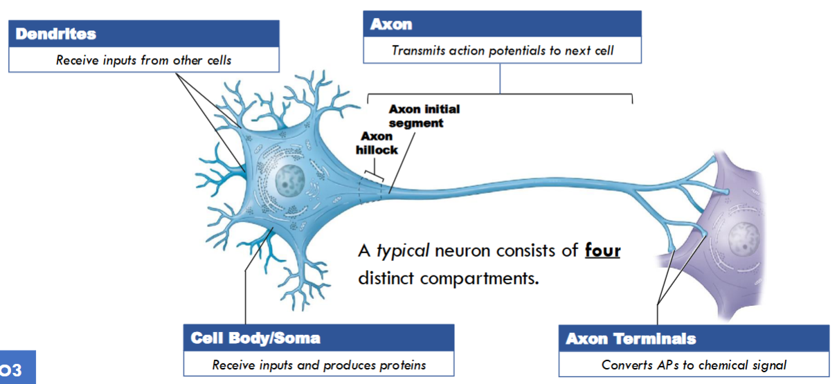
Identify the four compartments of a neuronal cell, explain the basic functions of each compartment in terms of information flow, and identify which neuronal compartments are associated with white vs gray matter.
Dendrites
Receive inputs from other cells
Associated with gray matter
Axon
Transmits action potentials (AP) to next cell
If myelinated, belongs to white matter
Cell Body (Soma)
Receives inputs and produces proteins
Axon Terminals
Converts APs to chemical signal
Associated with gray matter
Compare and contrast between involuntary neural responses (including reflexes) and voluntary responses.
Reflex is a rapid, automatic response triggered by specific stimuli
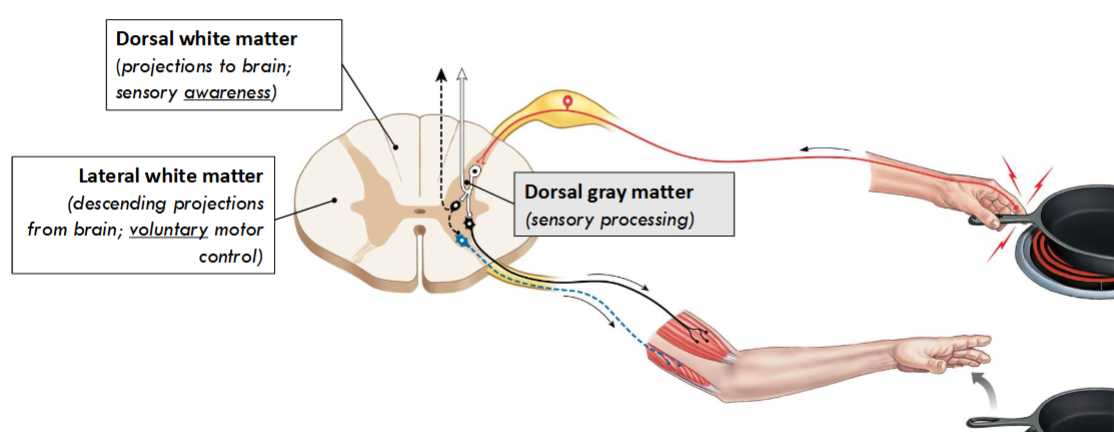
Spinal reflexes are controlled in the spinal cord, functioning without any input from brain; grey matter is important for this like a withdrawal reflex
Withdrawal Reflex, when you touch something hot it is involuntary your spinal cord will move your arm away but you may voluntarily say something in response
Stretch/Jerk Reflex, involuntary stimulation of a motor neuron in the knee or elbow vs voluntary as to why you would do that
Use information about the organization of nerves, white and gray matter to make simple predictions about neurological symptoms arising from damage to peripheral nerves or different locations in the spinal cord (e.g. sensory/motor, voluntary/involuntary).
Circuits in spinal cord grey matter are necessary and sufficient for simple (spinal) reflexes, such as the withdrawal reflex
Dorsal gray matter (sensory processing)
Ventral gray matter (motor neuron somata)
Ex: touching a hot pan, dorsal gray matter detects pain and ventral gray matter lifts hand off
Spinal white matter transmits neural signals between the spinal cord and the brain
Dorsal white matter (projections to brain; sensory awareness)
Lateral white matter (descending projections from brain; voluntary motor control)
Ex: dorsal white matter would help with determining that a pan is hot, lateral white matter helps with voluntary control of hand/arm (to hopefully avoid the hot pan)
Compare and contrast the capacity of neurons in the CNS and PNS for repair, and the roles glial cells play in neuronal repair.
If a neuron dies, it will not be replaced
All neurons are post-mitotic meaning they cannot return to the cell cycle
In PNS if an axon is damaged, it can repair and regrow with help of Schwann cells
In CNS if an axon is damaged, it typically cannot repair or regrow
Instead Astrocytes inhibit axon sprouting
Without active axon terminals, the neuron dies through apoptosis
Many parts of the CNS are able to reorganize their synapses and make new local connections between their remaining neurons
Describe how diffusive forces and electrical forces on individual ions create an electrochemical gradient, and explain how this relates to an ion’s equilibrium potential.
If K+ was the only permeable ion diffusion of K+ out of the cell creates a net negative charge inside the cell, pushing some K+ back inward
If K+ was the only permeable ion, net K+ movement would cease when its two opposing forces are balanced (equal and opposite)
Equilibrium potential, an ion’s electrical and chemical gradients are in balance, and there is no net movement of ions across the membrane
Net movement in and out of cell are the same, no net force on that ion
Indicates an ion’s contribution to the resting membrane potential
Explain how a neuron’s transmembrane potential arises from ions with different equilibrium potentials and different relative permeability, and make simple predictions about how varying ion permeability can lead to changes in membrane potential using equilibrium potentials.
All potentials (even the resting membrane potential (RMP)) are created by the movement of ions through diverse ion channels
RMP arises from the unequal permeability of ions due to different leak channel abundance
Unequal permeability interacts with the unequal distribution of ions across the membrane
Transmembrane potential (Vm), because of selective permeability to different types of ions, all cells have a transmembrane potential; which is negative at rest
When a neuron is at rest, K+ is the most permeable ion, but not the only permeable ion
Each ion’s contribution to Vm is directly proportional to its permeability

Explain at least one reason why ion channels have a primary role in creating membrane potential, while ion pumps contribute indirectly through maintaining concentration gradients.
The transmembrane potential is maintained by ion pumps (especially the Na/K-ATPase)
Ion concentration differences are critical for the membrane potential; these differences are maintained by active transport
Ion pumps work over minutes to hours to keep ionic gradients stable
They only make a small direct effect on RMP
Ex: Na+ and K+ gradients: maintained by primary active transport (Na-K-ATPase pump)
Cl- gradient: maintained by secondary active transport (co-transport with K+)
Explain the difference between a gated and non-gated (leak) ion channel, identify which of these classes of channel are involved in establishing the resting membrane potential, and give examples of where channels with different types of gating could be found in neurons.
Recall that ion channels are diverse, they are categorized by their gating and by ion selectivity
RMP is created by leak channels, which are always open
Chemically-gated ion channels and mechanically-gated ion channels are found at the dendrites of the neuron
Voltage-gated ion channels are in the axon of the neuron

Identify at least two different factors that can alter the size of a graded potential, or predict the effect on graded potential amplitude or duration of defined manipulations of ion-channels or the ligands that affect them.
Graded potentials typically last for tens of milliseconds and can have different polarity (direction) and size
The amplitude of a graded potential can vary depending on the number of ion channels that open
Affected by:
Stimulus intensity
Receptor potential (mV)
Recall that transmembrane potential (Vm) is determined by permeability – more channels open, more ions flow to change the membrane potential!
Membrane potential changes spread away from the site where they are first induced, but get weaker the further they spread
Identify the four key features of action potentials (threshold, stereotypy, propagation without decrement and the refractory period) and explain how these differ in graded potentials.
All-or-none principle, given stimulus either triggers a typical action potential or triggers none at all
All-or-none (Threshold)
When the membrane potential reaches a threshold voltage (VTh) at the axon hillock, it triggers an action potential
In graded potentials, they take place in the cell body and dendrites; are analog, short-lived, and local changes in membrane potential
Stereotyped Dimensions
Action potentials are specialized membrane potential signals with a characteristic spiky shape (depicted on AP charts)
In graded potentials, amplitude can be added together temporarily over time or spatially from multiple sites
Propagation, describes the movement of an action potential down an axon
Action potentials can be rapidly transmitted long distances through an axon membrane without degrading
Action potentials propagate (spread long distances) by replicating themselves at each new section of the axon; in only one direction away from initial segment
In graded potential, they are strength varies with size of stimulus and decreases over distance
All-or-none (Refractory)
Action potentials always have a rapid sequence of depolarization and repolarization with refractory periods
Absolute refractory period, during this period, the membrane cannot respond to further stimulation; no second AP no matter how strong the stimulus
Relative refractory period, membrane can respond only to a larger than normal stimulus that can trigger a second AP
In graded potentials, can be depolarizing by EPSPs or hyperpolarizing by IPSPs
Explain how the different properties of voltage-gated sodium and voltage-gated potassium channels in an axon connect to the three different phases of an action potential, and to the action potential refractory period.
Voltage-gated sodium channels (VGNCs) open rapidly after a suprathreshold depolarization, then close through inactivation
VGNCs have two distinct gates:
Activation gate: opens by depolarization
Inactivation gate: open at rest, closes after a delay
Voltage-gated potassium channels open very slowly after a suprathreshold depolarization, and slowly close after the membrane repolarizes
Voltage-gated potassium channels have only one gate (activation gate)
The gate responds to transmembrane potential (Vm) crossing threshold, but only after a delay

Describe the anatomy of an axon terminal, and explain the role of voltage-gated calcium channels in neurotransmitter release.
Axon terminals are compartments that are specialized to release neurotransmitter from synaptic vesicles in response to an action potential
Neurotransmitter released into the synaptic cleft will bind to post-synaptic neurotransmitter receptor proteins
Voltage Gated Calcium Channels (VGCC) convert the AP depolarization into vesicular release of chemicals

Explain the term ‘post-synaptic potential’ (PSP), and define or correctly apply the terms inhibitory and excitatory within the context of PSPs in the nervous system.
Recall that graded potentials vary in many dimensions but they all degrade
Postsynaptic potentials are graded potentials that develop in the postsynaptic membrane in response to a neurotransmitter; two major types develop at neuron-to-neuron synapses
Amplitude of postsynaptic potential is affected by number of ion channels that open and thus pass current
Excitatory postsynaptic potentials (EPSP) is a graded depolarization caused by arrival of a neurotransmitter at the postsynaptic membrane that shifts the membrane potential closer to the threshold
This facilitates the membrane, the larger the degree, the smaller the additional stimulus needed to trigger an action potential
Inhibitory postsynaptic potentials (IPSP) is a graded hyperpolarization of the postsynaptic membrane
When membrane hyperpolarization occurs, the membrane is inhibited because a larger-than-usual depolarizing stimulus must be provided to bring the membrane potential to threshold
Define or correctly apply the terms agonist and antagonist within the context of PSPs in the nervous system.
Agonists, a drug that mimics the action of an endogenous neurotransmitter
Typically enhances cellular activity
Antagonists, drug that blocks the activation of a neurotransmitter receptor
Typically blocks cellular activity

Identify at least three different factors that can alter the size of a PSP, including neurotransmitter release, recycling, and receptor numbers, and predict the effects of drugs or toxins with a specific action on a process in a synapse.
Different sizes (Amplitude)
Directly proportional to the concentration of neurotransmitter molecules released into the synaptic cleft
The amplitude of a post-synaptic potential is affected by the number of ion channels that open and thus pass current
Larger potentials can be generated in two main ways:
More receptors available
Higher concentration of ligand available
Propagation
As PSPs are graded potentials that propagate along the dendrites and soma, strength varies with size of stimulus and decreases over distance
Recycling
Both synaptic vesicles and neurotransmitters are recycled by the axon terminal
Synaptic vesicles are reformed through endocytosis
Tetrodotoxin (Fugu - pufferfish): blocks VGNC activation; antagonist
Charybtoxin (Deathstalker scorpion): blocks VGKC activation; antagonist
Latrotoxin (black widow): inserts artificial Ca channels into the axon terminal; agonist
Botulinum toxin (Clostridium bacteria): destroys vesicle releasing machinery proteins; antagonist
Identify the features of ion channels that lead to a PSP being considered excitatory or inhibitory.
Recall that ion channels are diverse, they are categorized by their gating and by ion selectivity
Excitatory channels are specifically permeable to positive ions like Na+
EPSPs are produced when neurotransmitter binds to receptors that increase Na+ permeability (Eion = +70mV); this moves Vm toward AP threshold
Inhibitory channels are selectively permeable to negative ions like K+ and Cl-
IPSPs are produced when neurotransmitter binds to receptors that increase K+ permeability (Eion = -90mV) or Cl- permeability (Eion = -65mV); this keeps Vm below AP threshold
Explain what is meant by summation of synaptic potentials, or identify, describe or draw examples of temporal and spatial summation.
Summation, is integration of the effects of graded potentials on a segment of the plasma membrane
EPSPs and IPSPs result from activation of different types of chemically gated ion channels; if two different neurotransmitters arrive simultaneously and both channels open, net effect may be no change in membrane potential
Two forms of summation that can generate an action potential:
Temporal Summation occurs when a single synapse is active repeatedly
Ex: typical EPSP lasts about 20 msec but under maximum stimulation, action potential can reach axon terminal each millisecond; when a second EPSP arrives before effects of first EPSP have disappeared, another group of vesicles discharges ACh into synaptic cleft, more ACh molecules arrive at postsynaptic membrane and degree of depolarization increases
Spatial Summation involves multiple synapses that are active at the same time; effects on membrane potential are cumulative because each active synapse opens gated channels that allow ion movement into or out of the cell
Ex: active synapses generating EPSPs and opening gated channels that let sodium enter, effects are cumulative and degree of depolarization at initial segment depends on how many excitatory synapses are active at any given moment; and how far they are from the initial segment
Discuss how myelin can affect action potential conduction speed and the ATP usage of a neuron.
Myelin provides a form of electrical insulation, enhancing the spread of potentials by increasing conduction speed and producing saltatory propagation
Saltatory propagation is when an action potential appears at the initial segment of a myelinated axon, the local current skips the internode and depolarizes the closest node
Through this process, myelin decreases ATP usage by restricting ion exchange and ionic leakage to the uninsulated nodes of Ranvier
Conduction speed is the time it takes an action potential to travel a given distance along an axon (usually measured in m/s)
Analyze simple neural circuit diagrams (e.g. of spinal reflexes) in terms of information flow through a circuit, including correctly applying the terms excitatory, inhibitory, convergence, divergence and recurrence.
Reflex arc
Stimulation of a Receptor -> Activation of a Sensory Neuron -> Information Processing in CNS
-> Activation of a Motor Neuron -> Response of Peripheral Effector
Information processing begins when sensory neuron releases excitatory neurotransmitters at the postsynaptic membrane of interneuron; neurotransmitter produces an EPSP integrated with other stimuli arriving at postsynaptic cell
Excitatory postsynaptic potentials (EPSP) is a graded depolarization caused by arrival of a neurotransmitter at the postsynaptic membrane that shifts the membrane potential closer to the threshold
This facilitates the membrane, the larger the degree, the smaller the additional stimulus needed to trigger an action potential
Inhibitory postsynaptic potentials (IPSP) is a graded hyperpolarization of the postsynaptic membrane
When membrane hyperpolarization occurs, the membrane is inhibited because a larger-than-usual depolarizing stimulus must be provided to bring the membrane potential to threshold
Convergence, one post-synaptic neuron receives and summates many different inputs
Divergence, one pre-synaptic neuron shares its outputs with more than one neuron
Reverberation, collateral branches of axons somewhere along the circuit extend back toward the source of an impulse and further stimulate the presynaptic neurons
Is a positive feedback loop involving neurons
Describe the roles of the skeleton, meninges/CSF, and the BBB in the protection and maintenance of the CNS.
Skull and vertebral column, meninges, and cerebrospinal fluid protect the CNS
Meninges are protective tissue layers covering the brain and spinal cord
Cerebrospinal fluid (CSF) circulates from the ventricles into the subarachnoid space
CSF cushions impact between CNS and skull
The Blood Brain Barrier (BBB) restricts the transport of water and molecules between the blood and CNS
The blood brain barrier is a tight epithelium, which means only lipophilic molecules OR hydrophilic molecules with dedicated transport proteins can reach the CNS
Astrocytes in the BBB control blood flow to the local area in response to metabolic demands and neural activity

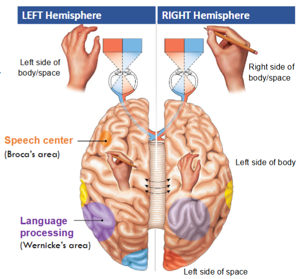
Define what is meant by a ‘primary cortical area’ for both motor and sensory processing, and explain key organizational principles of primary cortical areas, especially topographic mapping and crossed projections.
Primary cortical areas are connected to spinal neurons on the opposite side of the body
Lateralization of function arises because projections between spinal cord and brain decussate (cross the midline)
Most of the neuron cell bodies in the human brain are found within the 4-5 lobes of the cerebral cortex
Primary cortical areas are usually organized as topographical maps of the body or the environment
The brain is lateralized especially in terms of primary sensory and motor function
Primary cortices process information from the other side of the body/space
Some special tasks are processed only/primarily on one side of the cerebral cortex (e.g. language)

Use information about the organization & function of primary cortical areas and ascending and descending spinal pathways to make simple predictions about the relationships between the location of damage and the type of symptoms experienced.
Ex: damage to right side primary somatosensory cortex would affect the sensation of feeling (numbness) in the left hand/fingers based on info from topographical map of brain and lateralization.
Compare and contrast what is measured in an EEG and an fMRI, and how different activities, brain states, or disease states can affect these measurements.
A functional MRI (fMRI) scan estimates activity in different CNS locations by measuring the relative amount of local blood flow under different conditions
More blood flow = area ‘lights up’ on scan
Strokes occur when nervous tissue is damaged due to disruptions to blood supply to part of the brain; fMRI can map how the brain reorganizes after stroke, tracking motor and cognitive recovery, predicting long-term outcomes
Printed recording of the electrical activity of the brain is called an electroencephalogram (EEG); electrical patterns are called brain waves
EEG recordings can be used to separate different brain states, such as the stages of sleep
Alpha waves occur in the brains of healthy awake adults who are resting with their eyes closed
Alpha waves disappear during sleep but they also vanish when the individual begins to concentrate on some specific task
Beta waves are higher-frequency waves that appear in people who are either concentrating on a task, under stress or in a state of psychological tension
Theta waves may appear transiently during sleep in normal adults but are most often observed in children and in intensely frustrated adults
Presence of theta waves under other circumstances may indicate the presence of a brain disorder, such as a tumour
Delta waves are large amplitude low frequency waves, normally seen during deep sleep in people of all ages
Also seen in the brains of infants (whose brains are still developing) and in awake adults when a tumour, vascular blockage, or inflammation has damaged portions of the brain
Seizures (abnormal overactivity in neural circuits) of all kinds can be recorded and tracked by an EEG