Protein Structure
 Call Kai
 Learn
 Practice Test
 Spaced Repetition
 Match
 Flashcards
 Knowt Play
1/31

There's no tags or description
Looks like no tags are added yet.
32 Terms
Protein Structure Types
Fibrous
Membrane
Globular
Intrinsically Disordered
Need to know the general principals of each type of structure to understand health and disease related structure-function relationships

Fibrous Proteins
Function: Provide support, shape and structure
Structure:
Polypeptide chains arranged in long segments or sheets
Usually consists of a single type of secondary structure (alpha-helix/beta sheet)
Usually have a simple, repeating primary sequence pattern

Proline
Ring structure provides hindrance to the alpha-helical conformation
Disfavoured in both α-helices and β-sheets because it has no backbone HN to participate in hydrogen bonds
So, proline fits well in the first turn of an α-helix but anywhere else it usually produces a significant bend in the helix
Called a “helix-breaker”
Glycine
Also a “helix breaker”
Its high conformational flexibility makes it entropically expensive to adopt the relatively constrained α-helical or β-sheet structure

α-Keratin
Found in hair, wool, skin, hooves
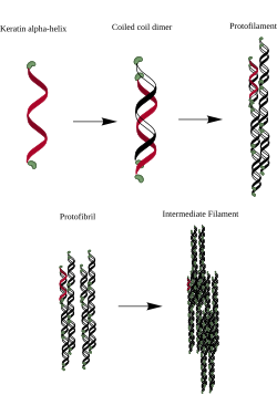
The two-chain coiled coils combine into higher order structures called protofilaments and protofibrils
The two helices in the coiled coil are held together by hydrophobic interactions between hydrophobic residues
Same pattern of 7 residues repeats: HPPHPPP (H=hydrophobic, P=polar)
Coiled coils found in many kinds of proteins
Epidermolysis bullosa simplex (EBS)
One α-keratin mutation that leads to a blistering skin condition
R211P
The notation means that an R (arginine), at amino acid 211 in the sequence, is mutated to a P (proline)
Proline causes kinks in α-helices - so disrupts the helical secondary structure of α-keratin found in skin
Disease-Related Mutations in Coiled-Coils of other Proteins
Common
e.g. familial hypertrophic cardiomyopathy
Coiled-coil of myosin
Fibroin
Consists of layers of antiparallel beta sheets
Parallel and anti-parallel
Hydrogen bonding occurs between beta-strands
Side chains alternate up and down

Silk Fibroin
Protein made by silk moth (Bombyx mori)
Primary structure → ~3000-4000 amino acids
45% glycine
30% alanine
12% serine
No cysteine
87% of the protein is composed of only 3 AA
Repeating unit → -(gly-ser-gly-ala-gly-ala)n-
Glycine and alanine have small side chains
Sheets stack with glycines facing each other, and ala/ser facing each other - stabilized by noncovalent interactions
How does having small glycine sidechains make silk fibroin strong?
Close packing of the β-sheets and an interlocking arrangement of R groups

β –Sheets, Amyloid β Peptide and AD Pathogenesis
Amyloid beta (Aβ) peptides are aggregation-prone peptides
These peptides self-assemble into β-sheet–rich fibrillar structures

Collagen
Main constituent of bones, tendons, ligaments, and blood vessels of higher organisms
Consists of a repeating tripeptide in which every third residue is a glycine (GXY)
X & Y are usually proline (although lysine occurs sometimes)
Lots of the prolines are post-translationally modified to be hydroxyproline
Secondary structure → polyproline helix:
Stabilized by repulsion between prolines/hydroxyprolines

Collagen building blocks:
Collagen requires the amino acids proline and lysine to be chemically modified through hydroxylation to become hydroxyproline and hydroxylysine
Collagen enzyme activation:
The enzymes prolyl hydroxylase and lysyl hydroxylase carry out these reactions
Vitamin C acts as a necessary cofactor and electron donor to keep iron (Fe²⁺) in its active ferrous state
Collagen and structural stability:
Without vitamin C, proline cannot be efficiently converted into hydroxyproline
Hydroxyproline is vital for forming and stabilizing the collagen triple helix
Membrane Proteins
Found in the plasma membrane that surrounds a cell
Found in the membranes of organelles
~30% of all genes in eukaryotes code for membrane proteins
~>> 30% of drug targets
Because membrane proteins exist (at least in part) in a hydrophobic lipid environment, the organization of membrane proteins follows different rules from those of soluble proteins which exist in a polar environment
The surface of the transmembrane regions of membrane proteins are composed primarily of amino acids with hydrophobic side-chains
Hydrophilic residues are primarily located in the segments that protrude from the hydrophobic bilayer interior

Functions of Membrane Proteins
Junctions → serve to connect and join two cells together
Enzymes → fixing to membranes localizes metabolic pathways
Transport → responsible for facilitated diffusion and active transport
Recognition → may function as markers for cellular identification
Anchorage → attachment points for cytoskeleton and extracellular matrix
Transduction → function as receptors for peptide hormones
Two Types of Membrane Proteins
Peripheral Membrane Protein
Integral Membrane Protein
Peripheral Membrane Proteins
Temporarily associated with the membrane
They can be easily removed, which allows them to be involved in cell signaling
Can be attached to integral membrane proteins, or they can stick into a small portion of the lipid bilayer by themselves
Often associated with ion channels and transmembrane receptors Most peripheral membrane proteins are hydrophilic.

Integral Membrane Proteins
Permanently embedded within the plasma membrane
Can be classified according to their relationship with the bilayer:
Integral monotopic proteins
Span only one leaflet of the bilayer
Transmembrane proteins
Span the entire plasma membrane

Integral Membrane Proteins: Transmembrane Proteins
Transmembrane proteins
Span the entire plasma membrane : Bitopic or Polytopic
Bitopic Integral Membrane Protein
cross the membrane once (single-pass)
Polytopic Integral Membrane Protein
cross the membrane more than once (multi-pass)
either an alpha-helical protein or a beta-sheet protein
Membrane Protein Organization
The thermodynamic cost of transferring charged or highly polar uncharged compoundsi nto the non-polar hydrocarbon interior of bilayer membranes is very high.
Two consequences:
Most of the amino acid sidechains of transmembrane segments must be non-polar
The very polar peptide bonds of transmembrane segments must participate in hydrogen bonds in order to lower the cost of transferring them into the hydrocarbon interior
This hydrogen bonding is most easily accomplished with α-helices, for which all peptide bonds are hydrogen-bonded internally
It can also be accomplished with beta-sheets provided that the β-strands form closed structures such as a β-barrel
Computational Studies: Predicting Membrane Protein Structure
Transmembrane helices in integral membrane proteins are composed of stretches of about 20 predominantly hydrophobic residues
Separated by polar connecting loops
Transmembrane helix predictions make use of a hydrophobicity scale
Hydrophobicity scales are composed of experimentally determined transfer free energies for each amino acid
There are several published scales that all vary from each other a little bit - some take into account just the sidechain, some backbone plus side chain, some measure transfer to an organic solvent, some to an actual bilayer
If there’s a non-hydrophobic sidechain in a transmembrane helix…it’s there for a reason:
Enzymatic Activity and Ion Transport
Gly and Ser residues often form hydrogen bonds that stabilize helix-helix interactions
In general, seeing a polar, hydrogen-bonding capable side chain in the middle of a transmembrane helix is a big clue that it’s doing something important
Globular Protein Organization
Globular proteins (water soluble) are arranged so that most of the hydrophobic residues (e.g. I,L,V, F, etc.) are on the inside, and most of the hydrophilic residues are on the outside
Mutating a hydrophobic residue from the core of the protein to a polar or charged residue usually completely disrupts the structure and function of the protein

How do we use sequence data (e.g. the sequences of several protein homologs) to identify the likely hydrophobic core amino acids (even if we don’t have a 3D structure)?
Align the sequences of the homologs and look for conserved hydrophobic residues
Hemoglobin and Sickle Cell Anemia
Glu6 → Val6 Mutation
The mutation creates a hydrophobic patch on the surface of the beta subunit that causes hemoglobin tetramers to polymerize into long fibrils
The mutation also confers resistance to malaria (which is why it has persisted) - sickling increases the permeability of the red blood cell to potassium ions, the concentration of potassium inside the cell decreases, this decreases kills the parasite
The long fibrils distort the shape of the red blood cell (into a “sickle” shape)
The distorted cells don’t fit through the capillaries and can block them and hence interrupt the delivery of oxygen to the tissues
Homology
Refers to biological features including genes and their products that are descended from a feature present in a common ancestor. Homologous features such as genes are referred to as homologs (or homologues)
Homologous genes become separated in evolution in two different ways:
Separation of two populations with the ancestral gene in two species → orthologs
Gene duplication of the ancestral gene within a lineage → paralogs
What’s the term for two related proteins from the same organism?
Paralogs
Arg 1 (Arginase 1):
A cytosolic enzyme predominantly expressed in the liver
Catalyzes the final step of the urea cycle—hydrolyzing L-arginine into L-ornithine and urea—to detoxify ammonia
Deficiency in Arg 1 leads to hyperargininemia and urea cycle impairment
Arg 2 (Arginase 2):
A mitochondrial enzyme found mainly in extrahepatic tissues (such as the kidneys)
Primarily involved in regulating L-arginine availability, nitric oxide (NO) metabolism, and the synthesis of polyamines rather than the core hepatic urea cycle
Why make a multiple sequence alignment?
Figure out which residues have been conserved over evolutionary time-spans and thus critical for structure and function
Infer evolutionary relationships [More a biology aspect than a human health and disease aspect]