Respiratory and lymphatic system
 Call Kai
 Learn
 Practice Test
 Spaced Repetition
 Match
 Flashcards
 Knowt Play
1/157

There's no tags or description
Looks like no tags are added yet.
158 Terms
what is the nasal vertibule
enterance to nose
pharynx is made up of 3 structures. what are they
nasopharynx
oropharynx
laryngopharynx
nasopharynx is the
most superficial
laryngopharynx is
inferior part of the pharynx
upper respiratory tract
• Nose
• Nasal vestibule
• Nasal cavity
• Nasopharynx
• Oropharynx
• Uvula
• Laryngopharynx
• Epiglottis
• Larynx
• Glottis
what are the 3 nonkeratinized stratified squamous epithelium structures of the upper respiratory tract
nasal vestibule
oropharynx
laryngopharynx
what are the 2 structures that are ciliated pseudostratifeid columnar epithelium with goblet cell
nasal cavity
nasopharynx
lower respiratory tract
• Trachea
• Carina
• Primary or principal bronchi
• Secondary or lobar bronchi
• Tertiary or segmental bronchi
• Bronchioles
• Terminal bronchiole
• Respiratory bronchiole
• Alveolar sac, alveolar duct (cuboidal epithelium), alveolus (simple squamous epithelium)
the alveolar duct what type of epithelium
cuboidal epithelium
alveolus epithelium
simple squamous epithelium
conducting zone
all parts until up to the respiratory bronchiole
respiratory zone
• Respiratory bronchiole
• Alveolar sac with alveolar ducts and alveoli
external structures anterior to larynx
• Hyoid bone
• Thyroid cartilage
• Cricoid cartilage
right lung
• Upper, middle, lower lobes
• Horizontal and oblique fissures
left lung
• Upper and lower lobes
• Oblique fissure
• Cardiac notch (impression)
pleural cavity
parietal pleura
visceral pleura
sinuses
• Frontal (2)
• Maxillary (2)
• Ethmoid (2)
• Sphenoid (1)
inspiration
• External intercostals
• Diaphragm (phrenic nerve, C3. 4, 5)
• Accessory muscles: sternocleidomastoid, scalenes
diaphragm innervated by
(phrenic nerve, C3. 4, 5)
accessory muscles of inspiration
sternocleidomastoid
scalenes
expiration
• Internal intercostals
• Diaphragm
• Accessory muscles: abdominal muscles
accessory muscles of expiration
abdominal muscles
Lymphatic organs
• Pharyngeal tonsils (adenoids)
• Palatine tonsils
• Lingual tonsils
• Thymus
• Spleen
• Appendix
• Lymph
• Lymph nodes
• Thoracic duct: drains lymph from the left body and most of the right
• Right lymphatic duct: drains the right face, right arm and right chest and upper abdom
thoracic duct
drains lymph from the left body and most of the right
right lymphatic duct
drains the right face, right arm and right chest and upper abdomen.
hard palate
top of mouth opening
soft palate
deeper extension from hard palate
uvula
extension of from the soft palate
epiglottis
lid of larynx and not part of larynx
glottis
part of larynx
consists of:
vocal cords and space between them
what does the glottis consist of
vocal cord and space between them
when inhaling air it goes through

if epiglottis doesn’t close when swallowing food
it could go down into windpipe (larynx→ trachea→ lungs)
processing of eating

certain structures anterior to larynx
hyoid bone
thyroid cartilage
cricoid cartilage
2 type of cells of epithelial cells in the respiratory system
1, non keratinized stratified squamous epithelium
ciliated pseudo stratified columnar epithelium with goblet cells
what structures in the respiratory are non keratinized stratified squamous epithelium
nasal vestibule
oropharynx
laryngopharynx
what structures in the upper respiratory tract are ciliated pseudo stratified columnar epithelium with goblet cells
nasal cavity
nasopharynx
carina
where 2 branches branch off
pathway air takes as it travels from the trachea into the lungs
trachea
primary bronchi
secondary bronchi
tertiary bronchi
bronchioles
terminal bronchioles
respiratory bronchioles
conducting zone:
trachea → terminal bronchioles (moves air)
respiratory zone
respiratory bronchioles → alveolar ducts → alveolar sacs (gas exchange)
alveolar ducts
tiny ducts that connect respiratory bronchioles to avelar sacs; cuboidal epithelium
what type of epithelium is the alveolar ducts
cuboidal epithelium
alveolar sacs
filled with spiral called alveoli
alveoli
functional unit of lung
alveoli epithelium
simple squamous
pulmonary blood flow pathway
RV
pulmonary trunk
pulmonary arteries
pulmonary arterioles
alveoli
pulmonary venules
pulmonary veins
left atrium
conducting zones general idea
-where is conducted when we are breathing
all parts up to respiratory bronchiole
respiratory zone
consists of:
-respiratory bronchiole
(alveoli )
what are the 2 layers of the lining of the lungs
visceral pleura
parietal pleura

what is #1
visceral pleura
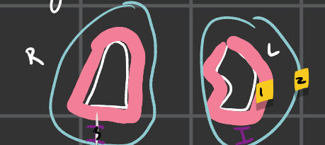
#2
parietal pleura

#3
pleural cavity
what is visceral pleura
layer superficial to lung
what is -parietal pleura
outer layer that connects to the thoracic wall itself along w/ diaphragm
what is the pleural cavity
space btw visceral and parietal pleura
sphenoid sinus
only sinus where there’s not a pair
what are the 4 sinuses
sphenoid sinus
ethmoid sinus (2)
frontal sinus (2)
maxillary sinus (2)
function of sinuses
produce mucus to moisture skull
lighten skull
2 types of breathing
inspiration
expiration
inspiration, what is it
taking a breath; upper chest breathing
what muscles are part of inspiration
diaphragm
external intercostal
accessory muscle
sternocleidomastoid + scalene
diaphragm gets innervated by
C345
external intercostal
in between ribs
expiration, what is it
passive breathing
what muscles involved in expiration breathing
diaphragm
internal costal
accessory muscle: abdominal muscles
lymphatic system
-vessels, cells, organs
function of lymphatic system
carry excess fluid to bloodstream
filter pathogens form blood
examples of lymphatic system
lymph nodes swell with infections
lymphatic vessels transport lymphocytes
lymph
-colorless fluid
-contains wbc
bathes tissue
drains through lymphatic system into bloodstream
lymph circulation
blood capillaries
lymphatic vessels
lymphatic ducts
blood stream
lymphatic vessels travel through
lymph nodes
right lymphatic duct
going to drain into the right subclavian veins
thoracic duct
drain into left subclavian vein
other lymph structures
thymus
spleen
appendix
lymphoid nodules
help children bodies recognize, destroy and developed immunity to pathogen
tonsils
lymphoid nodules
located along inner surface of pharynx
3 important things that pass through diaphragm
Inferior vena caca
esophagus
aorta
once descending aorta crosses diaphragm it becomes
abdominal aorta
respiratory system uses
proximal/ distal
why is the hyoid bone important
an attachment place for things like vocalization, talking, and swallowing
adam’s apple
protuberance on cartilage
carina is where
trachea splits into 2 different tubes
thyroid gland sits
on top and closer to cricoid cartilage
larynx is a connection point in between
glottis and trachea

#1
diaphragm
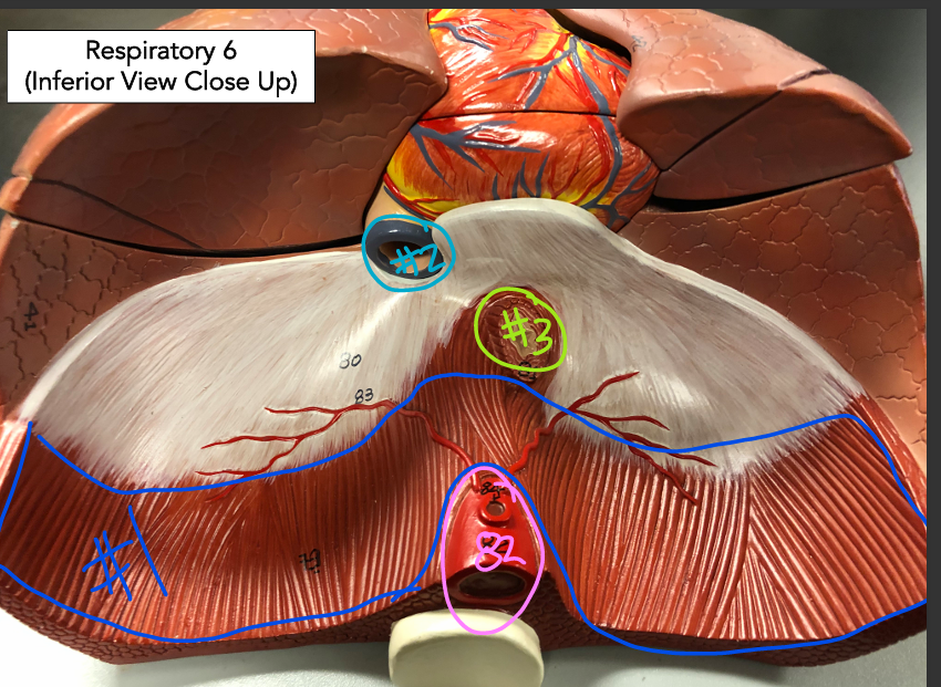
#2
inferior vena cava

#3
esophagus

#8
aorta

#1
hyoid bone

#3
laryngeal prominence (adam’s apple)

#2
thyroid cartilage

#4
cricoid cartilage
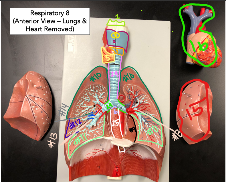
#5
thyroid gland
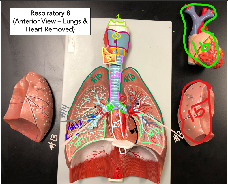
#6
carina

#7
secondary bronchus

#8
tertiary bronchi
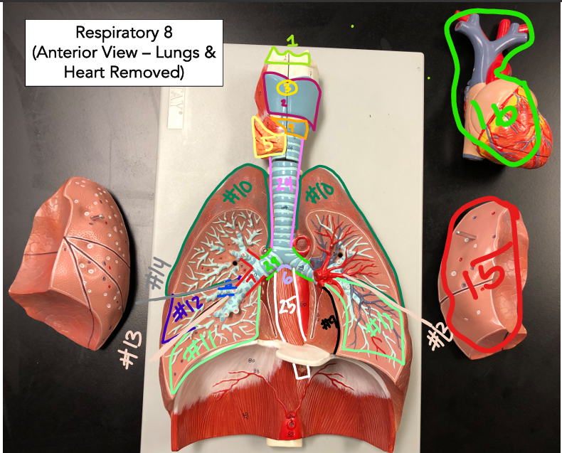
29
primary bronchus

#9
cardia notch/ impression