Chp 9- review
 Call Kai
 Learn
 Practice Test
 Spaced Repetition
 Match
 Flashcards
 Knowt Play
1/26

There's no tags or description
Looks like no tags are added yet.
27 Terms
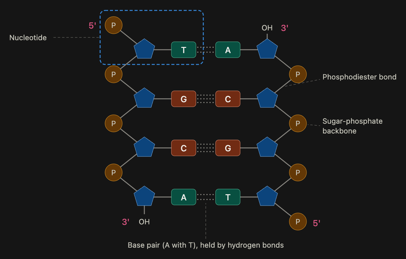
Know where the sugar-phosphate backbone, the 5' and 3' ends of each strand, a base pair, and a phosphodiester bond are on the DNA diagram. [Image 1]
The sugar-phosphate backbone is the two outer rails, made of alternating sugar and phosphate groups.
The left strand has its 5' end at the top (a phosphate) and its 3' end at the bottom (a free OH on the sugar). The right strand is flipped, with 3' (OH) at the top and 5' (phosphate) at the bottom, which is what antiparallel means.
A base pair is one base from each strand joined across the middle (A with T, or G with C).
A phosphodiester bond is the covalent bond that links one nucleotide to the next: a phosphate connects the 3' carbon of one sugar to the 5' carbon of the next sugar on the same strand.

What does the dashed blue box enclose on the DNA diagram? [Image 1]
It encloses one nucleotide, the building block of DNA
What types of bonds form between complementary bases?
Hydrogen bonds: 2 between A and T, and 3 between G and C. Each one is weak, so the strands can be unzipped for copying, but together they hold the helix together.
What does it mean when we describe DNA replication as being semiconservative?
Each new DNA molecule is half old and half new: both parent strands are kept, and each one is the template for a new partner strand. It's like unzipping a zipper and giving each half a brand-new matching half.
What is the complementary DNA sequence of 5'-ATC GGC TAC GTT CAC-3'?
3'-TAG CCG ATG CAA GTG-5', which is the same thing written 5' to 3' as 5'-GTG AAC GTA GCC GAT-3'. Pair A with T and C with G, and the new strand runs antiparallel to the given strand.

Know where DNA pol III, helicase, primase, DNA pol I, DNA ligase, the leading strand, the lagging strand, and a primer are on the replication diagram. [Image 2]
Helicase sits at the fork and unzips the parent DNA.
Primase sits just behind it on the lagging template and lays down short RNA primers.
A primer is the short RNA piece that gives DNA polymerase a place to start.
DNA pol III extends the primers, adding nucleotides 5' to 3' on both strands.
The leading strand is built continuously toward the fork.
The lagging strand is built in separate pieces (Okazaki fragments) away from the fork.
DNA pol I (farther from the fork, on the older fragments) replaces the RNA primers with DNA, and DNA ligase seals the gaps between fragments.
What is the enzyme (not pictured) that is involved in the supercoiling of a prokaryotic chromosome?
DNA gyrase, a topoisomerase. It twists (supercoils) the circular chromosome so it fits in the cell, and it relieves twisting stress ahead of the replication fork.
What is the name of the small fragments of 100-200 nucleotides synthesized on the lagging strand during DNA replication?
Okazaki fragments.
Why does the lagging strand have to be replicated in these short pieces?
DNA polymerase can only add nucleotides in the 5' to 3' direction.
The lagging template runs the opposite way, so the new strand has to be built backward, away from the fork, in short chunks as the helix unwinds. Each chunk needs its own RNA primer, and the pieces get stitched together afterward by pol I and ligase.
A gene has the template sequence 3'-TTC AGC TAC CCA GCA CCG ACG GCT AAA TCA GCT ATT CCG GGT TCA-5'. What is the sequence of the mRNA?
Swap each base for its RNA partner (A to U, T to A, G to C, C to G): 5'-AAG UCG AUG GGU CGU GGC UGC CGA UUU AGU CGA UAA GGC CCA AGU-3'. The given strand is the template, so the mRNA is its complement, read 5' to 3'.
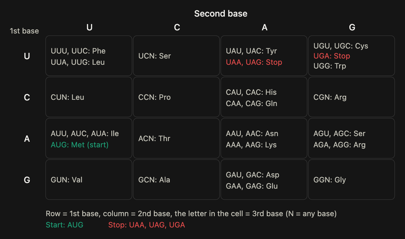
Know how to read the codon chart, including where the start codon and the stop codons are. [Image 3]
Find the first base of the codon in the left column (row), the second base across the top (column), and the third base inside the cell. AUG is the start codon and also codes for Met. UAA, UAG, and UGA are the stop codons, and they code for no amino acid. Many amino acids have several codons, which usually differ only in the third base.
What is the amino acid sequence of the protein made from that mRNA (remember translation starts at a specific codon)?
Translation starts at the first AUG (the third codon), so AAG and UCG are skipped. Reading from AUG: AUG = Met, GGU = Gly, CGU = Arg, GGC = Gly, UGC = Cys, CGA = Arg, UUU = Phe, AGU = Ser, CGA = Arg, then UAA is a stop. Protein: Met - Gly - Arg - Gly - Cys - Arg - Phe - Ser - Arg (9 amino acids).
What will happen to the protein made from the new sequence created by inserting TT into 3'-AAA TGA TAC CAA TCT ACC GGC AGA ATT TGA CTA-5'?
A shorter protein will be made because the insertion created a premature stop codon. Original: the mRNA is UUU ACU AUG GUU AGA UGG CCG UCU UAA…, so the protein is Met - Val - Arg - Trp - Pro - Ser (stop at UAA). New: the template reads TCT ATT CCG…, so the mRNA is UUU ACU AUG GUU AGA UAA GGC…, which gives Met - Val - Arg and then an early stop at UAA. The start codon (AUG) is untouched, so translation still begins, and the sequence changed, so it is not the same protein.
What type of mutation occurred in the TT insertion scenario?
Nonsense, because the change created a premature stop codon (UAA) that cuts the protein short. (Since two bases were inserted, it is also a frameshift, but nonsense is the choice listed.)
Why are inserted or deleted bases known as a frameshift, and why do frameshift mutations almost always result in a nonfunctional protein?
Bases are read in groups of three (codons), so inserting or deleting bases that aren't a multiple of three shifts the "frame" those groups are read in. Every codon after the change is regrouped, so all the downstream amino acids are different, and a premature stop codon often appears. Picture a sentence with one extra letter added: "The fat cat ate the sad rat" becomes "Tah efa tca tat eth esa dra t," gibberish from that point on. The whole rest of the protein is scrambled, so it almost never works.
What is an operon?
An operon is a group of genes in the same pathway that are controlled together by one switch (a promoter and an operator), so the cell only makes those proteins when it needs them, which saves energy.
What is the difference between inducible and repressible operons?
An inducible operon is normally OFF and is turned ON by a substrate (like lactose in the lac operon); these are used to break nutrients down. A repressible operon is normally ON and is turned OFF by its product (like arginine in the arginine operon); these are used to build things.
Describe what happens when the lac operon is "off." What is absent in this scenario? [Image: lac operon picture]
Lactose is absent. With no lactose, the repressor (made by lacI) sits on the operator and blocks RNA polymerase, so lacZ, lacY, and lacA are not transcribed and no lactose-digesting enzymes are made.
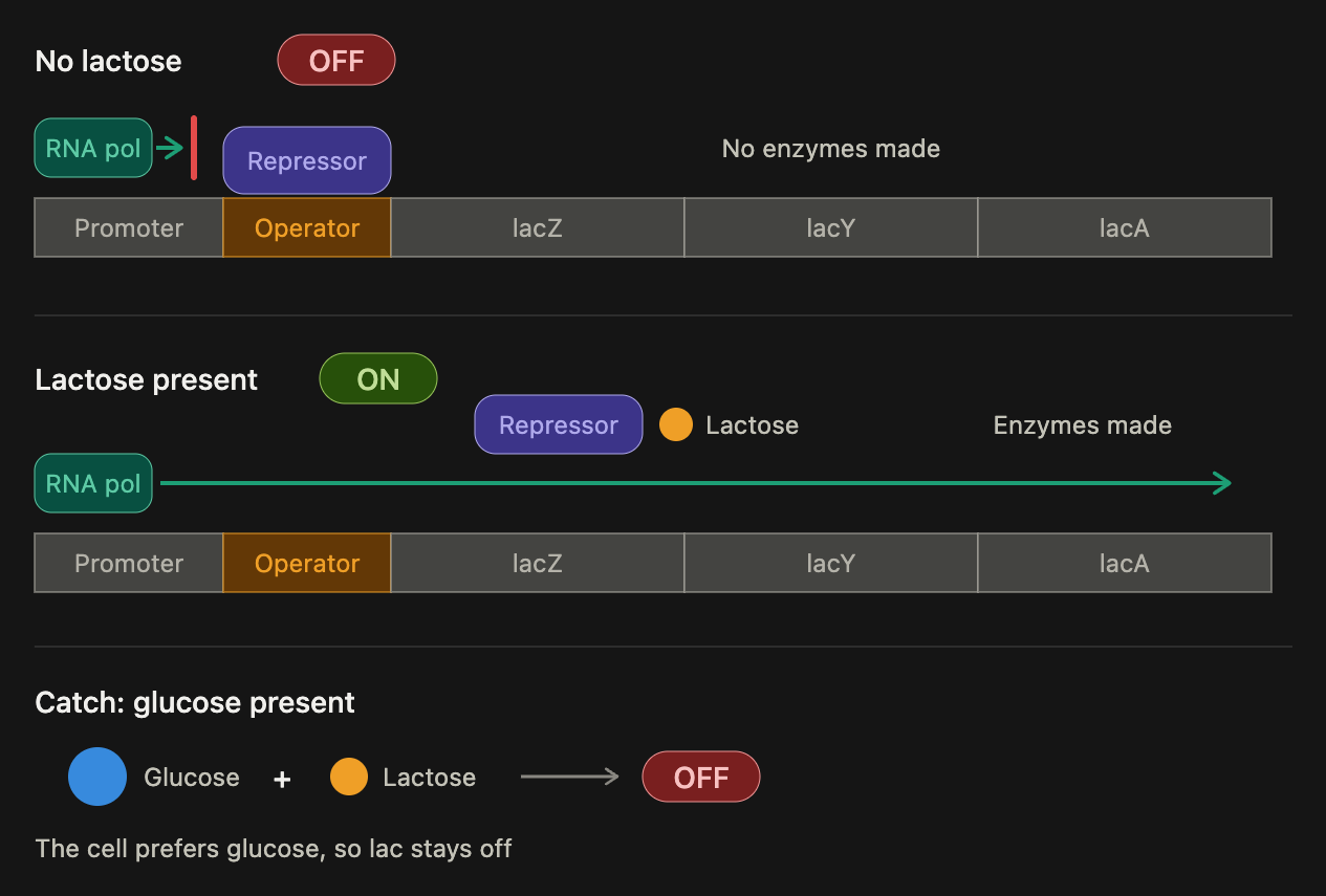
Describe what happens when the lac operon is "on." [Image: lac operon picture]
Lactose is present, so it acts as the inducer: it binds the repressor and changes its shape, so the repressor can no longer bind the operator. RNA polymerase transcribes lacZ, lacY, and lacA, and the cell makes beta-galactosidase (splits lactose), permease (brings lactose in), and transacetylase. When the lactose is used up, the repressor goes back on the operator and turns the operon off. (It only works when glucose is not available.)
List the ways genetic material can be transferred from one bacterial cell to another.
Conjugation (two cells connect by a pilus and pass DNA), transformation (a cell picks up free DNA from a dead cell), and transduction (a bacteriophage carries DNA between cells). Transposons ("jumping genes") can also move resistance genes between a chromosome and a plasmid, which helps them spread.
With regard to the vanA operon, does vancomycin act as a repressor or an inducer? Explain your answer.
Inducer. The vanA operon (the vancomycin resistance genes) is normally OFF and is turned ON when vancomycin is present, just like lactose turns on the lac operon. So the cell only makes the resistance proteins when it is exposed to the drug.
Why do we worry that over-prescription of vancomycin will lead to more cases of vancomycin resistance?
Every dose of vancomycin kills susceptible bacteria and leaves resistant ones to survive and multiply (selection pressure), so more use favors resistant bacteria. Resistance genes like vanA can also be carried on plasmids and transposons and passed to other bacteria (for example by conjugation), which is how Staphylococcus aureus became vancomycin-resistant (VRSA). Vancomycin also switches the vanA operon on in bacteria that carry it.
Why are patients with chronic conditions like diabetes, kidney failure, or cancer at greater risk of developing infections with drug-resistant organisms like VRSA?
Their immune systems are weakened, so infections are harder to clear and take hold more easily. They are also in and out of healthcare settings more (hospital stays, dialysis, catheters, wounds), where resistant bacteria are common, and they take repeated courses of antibiotics, which select for resistant bacteria.
Describe the general stages in the multiplication of double-stranded DNA viruses.
(1) Attachment: the virus binds a receptor on the host cell. (2) Penetration and uncoating: the virus enters (by fusion or endocytosis) and releases its DNA. (3) Synthesis: the viral DNA usually enters the nucleus, where host enzymes transcribe viral genes into mRNA; the host ribosomes make viral proteins (early enzymes, then capsid proteins), and the viral DNA is copied. (4) Assembly: new capsids form and package the new genomes. (5) Release: the new viruses leave by lysing the cell or by budding.
Explain why viruses like HBV (hepatitis B), herpesviruses, and papillomaviruses are called oncogenic.
They can turn a host cell cancerous. Their DNA or proteins disrupt the cell's growth controls, for example by switching on growth-promoting genes (oncogenes) or switching off tumor-suppressor genes (HPV proteins block p53 and Rb), so the cell divides uncontrollably. Examples: HBV can lead to liver cancer, HPV to cervical cancer, and herpesviruses like EBV to certain lymphomas.
How can viruses like HIV remain "latent" in host cells for years?
HIV is a retrovirus: reverse transcriptase copies its RNA into DNA, and that DNA inserts into the host chromosome as a provirus. A provirus can sit silent, making little or no viral protein, and it gets copied along with the host DNA each time the cell divides. Because the cell shows no viral proteins, the immune system can't spot it, until a signal reactivates it.
Explain why some viruses must enter the nucleus to replicate whereas others do not.
Most DNA viruses need the nucleus because the host's DNA and RNA polymerases and nucleotides for making DNA and mRNA are there, and the virus can't copy its DNA on its own. Most RNA viruses replicate in the cytoplasm because they carry or make their own RNA-dependent RNA polymerase and need nothing from the nucleus. Exceptions: retroviruses (HIV) insert their DNA into the nuclear chromosome, influenza (an RNA virus) replicates in the nucleus, and poxviruses (DNA viruses) stay in the cytoplasm because they bring their own enzyme