Exercising Testing + Prescription (Exam 2)
 Call Kai
 Learn
Learn Practice Test
 Spaced Repetition
 Match
Match Flashcards
 Knowt Play
1/71

There's no tags or description
Looks like no tags are added yet.
Name | Mastery | Learn | Test | Matching | Spaced | Call with Kai | Chat |
|---|
No analytics yet
Send a link to your students to track their progress
72 Terms
Myocardium Contractile Cells..
What are the properties of myocardial contractile cells?
Make up ~90–95% of heart mass
Contractile → generate force to pump blood
Excitable → respond to an electrical stimulus
Conductive → move electrical charge from one cell to another
What are the key features of cardiac contractile muscle cells?
Striated
Many large mitochondria → produce ATP
Lots of myoglobin → stores/carries O₂ to mitochondria
Intercalated discs → connect cardiac cells together
Electrical connections allow cells to contract together
Highly fatigue-resistant / essentially does not fatigue under normal conditions
How are cardiac muscle fibers oriented, and how does the heart contract?
Cardiac fibers have a unique spiral/helical orientation
Contraction produces a squeeze + twist motion
No summation/tetanus in cardiac muscle
This organization helps efficiently eject blood from the ventricles

key cell electrophysiology…
Charge
Electrical potential measured in millivolts (mV)
Polarized
Separation of charge across the plasma membrane → negative inside, positive outside
Depolarization
Internal charge becomes less negative due to influx of positive ions
Repolarization
Polarized voltage is restored due to efflux of positive ions
Myocardium Conductive Cells…
What percentage of heart mass is made up of conductive cells?
5–10%
Automaticity
Generates spontaneous electrical charge without external stimulation
Pacemaker cells only
Excitability
Ability to respond to an electrical stimulus
All cardiac cells
Conductivity
Ability to move electrical charge from one cell to another
All cardiac cells
Contractility
Ability to cause cardiac muscle contraction
Myocardial cells only
What are key features of myocardial conductive cells?
Unique ion channels
Intercalated discs
Regulated by the autonomic nervous system
What are the ion movements during the conductive cell action potential?
Prepotential → slow Na⁺ influx
Depolarization → rapid Ca²⁺ influx
Repolarization → K⁺ efflux

What is the prepotential (pacemaker potential)?
Slow spontaneous depolarization caused mainly by Na⁺ influx through leaky/funny (If) channels → brings cell to threshold.
What happens once a conductive cell reaches threshold?
Ca²⁺ channels open → rapid Ca²⁺ influx → depolarization.
What causes repolarization in conductive cardiac cells?
K⁺ efflux → membrane potential becomes negative again.
Why do conductive cardiac cells have automaticity?
They do not maintain a stable resting membrane potential → prepotential spontaneously brings them to threshold → repeated action potentials.
The conduction system of the heart…
What is the normal electrical conduction sequence of the heart?
SA node → Bachmann’s bundle + AV node → AV bundle (Bundle of His) → right & left bundle branches → Purkinje fibers
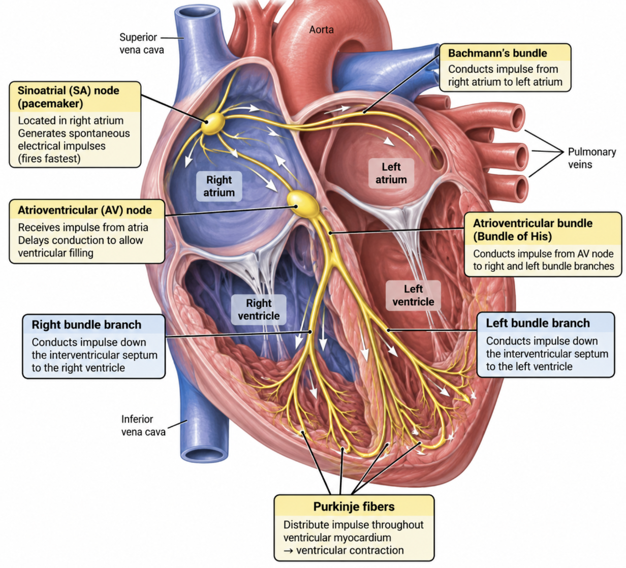
What is the SA node?
Pacemaker of the heart → fires fastest because it has the most active pacemaker channels.

Where is the SA node located?
Right atrium.
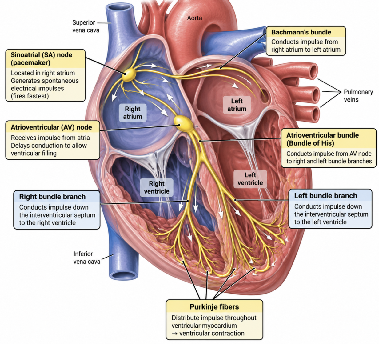
What is the function of Bachmann’s bundle?
Conducts the electrical impulse from the right atrium → left atrium.

What is the function of the AV node?
Receives the atrial electrical impulse and conducts it toward the ventricles.

What is the AV bundle also called?
Bundle of His.
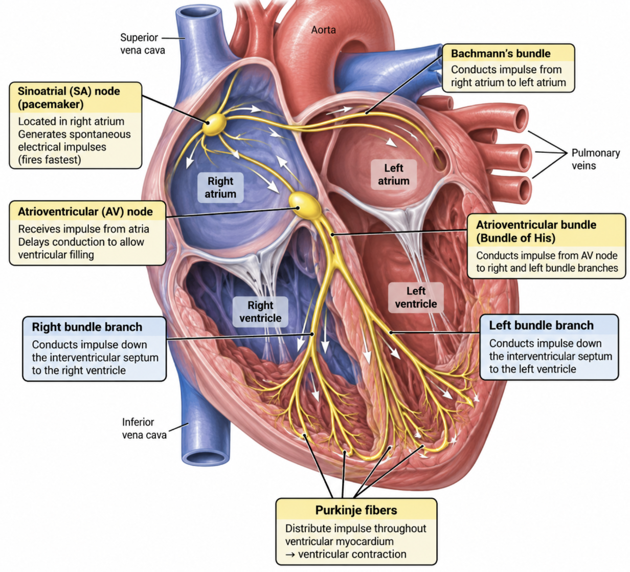
Where does the AV bundle conduct the electrical impulse?
From the AV node → right and left bundle branches.

What do the right and left bundle branches do?
Conduct the electrical impulse down the interventricular septum toward the apex.

What do Purkinje fibers do?
Distribute the electrical impulse throughout the ventricular myocardium → ventricular contraction.
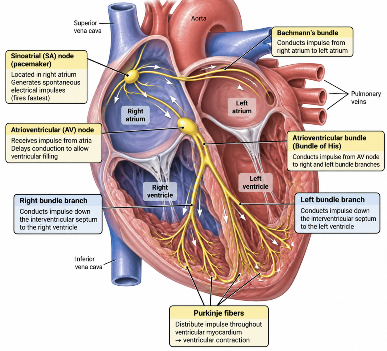
What is the overall pathway of blood through the right side of the heart?
Right atrium → tricuspid valve → right ventricle → pulmonary valve → lungs

What type of circulation does the right ventricle supply?
Pulmonary circulation → sends blood to the lungs.
What is the overall pathway of blood through the left side of the heart?
Left atrium → bicuspid/mitral valve → left ventricle → aortic valve → body
What type of circulation does the left ventricle supply?
Systemic circulation → sends blood to the body.
What are the atria primarily considered?
Receiving chambers.
Which valve is between the right atrium and right ventricle?
Tricuspid valve.
Which valve is between the left atrium and left ventricle?
Bicuspid (mitral) valve.
Which valve does blood pass through when leaving the left ventricle?
Aortic valve.
ECG…
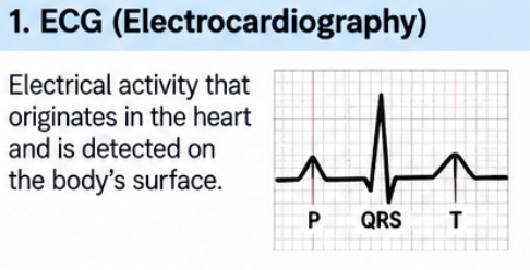
What is electrocardiography (ECG)?
Records the heart’s electrical activity detected at the body’s surface
Skin electrodes measure voltage changes produced by the heart
Voltage changes are amplified and visually displayed/recorded as an ECG tracing
What is the isoelectric line?
The flat baseline of the ECG when cardiac tissue is in its resting polarized state.

What does deflection direction describe on an ECG?
Direction the tracing moves from the isoelectric line.
Positive (+) = upward
Negative (−) = downward

What is a wave on an ECG?
A positive or negative deflection away from the isoelectric line.
Example: P wave or T wave.

What is a complex on an ECG?
Several waves occurring together.
Example: QRS complex = Q + R + S waves.

What is a segment on an ECG?
The line between waves or between a wave and a complex.
Examples: PR segment, ST segment.

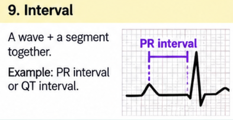
What is an interval on an ECG?
A wave + a segment measured together.
Example: PR interval = P wave + PR segment.
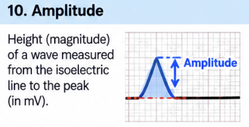
What is amplitude on an ECG?
Height or depth of a wave from the isoelectric line.
Directly related to the mass of tissue electrically active at that time.
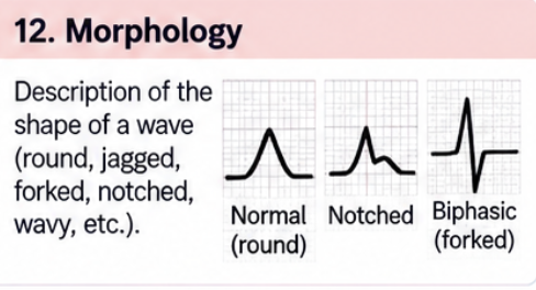
What is morphology on an ECG?
Description of the shape of a wave.
Examples: round, jagged, forked, wavy.
What is duration on an ECG?
ime from the start to the end of a wave, segment, or interval.


ECG Primer basics 8-12…
What are the two cardiac cell types and their main functions/properties?
Myocardial (working) cells → contraction/relaxation; contractility, excitability, conductivity.
Pacemaker cells → spontaneously generate impulses; automaticity, excitability, conductivity.
All cardiac cells share excitability + conductivity.
What are the key electrolyte/ion facts in cardiac cells?
Electrolyte → dissociates in water into charged ions.
Cation = +; anion = −.
K⁺ = primary intracellular ion.
Na⁺ = primary extracellular ion.
Resting/polarized cell → inside is more negative than outside.
What controls ion movement across cardiac cell membranes?
Membrane channels → may be always open, gated, or selective.
Concentration gradient → high → low.
Electrical gradient → like charges repel; opposites attract.
Na⁺/K⁺ pump → actively transports ions against their electrochemical gradients.
What happens during depolarization and repolarization according to the primer?
Depolarization → K⁺ begins leaving → Na⁺ permeability ↑ → Na⁺ rushes in → inside becomes positive.
Depolarization = electrical event; contraction = mechanical event.
Repolarization → primer describes Na⁺/K⁺ pump moving Na⁺ out + K⁺ back in → resting state restored.
What should I know about the SA node and backup pacemakers?
SA node → upper right atrium near SVC; 60–100/min; highest automaticity → normal pacemaker.
AV junction → 40–60/min.
Ventricles → 30–40/min or less.
Farther from SA node → slower intrinsic rate.
How does the impulse travel from the SA node through the atria?
SA node fires →
Bachmann’s bundle → left atrium
Internodal tracts → right atrium
→ atrial depolarization/contraction → AV node.
Where is the AV node and what are its 3 main functions?
Location → lower right atrium near interatrial septum.
Only normal conduction pathway between atria and ventricles.
Functions:
Delays conduction → allows atrial kick.
Backup pacemaker at 40–60/min.
Blocks some impulses during rapid atrial rates.
Trace conduction from the AV node to the ventricular muscle.
AV node → Bundle of His → R/L bundle branches → Purkinje fibers → ventricular muscle.
Right bundle → RV.
Left bundle → anterior + posterior fascicles → anterior/posterior LV walls.
What are the key conduction-speed and escape-rhythm facts?
Slowest conduction → AV node.
Fastest → His-Purkinje system (His + bundle branches + Purkinje).
Escape pacemaker → slower secondary pacemaker takes over if faster pacemaker fails.
Escape rhythm named by site of origin.
What does the P wave represent, and what happens to atrial repolarization?
P wave → atrial depolarization.
Atrial repolarization usually isn't visible because it occurs during ventricular depolarization and is hidden in the QRS complex.
What is the difference between the PR interval and PR segment?
PR interval → onset atrial depolarization → onset ventricular depolarization.
PR segment → end P → beginning QRS; short isoelectric line.
PR segment used as baseline to assess ST elevation/depression.
What do QRS, ST, T, and U represent?
QRS → ventricular depolarization.
ST segment → early ventricular repolarization.
T wave → ventricular repolarization.
U wave → late ventricular repolarization; not always present.
What are the QT interval and R-R interval?
QT → total ventricular activity; beginning QRS → end T.
One PQRST = one cardiac cycle/heartbeat.
R-R interval → one R wave to next R wave; assesses rhythm regularity.
How are ECG deflections classified relative to the isoelectric line?
Isoelectric line → flat baseline; electrical activity absent.
Above baseline → positive deflection.
Below → negative deflection.
Both positive + negative → biphasic deflection.
How does current direction affect ECG deflection?
ECG lead views activity between one + pole and one − pole.
Current toward + pole → positive deflection.
Toward − pole → negative deflection.
Away from poles → biphasic deflection.
What determines ECG waveform size, and why is QRS larger than P?
Deflection size depends on magnitude of electrical current/voltage generated by depolarization.
Ventricles have greater muscle mass → generate more voltage → QRS normally larger than P wave.
What happens during the absolute refractory period?
Onset QRS → peak T.
Cells cannot respond to any stimulus because they haven't repolarized to threshold potential.
Myocardial cells cannot contract; conduction cells cannot conduct another impulse.
What happens during the relative refractory period?
Peak T → end T.
Cells can respond to a strong stimulus.
Also called vulnerable period of repolarization.
Strong stimulus may take over pacemaker control; e.g., PVC → ventricular tachycardia.
What happens during the supernormal period?
Occurs during a short portion near the end of the T wave, just before complete repolarization.
Cells can respond to a weaker-than-normal stimulus.
What do the horizontal and vertical ECG boxes measure?
Horizontal = time.
1 small box = 0.04 sec.
2 boxes = 0.08 sec; 3 boxes = 0.12 sec.
Vertical = voltage/amplitude.
1 small box = 1 mm.