Lecture 2a: Cell Structure and Function
 Call Kai
 Learn
 Practice Test
 Spaced Repetition
 Match
 Flashcards
 Knowt Play
1/26

There's no tags or description
Looks like no tags are added yet.
27 Terms
magnification
ability to make an object larger
resolution
the ability to distinguish small objects that are close together as separate and distinct
Simple staining
One dye used to color specimen
Simple stain - basic dye
Positively charged chromophore, which binds to negatively charged cellular components
simple stain - acidic dye
- charged chromophore, binds to + charged cellular components.
The gram stain
separates bacteria into 2 groups based on cell wall structure
Gram positive cells are ____ because they….
purple , retain the primary stain
Gram negative cells are ___ because they…
pink/red, lose the primary stain and take color of counterstain
acid fast stain, how does it work and what is the result?
Detect mycolic acid in the cell of of the genus Mycobacterium
Mycobacterium retain the primary stain (pink), and everything else takes on counterstain (blue)

Endospore stain, how does it work and what is the result?
Endospores retain primary color (green) and cells are counterstained (pink)

phase-contrast microscopy: How, Image, Key points
Phase ring amplifies difference in refractive index of cell and surroundings
Dark cells on a light background
No stain needed, can see live samples
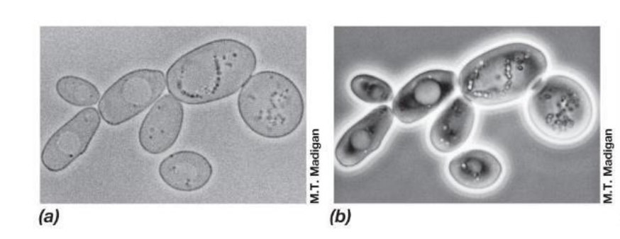
Bright-field microscopy: How, Image, Key Points
Contrast between specimen and surroundings
normal magnified image
basic scope

Dark field microscopy: how, image, key points
Hollow cone of light, only refracted light enters objective
bright specimen on light background
good for bacteria that doesn’t stain well
Fluorescence microscopy: how, image, key points
specimen emits one colour when lit by another
glowing specimen
cells can have natural fluorescence or we use fluorescent dye

Differential interface contrast (DIC) microscopy
uses a polarizer to create 2 beams of light
gives structures a 3D appearance
confocal scanning laser (CSL) microscopy
uses a computerized scope and laser
focuses on single layers, then compiles layers into 3D image
electron microscopy
uses electrons instead of light photons
electron wavelength shorter than light therefore higher resolution
Transmission electron microscope (TEM): how, image, key points
electrons pass through specimen
can see internal structures
specimen must be very thin and stained with metals
Scanning electron microscopy (SEM): how, image, key points
electrons scan specimen
3D image of specimen surface
specimen is coated with a thin film of heavy metal
how can selective forces shape cell morphology?
small cells/high SA:V - nutrient uptake
helical/spiral shape - swimming motility
filamentous - gliding motility
Advantages to being a smaller cell
small cells have more surface area relative to cell volume
support greater nutrient uptake exchange per unit cell volume
grow faster than larger cells
plasma membrane: what is it and function
thin structure that surrounds the cells
highly selective permeable barrier. concentrates nutrients and excretes waste
how is the cytoplasmic membrane stabilized ?
Mg and Ca by forming ionic bonds w negative charges on the phospholipids
hydrogen bonds and hydrophobic interactions
what kind of linkages do archaeal membranes have? bacterial?
archaeal has ether, bacterial has ester

what makes archeal membranes so heat resistant?
lipid monolayer

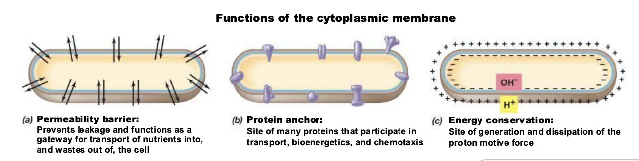
list 3 membrane functions
permeability barrier
protein anchor
energy conservation
how do archaeal membranes differ from bacterial membranes?
lack fatty acids, have isoprenes instead
instead of phospholipids, have glycerol diethers/ tetraether
can exist as monolayer, bilayer or a mix of both