physio e3l4: ch 12 muscle physiology
 Call Kai
 Learn
Learn Practice Test
 Spaced Repetition
 Match
Match Flashcards
 Knowt Play
1/35

There's no tags or description
Looks like no tags are added yet.
Name | Mastery | Learn | Test | Matching | Spaced | Call with Kai | Chat |
|---|
No analytics yet
Send a link to your students to track their progress
36 Terms
skeletal muscle:
where is it
3 exceptions
connected to two or more bones by tendons
3 exceptions
facial = to skin
larynx = to cartilage
other muscles like sphincters
3 connective tissues and what layer they surround
epimysium = continuous w/ tendon connective tissue
perimysium = divides muscle into fascicles (100s, 100s of myofibers/musclefibers)
endomysium surrounds muscle fibers

components of a muscle fiber (myofiber):
what is it
made of
sarcomere
how nucleated
sarcolemma
Transverse (T) tubule
a muscle cell
many myofibrils
smallest contractile unit of a muscle fiber
multinucleated and extend length of the muscle
sarcolemma = PM that surrounds muscle fiber
T Tubules = groove in the sarcolemma going down
components of a muscle fiber/cell (myofiber):
sarcoplasm
mitochondria
sarcoplasmic reticulum/lateral sacs
Triad
cytoplasm of muscle fiber
a lot for ATP
smooth ER that stores Ca+
T tubule groove b/t 2 sarcoplasmic reticulum (lateral sacs)

myofibers:
what is it and 2 parts
what do the 2 parts form
give what appearance to what two muscles
arrangement of thick and thin filaments, which are contractile proteins
thick filament = myosin
thin filament = actin
thick and thin filaments form sarcomeres
give striated appearance to skeletal and cardiac muscle

structure of a sarcomere
red and blue?
A band
H zone
M line
I (pronounced “eye”) band
z line
1 sarcomere = Z- line → Z-line
red = myosin (thick), blue = actin (thin)
A band = has overlap of actin/myosin, dark band is the thick filament
H zone = myosin only
M line = anchors myosin, perpendicular to long axis
I band = actin only, light band
Z line = anchors actin, perpendicular to long axis

myosin (thick myofilament) structure:
2 parts
orientation
binding sites
what does it look like
myosin tail = towards M line
myosin head = towards I band
actin binding site
nucleotide binding site for ATP and ATPase

myosin (thick myofilament) structure:
2 forms of myosin head?
how to move b/t them?
how is myosin arranged?
myosin head 2 forms
high E = when connected to ADP + P
low E = when ATP
low E → high E via ATP hydrolysis by ATPase
2 myosin bound at tails = m line crossbridges
actin (thin filament) structure:
3 parts structure
G-actin mC = each contains myosin binding site
F actin = fibrous string made of G actin mC’s
double helical actin strands = made of F actin
anchored at Z line
tropomyosin and troponin:
what do they both do
what are they/purpose of each
both are myofiber regulatory proteins
tropomyosin = overlaps binding sites on actin for myosin
covers myosin binding site on G actin mCs
troponin complex
attaches to tropomyosin
binds Ca+ reversibly = regulate skeletal muscle contraction

titin:
what is it
what does it do
why important
what does it look like
myofiber structural protein
anchors thick filaments to thin filaments
provides sarcomere structural support and elasticity
2 parts muscle force generation
excitation-contraction coupling → cross bridge cycle
excitation-contraction coupling = how muscle contractions turned on/off
cross bridge cycle = how muscles generate force
what is muscle contraction
why?
what is happening at:
A band
I band
H zone
how slide?
shortening of sarcomeres
overlapping actin and myosinn filaments slide past each other
sections
A band = no change
I band = shortens
H zone = shortens
sliding due to cyclical formation and breaking of cross bridges = crossbridge cycle
excitation contraction:
where
3 steps (w/o details)
ryanadine receptor acts like
what comes after excitation contraction?
neuromuscular junciton
steps
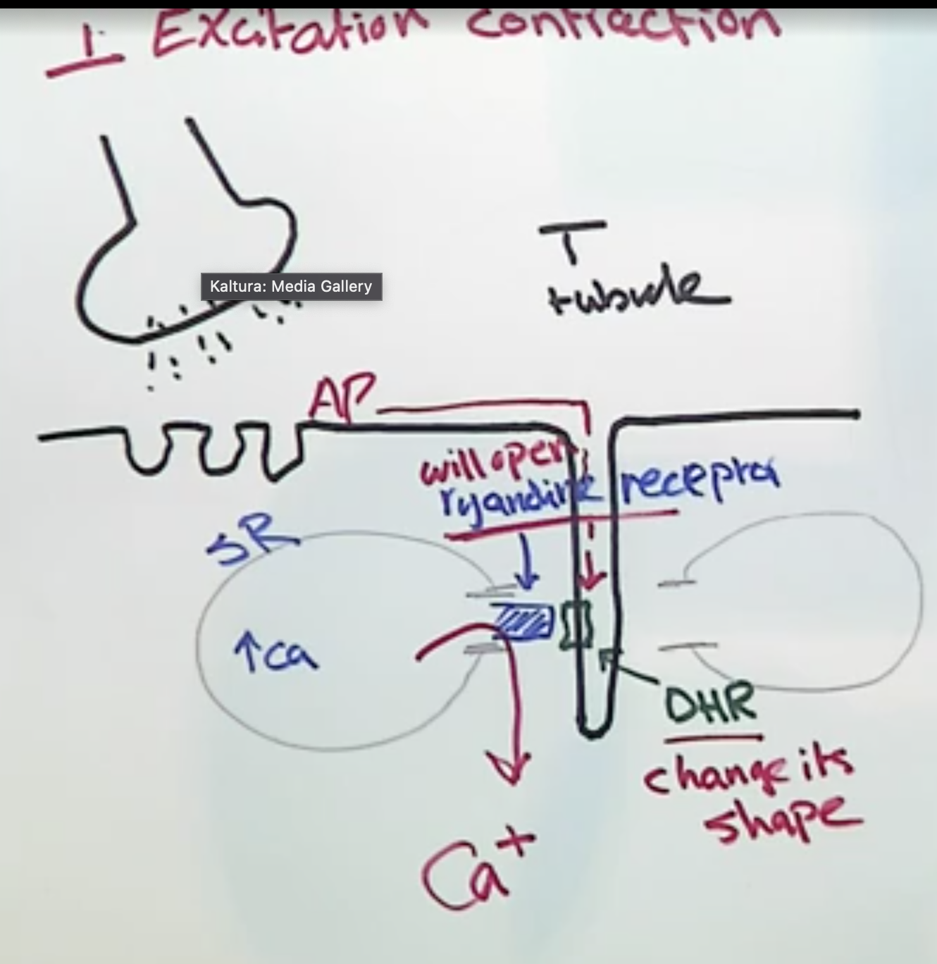
neurotransmitter activates muscle → AP travels down T tuble until it reaches voltage gated dihydrothymidine receptor (DHR)
AP causes DHR to change shape → ryandine receptor to open (mechanically gated)
ryandine receptor opens → Ca+ exits lateral sacs of sarcoplasmic reticulum
gate on sarcoplasmic reticulum
cross-bridge cycle
cross-bridge cycle:
3 things present at the beginning
6 ateps
why does rigor mortis happen
why does rigor mortis eventually relax?
beginning
high Ca+ from SR/excitation contraction
myosin in low E form (ATP)
G actin bound to tropomyosin and troponin
cycle
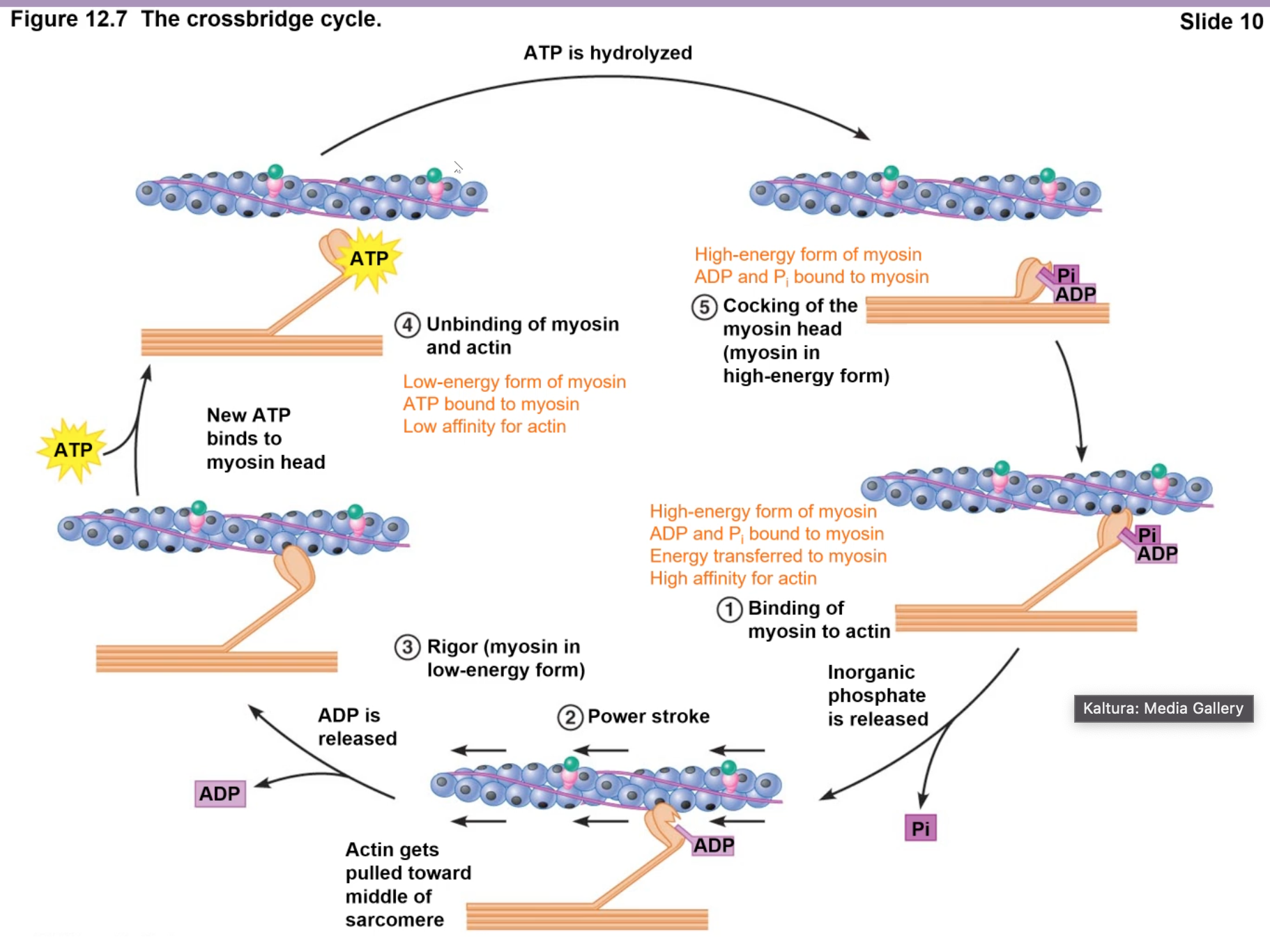
low E myosin (ATP) -(hydrolysis)→ high E myosin (myosin head conformation change)
Ca+ binds troponin → troponin pulls/shifts trotropomycin, exposing the myosin binding site on actin
exposed myosin binding sites bind High E myosin (ADP+P) → releases P
releasing P → “Power Stroke” = myosin pulls actin
after power stroke, ADP released = myosin and actin STUCK together in contraction, no movement (cross-bridge)
rigor mortis = peak 12 hours, lasts 24 hrs, then will begin relaxing, fully relaxed 48 hours
contraction stops when new ATP added to myosin (Low E) and separates from actin
enzymes break myofibers
how is the cross bridge cycle analogous to rowing a boat:
cross bridge or myosin head =
linking of myosin head to actin =
power stroke =
release of cross bridge =
oar
oar contact w/ water
oar pulling through water
oar out of water and repositioning
3 ways how to stop contraction? how pump?
stop APs = stops excitation
release Ca+ from troponin = tropomyosin covers myosin binding sites on actin
remove Ca+ from cytosol bay pumping it back into sarcoplasmic reticulum (storage)
Ca+ - ATPase pump in the sarcoplasmic reticulum uses ATP
muscle activity across a joint:
stationary vs mobile
origin and insertion
muscles can only ___ not ___
how antagonistic
most muscles connect two bones via tendons. when muscle contracts, one bone is stationary, one bone moves
origin = muscle’s point of attachment POA to the ststationary bone
insertion = muscle’s POA to moveable bone
pull not push
combination of contraction and relax = flexor flexes, extensor relaxes
motor unit stim and motor unit inhibition
2 types of muscle fibers?
what are they
what do they do?
how innervated?
extra fusal fibers
contractile cells of the muscle, outer
generate force/skeletal muscle contraction
innervated by alpha motor neurons
intrafusal fibers
contractile cells of the muscle spindle, inner
adjust sensitivity of muscle to stretch
innervated by gamma motor neurons
muscle spindle:
what does it do
how controlled
AP frequency when:
stretched
relaxed
contracted
detects changes in muscle length
sensitivity of sensory endings adjusted by action of intrafusl fibers
AP freq when:
stretched = a lot of AP bc there’s load on the muscle
relaxed = some AP bc of muscle tone=
contracted = none
alpha and gamma motor neurons are _____
why?
coactivated
intrafusal fibers can’t detect stretch when they aren’t being activated/contracted
intrafusal fibers aren’t being stretched during a muscle contraction, so can’t detect stretch when flexing
ability to both flex + detect stretch recovered by coactivation of gamma (intrafusal activation) with alpha (extrafusal activation)
Golgi tendon organs (GTOs):
what are they
how do they work
why important
sensory capsules within tendons that detect passive tension/stretch
tendon stretch activates the GTO
provide reflex inhibition/relaxation of muscle to prevent injury from too much stretch/force
smooth muscle:
where is it found
what NS
3 parts of structure
internal organs and BVs
autonomic
small spindle-shaped cells
nonstriated
gap junctions
varicosities on surface bc have gap junctions
smooth muscle:
how contract
3 contraction structures different
range/axes of contraction?
3 reasons why?
sliding-filament mechanism of contraction
non-striated, no sarcomeres, has dense bodies that also have thick and thin filaments
longer range of contraction and multiple axes of contraction
longer actin/myosin
myosin heads whole length
multiple directions of contraction
smooth muscle excitation contraction:
5 steps
Calcium dependent calcium release:
most Ca+ enters from extracellular via voltage gated Ca+ channel in PM
extracellular Ca+ triggers release of SR Ca+
Ca+ binds calmodulin
Ca+-malmodulin binds/activates myosin light-chain kinase (MLCK)
phosphorylated MLCK → myosin ATPase active → cross bridge cycle
phosphatase unphosphorylates MLCK → no myosin ATPase activity → no cross bridge cycle
smooth muscle excitation contraction:
2 differences from skeletal?
depends on extracellular Ca+
no T tubule → DHP → ryanadine receptor → Ca+ from SR
Ca+ binds calmodulin instead of troponin
3 ways relax smooth muscle.
contraction time in smooth muscle
phosphatase removes phosphate from myosin
Ca removed from cytoplasm via
Ca-ATPase or
Ca/Na counter transport (secondary active)
slower than skeletal
smooth muscle neural regulation:
excitatory or inhibitory?
what determines response?
neurotransmitter release from?
both
response depends on receptor type
varicosities
multi unit smooth muscle:
where?
gap junctions? synchronous?
each fiber acts ___
3 ways how
large airways, arteries, eye (ciliary/iris)
no gap junctions = not synchronous
individually
own innervation (varicosity touches every cell not just on surface)
no recruitment
no tone
single unit smooth muscle:
where?
gap junctions? synchronous? innervates how many cells?
each fiber acts ___
most common, intestine, blood vessels, respiratory tract
yes and yes, varicosity on the surface (innervates few cells) bc has gap junctions
contract together as a unit
single unit smooth muscle:
pacemaker cells
tone
graded contractions
stretch reflex
pacemaker cells = spontaneous depolarizations by itself
tone = level of contraction w/o stimulation
graded contractions = no recruitment
stretch reflex = sudden or prolonged stretch induces relaxation = protection
spontaneous demoralizations:
what muscle type
2 types
pacemaker potentials
why
slow wave potentials
why
smooth and cardiac
pacemaker potentials = spontaneous depolarizations to threshold
permeability to Na+, Ca+, K+
slow wave potential = cycles of graded potentials at Vm bc of Na+ permeability

cardiac muscle:
how “intermediate b/t skeletal and smooth?”
2 like skeletal
3 like smooth
1 like both
like skeletal
striated w/ sarcomeres
troponin-tropomyosin regulation
like smooth
gap junctions (within intercalated disks)
pacemaker cells
innervated by autonomic NS (varicosities)
like both
Ca+ from extracellular fluid and SR
cardiac muscle:
length of AP
summation? why?
what type of refractory period?
length of AP = length of contraction
no summation due to long refractory period
absolute (absolutely not going to get AP)
normal AP v. cardiac muscle AP?
compare steps (normal 3, cardiac3)
skeletal/smooth
depolarization = Na+ in
apex of AP = Na+ channels close
drop = K+ leaving leak channels
cardiac has a long refractory period
depolarization = same
apex = same
plateau = k+ leaving leak channels AND Ca+ entering