Cranial Nerves Part 2
 Call Kai
 Learn
 Practice Test
 Spaced Repetition
 Match
 Flashcards
 Knowt Play
1/71

There's no tags or description
Looks like no tags are added yet.
72 Terms
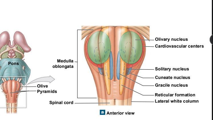
all communication between brain and SC passes thru the
medulla oblongata
what do the nuceli in the medulla do
act as relay stations for sensory/motor pathways
associated with cranial nerves
associated w autonomic control of visceral organs
what are the three relay stations of the medulla oblongata
gracile nucelus and cunerate nucleus
solitary nucelus
olivary nucli
what does the gracile and cuneate nucleus do with the medulla
pass somantic sensory info to the thalamus
what does the solitary nucelus do in the medulla
recieved visceral sensation from spinal nerves and cranial nerves
what does the olivary nuclei do
passes info from the cerebrum , SC, diencephalon and brainstem to the cerebellum
creates the olives of the medulla

label the parts of the medulla
what are the cranial nerves that contains sensory and motor nuclei? (5)
VIII
IX
X
XI
XII
what are reflex centers of autonomic nuclei
these are areas that recievie input from cranial nerves, cerebral cortex and brainstem
when we speak on the mid brain what is the two pairs of nuceli collectively called?
what is the surface called
two pairs of nuceli: corpora quadrigemina
surface: tectum
what is the midbrain or tectum (surface layer) responsible for
auditory and visual stimuli
what type of processing occurs in the inferior colliculi of midbrain and superior colliculi
inferior: auditory
superior:visual
the red nucelus of the mesencephalon does what
this pair of nuceli is involved in maintaining alertness, muscle tone and limb position.

label

the diencephalon contains of three things what are they?
epithalamus: pineal
thalamus: forms wall of 3 ventricle, egg shape
hypothalamus extens near optic chiasm,
label each parts of the thalamic nuceli:
part of limbic system
relay info to frontal love
relay info to parietal lobe
relay info to occipital lobe
adjust acitviyy in cingulate gyrus and parietal lobe
anterior nuceli
medial nuclei
ventral nuceli
posterior nuclei
lateral nuceli
here is a desciription what are we talking abt
subconcious control of skeeltal muscle
hr, bp, respiration, digestion
secretion of ADH
body temp
circadian rhythm
thirsy drive
hypothalamus
what seperates the anterior and posterior lobes of the cerebellum
primary fissure
what are purkinjie cells
large neurons in the cerebellum that help control balance, coordination, and smooth movements.
what does the arbor vitae of cerebellum do
Connects cerebellar cortex with cerebellar peduncles
when we label the cereellur pendunncles label each
what connects the cerebellum with mesencephalon, diencephlon and cerebrum
what is the part that communicates between the cerebellum and pons
what is the part that connects the cerebellum with the medulla obglonta
superior cerebellar peduncle
middle cerebellar penduncle
inferior cerebllar penduncle
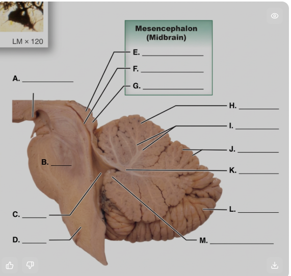
label

does the cerebrum contain basal nuclei and the limbic system
yes
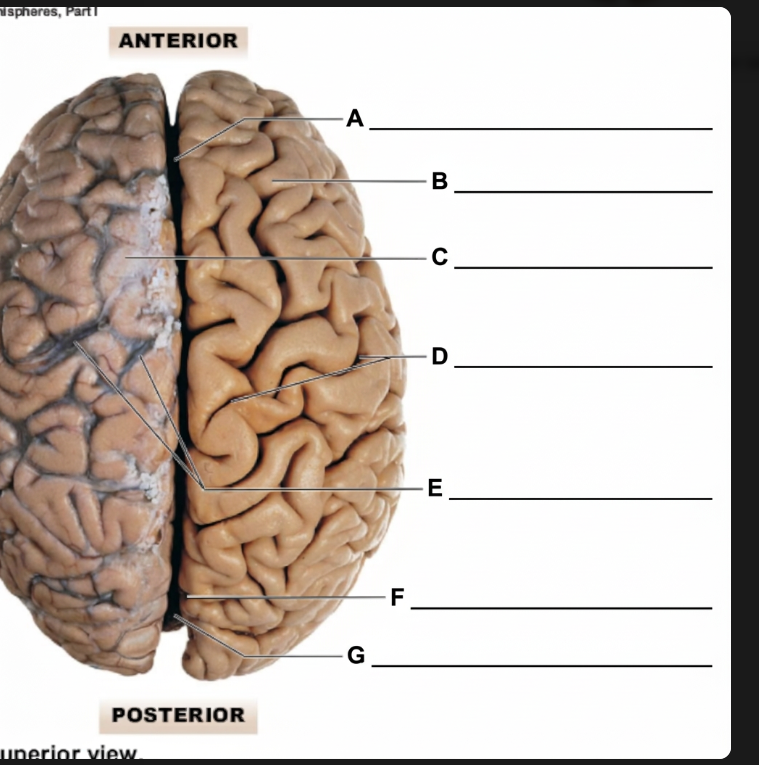
label

descrive function of each
frontal love
occipital
parietal
temporal
Frontal lobe: conscious control of skeletal muscles
• Occipital lobe: perception of visual stimuli
• Parietal lobe: conscious perception of touch,
pressure, vibration, pain, temperature, and taste
• Temporal lobe: conscious perception of auditory
and olfactory stimuli/deep is the insula

label

what area contains the primary motor cortex and pyramidal cells.
precentral cortex of the brain
what are consists of the primar somatosensory corte and Neurons receive somatic sensory information for
touch, pressure, pain, taste, and are associated with
visual cortex, auditory cortex, olfactory cortex,
and gustatory cortex
postcentral gyrus
label each
what is the area that allows to understand size, shapes, form
what is the area in memory and learned movements
what is the area that visually recongizrd objects
what are arecongizes sound
somatosneosry area
premotor cortex
visual association
auditory association
what are of the brain has high orer functions such as information processing
cerebrum
what is the wernickie area?
brocas area?
receptive speech area
werenickie: persoanity/analytical area
brocas: speech production
receptive speech: adjusts speech based on audiotry stimuli
the prefrontal cortex does what
and what is prefrontal lobotomy
this cortex performs complicated learning/reasoning
lobotmy: removal of the lobe
the left hemisphere of the brain is important for?
what about the right hemi?
let: speech, writing, language math
righ: touch, spatial visualization
the central white matter has three fibers that act as brain wiring such as
association
commissural
projection
what does each do
associaiton fiber
commissural fiber
porection fiber
Association fibers: Connect areas within the same brain hemisphere.
Commissural fibers: Connect the left and right brain hemispheres. (ex corpous callosum)
Projection fibers: Connect the cerebral cortex with lower brain areas and the spinal cord.

label

how do the brain wires help the brain communicate
Together, association, commissural, and projection fibers form communication pathways that connect areas within one hemisphere, between both hemispheres, and between the brain and the rest of the nervous system.
what is the basal nuceli ?what do they do
masses of gray matter embedded in the
white matter inferior to the lateral ventricles
Controls muscle tone in skeletal muslce subconsciously
Coordinates learned movements
Relays information from the cerebral cortex
what five things make up the basal nuceli
Caudate nucleus
• Lentiform nucleus
• Claustrum
• Putamen
• Globus pallidus
the caudate nucelus of basal nuclei does?
globus pallidus does?
Control the cycles of arm and leg movements when
walking (caudae)
adjusts mucle tone for walking (globus)
where is the limbic system located in the brain
between the cerebrum and the
diencephalon just superior to the corpus callosum
people w issues within the limbic system have issues with
mental state and personality
what is the overall function of the limbic system (4)
emotional staes
linkes concious function w unconcious functions
memory storage/retriveal
functional grouping
how many pairs of cranial nerves are there?
12
do the cranialnerves come from the spinal cord
no, they come directly off the brain and travel out to the head, neck, and organs.
what is the mnemoni to remeber the cranial nerves from anterior to posterior cranial nerves
Oh Oh Oh, Two Tired Asses Fell Very Gently Violently And Horrifically. Ah
what are the 12 cranial nerves
(I) O: olfactory nerve/bulb
(II) O: opic nerve
(III) O: oculomotor nerve
(IV) T: Trochelar Nerve
(V) T: Trimgeminal nerve
(VI) A: Aducens nerve
(VII) F: Facial nerve
VIII V: Versitivuochcolear or auditory nerve
(IX) G: Glossopharyngneal nerve
(X) V: Vagus nerve
12 XII H: Hypoglossal nerce
11 XI A: Acessory Nerve
11 and 12 are swithed in numbering but not anatomical position.
what is the function, and destination of the olfactory nerve
function: smell
destination: olfcytory bulbs
what is the function, and destination of the optic nerve
function: vision
destination: diencephlon then to occipital love
they continue to their destination via optic tracts and from the retina to optic chiasm
what is the blindspot of the eye
nerves enter retina
what is the function and destination of the oculomotor nerve
function: controls the extra ocular eye muscles. and will change the diameter of the pupil, shape of lens to focus on the retina
destination: extra ocular eye muscles
oriin: mesencephhlon
what is the function, origin and destination of the trochelar nerve
function: controls the superior oblique eye muscle/motor
origin: mesencephlon
destination: superior oblique muscle
smallest cranial nerve

know this image
what is the function of the trigenimal nerve
hint three divisions
mixed nerve with both sensory and motor functions
ophtalmis division: sensation from forehed, eyelid, nose
maxilarry: lower eyelid sensation, upper lip, cheek
mandibular: controls chewing
what is the origin of the trigeminal nerve
Ophthalmic division (sensory)/orbital structures,
nasal cavity, forehead, superior eyelid
• Maxillary division (sensory)/inferior eyelid, upper lip,
gums, and teeth, palate, part of pharynx
• Mandibular division (mixed)/lower gums, teeth, lips,
palate (sensory)/motor nuclei of pons (motor)
what is the longest cranial nerve
trigeminal nerve
what is the destination of the trigeminal nerve
opthalmtic and maxisally divisoon:
mandiublar division:
pons
muscle of mastication

know where the dimensions are in this image/divisions
what is the function, origin and destination of the abducens nerve
function: controlling eye movement/motor
origin: pons
destination: innervates the lateral rectus eye muscle
what is the function origin and destination for the facial nerve
function: mixed nerve sensroy: controls sesnations and taste of face. motor: controls muscle of face
origin: sensory: taste buds motor: pons
destination: sensory: pons/ motor : muscles of the face

what nerve do you see to the right of the pons
facial nerve
what is the function, origin and destination fo the vestivulocochlear nerve
function: balance/hearing
origin: receptors of cochlea and vestiblue
destination: pons and medulla
axons travel to the vestibular
nuclei of the medulla oblongata
axons synapse in the cochlear
nuclei of the medulla oblongata
verstibular nerve
cochlear nerve
what is the function, origin and destination of glossopharyngeal nerve
function: mixed . sensory: tongue pain motor: swallowin
origin: sesnory: 1/3 tongue mtoor: salivary gland
destination: sensory: medulla motor” phayrngneal muscles for swalloing
what is the function, origin and destintion of the vagus nerve
function: mixed nerve, sensory: infromation from organs.
motor: sends info to organs
origin: sensory: organs motor: medulla
Destination: sneosry: medulla motor: muscles or respirtatory cardiovascualr nad digestive organs
function of vagus nerve more in depth
The vagus nerve (X) is the main nerve of the parasympathetic nervous system — it handles the "rest and digest" side of things.
what is the function, original and destination of the acessory nerve
function: motor. it controlls the trapezius, palata, phayrnc, layrnx,a nd sternocleiodmastoid
origin: SC, and medulla
destination: internal branch of muscles of palata, phayrnx and larynx,
external branch: sternocleoid mastoid and trapezius msucle
what is the function destination and origin of the hypoglossal nerve
funtion: controls tongue movement
origin: medulla
destnation tongue muscles
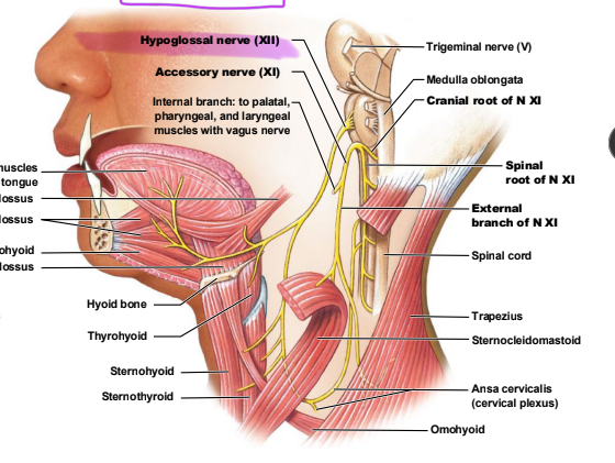
what cranial nerve is shown
hypoglossal nerve

what cranial nerve is shown in the circle
vagus
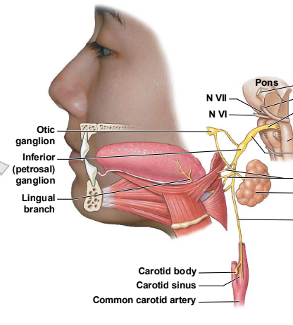
wha cranial nerve is seen right beneatht he ponts in yellow
glossopharyngeal nerve
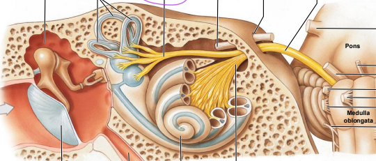
the yellow branching nerve on top is the
the seashell looking thing is the
yellow branching: verstibular nerve
seasheel: cochlea