Tumor of Nervous System
 Call Kai
 Learn
Learn Practice Test
 Spaced Repetition
 Match
Match Flashcards
 Knowt Play
1/46

There's no tags or description
Looks like no tags are added yet.
Name | Mastery | Learn | Test | Matching | Spaced | Call with Kai | Chat |
|---|
No analytics yet
Send a link to your students to track their progress
47 Terms
Intrinsic vs extrinsic tumor
Intrinsic = from the brain tissue
Extrinsic = from other source
Intrinsic brain tumors
Glioma = astrocytoma, oligodendroglioma; ependymoma, PCNSL, medulloblastoma, pineal gland tumor
Astrocytoma subtypes
grade I (pilocytic astrocytoma, subependymal giant cell astrocytoma)
grade II (astrocytoma)
grade III (anaplastic astrocytoma)
grade IV (glioblastoma)
low grade = I-II; high = III-IV
What is pilocytic astrocytoma
Tumor of CHILDHOOD (most common); cerebellum > optic nerves > brainstem; caused by BRAF mutation

What is this
Cystic lesion with enhancing mural nodule → Pilocytic astrocytoma
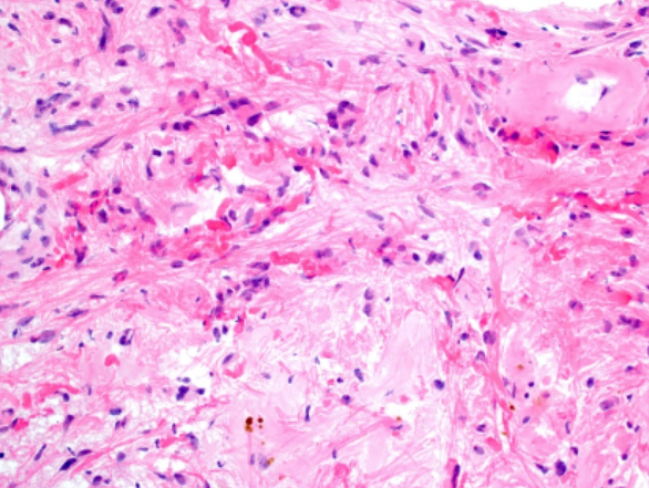
Describe this
Atatypical piloid glial cells (long glia), focal hemosiderin deposit, Rosenthal fibers (dark pink fibers) → Pilocytic astrocytoma

What is this
NON enhance tumor with increased T2/FLAIR signal → Grade II Astrocytoma; causes seizure in young adult
What is glioblastoma
Most common malignant primary tumor; 60-70 y/o Pt with headache, seizure + focal neuro deficit
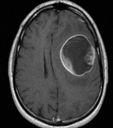
What is this
Ring enhancing lesion with central necrosis + edema, increased T2/FLAIR → glioblastoma

What is this
Butterfly tumor that CROSSES corpus callosum → Glioblastoma

Describe this
Hyperchromatic elongated astrocytes + irregular nuclear membrane; pseudopallisading necrosis surrounding central necrosis → Glioblastoma
What is Oligodendroglioma
15-20% of glioma which can either be grade II Oligodendroglioma or anaplastic Oligodendroglioma (AO) grade III

Describe this
Fried egg appearance + reticular blood vss growth → Oligodendroglioma

What is this
Non enhancing + calcification → Oligodendroglioma (better prognosis than pure astrocytic tumor)
What is ependymoma
From ependymal cell on ventricular surface; 5% childhood tumor from 4th vent in posterior fossa

What is this
Chronic supratentorial hydrocephalus from mass at floor of 4th vent → ependymoma
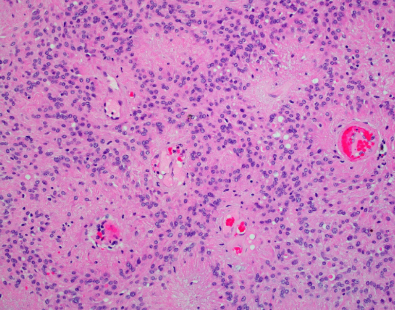
Describe this
The area surrounding vss = pseudorosettes → ependymoma
What is PCNSL
<3% primary brain tumor; common in immunocompromised Pt, EBV either earlier onset (31) compared to immunocompetent (60+)
PCNSL presentations
Subacute/chronic meningitis → CN and spinal nerve dysfunction → CSF and flow cytometry needed to confirm diag + stereotactic biopsy for histology

What is this
Periventricular lesion that CROSSES corpus callosum → PCNSL
Cancers that cross the corpus
Glioblastoma and PCNSL ONLY

Describe this
Diffuse infiltrate packed with mononuclear cell with scant cytoplasm
What is medulloblastoma
Most common malignant childhood tumor of periventricular → present with headache, ataxia, brainstem involvement signs

What is the difference between the left and right images
Lt lesion is in the ROOF of 4th vent; Rt lesion is in the FLOOR of 4th vent
Roof = medulloblastoma, floor = ependymoma

What are the differences between these three images on T2
Lt most has HYPERintense → pilocytic astrocytoma
The middle and rt is HETEROGENOUS enhancement → medulloblastoma and ependymoma that CANNOT be diff from this view

Describe this
Tumor cells around a central area (not lumen, canal or vss) filled with neurofibrillary process → TRUE rosette → medulloblastoma
What is Parinaud syndrome
Sign of pineal gland tumor → Signs of upward gaze palsy, convergence retraction nystagmus, light-near dissociation, and bilateral lid retraction.

What is this
Pineal gland tumor
Extrinsic benign tumors
Meningioma, Schwannoma, craniopharyngioma, hemangioblastoma
What is meningioma
35% of malignant primary brain tumor; Women and NF2 more common, found in dura mater commonly near cerebral convex (next to sagittal sinus) → present with headache, seizure + focal neuro deficit

What is this
In the DURA → Can tell because there is periarachnoid space between tumor and brain, dural tail present

Describe this
A points to whorl formation, B is psammoma bodies, C is nuclear pseudoinclusion → Meningioma features
What is Scwannoma
Tumor of cells that myelinate PERIPHERAL nerves (not CN I, II); most common are vestibular and acoustic neuroma (CN VIII) = hearing loss, tinnitus

What is this
Dense enhancement in internal auditory canal → Schwannoma

Describe this
The elongated TRUE pallisading nuceli = Antoni A and B with fibers between each pallisade = Verocay → sign of Schwannoma
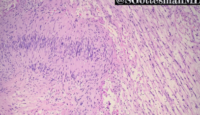
Describe this
Pallisade = Antoni → Lt = A = packed pallisade; Rt = B = loose pallisade
What is craniopharyngioma
Benign tumor from Rathke remnant → suprasellar, partial calcification, solid → present with headache, visual impairment, impaired growth in children + hypopituitary in adult

What is this
Craniopharyngioma
What is hemiangioblastoma
Benign, slow growing tumor with many vss found in cerebellum, brainstem, spinal cord; associated with VHL when found with retinal fibroma

What is this
Enhanced mural module → can present in many ways → Hemangioblastoma
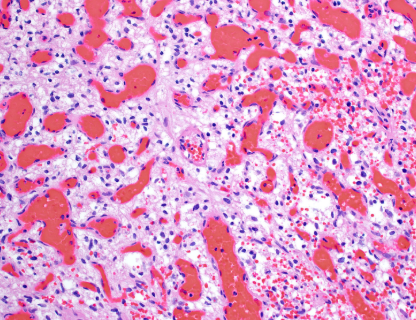
Describe this
Many vss + vacuolated stromal cell → hemangioblastoma
Common tumors that spread to brain
Lung, breast, melanoma is most common
What is leptomeningeal metastasis
Leukemia into subarachnoid space → BYPASS BBB
leptomeningeal metastasis presentation
Lumbar and cervical radiculopathies
Cranial neuropathies
Seizures
Confusion
Encephalopathy from hydrocephalus or raised intracranial pressure can be present
Note that weakness is atypical unless brain invasion is present.

What is this
Yellow arrow = visible CN V, VI, VII → do lumbar puncture
Brain metastasis management
Radiation, surgery, chemo
Leptomeningeal metastasis management
PALLIATIVE care; RT if there is nerve involvement, chemo for other malignancy