Cath - general
 Call Kai
 Learn
 Practice Test
 Spaced Repetition
 Match
 Flashcards
 Knowt Play
1/40

There's no tags or description
Looks like no tags are added yet.
41 Terms
What is the course of the LCx? How is it split into proximal/mid distal?
- Atrioventricular groove and goes posteriorly
- OM 1- 3
- Proximal: until OM1
- Mid
- Distal : Last OM branch until distal (goes to Apex)
What is the course of the LAD and how is it split into proximal/mid/distal?
- Interventricular groove
- Diagonals 1-4
- More septals from diagonals
- Proximal: until 1st branch (either septal branch - medial, or diagonal - lateral)
o There are 10 septal branches!
o Proximal septal branch supplies bundle of His
- Mid to end: divide by half
- Identify by : different movement from rest of vessels, presence of septal perforators
What are the branches of the RCA?
- Proximal: first RV branch
o Sinoatrial artery supplies Sinus node in RA > outwards (Supplied by LCx in some)
o Conus (supplies RVOT > inwards)
- Mid
o First RV branch
o Acute marginal after RV branch
- ?Atrioventricular branch
- Distal
o RPDA: Down interventricular septum (acute bend > goes up then goes down)
o RPL: posterolateral wall
What is barbaeu test?
oboth the radial and ulnar arteries are compressed simultaneously until the plethysmographic wave is lost. Then, the pressure on the radial artery is released, and the shape of the plethysmographic wave is evaluated (check pulse ox)
What are some relative C/i to radial access?
- Very small radial artery
- Known pathology in proximal vessel e.g. subclavian stenosis
- Raynauds/scleroderma/Kawasaki
- Need to use radial for CABG/used for CABG
o Left radial is preferred for access post CABG
How is TIMI flow graded?
Grade 0 No perfusion; no flow beyond the blockage.
Grade 1 Penetration without perfusion; contrast agent passes the blockage but doesn't fill the distal vessels.
Grade 2 Partial perfusion; contrast agent passes the blockage and opacifies the distal vessels, but slower than normal.
Grade 3 Complete perfusion; contrast agent flows as quickly and completely as normal, indicating full reperfusion.
When is DCB > DES?
Anything less than 2mm cannot put stent
Coronary ectasia/aneurysm
o If vessels are too huge there is risk of stent thrombosis
o Slow flow: also aspiration and balloon
Clot
Diffuse disease
High bleeding risk
Ostial disease/bifurcation disease
How is Ca severity graded? (OCT vs IVUS scoring system?)
Ca severity
- Arc
- Length
- Thickness
- Presence of nodule
OCT calcium scoring system
- Maximum calcium angle > 180: 2points
- Maximum calcium thickness > 0.5mm: 1point
- Calcium length > 5mm: 1point
- 4 or more high risk , need calcium modification therapy
IVUS scoring system
- Superficial calcium angle > 270spanning longer than 5mm
- 360 superficial circumferential calcium
- Presence of calcified nodule
- Vessel reference diameter < 3.5mm
- Score > /=2high risk of underexpansion
Management of calcified lesions?
- Cutting balloon
- Scoring balloon
- Rotational artherectomy: Rotablation
o C/I in saphenous venous graft (thrombus and microembolization with no reflow)
- Orbital artherectomy
- Intravascular lithrotripsy i.e. shock wave
Causes of coronary ectasia/aneurysm?
o Previous Kawasaki disease (25%)
o Artherosclerosis
o CTD
o Arteritis (Syphillis, takayatsu, Polyarteritis nodosa)
o Physiological adaptation
o Consider sepsis, endocarditis
o IGG4
Class guideilnes for A) CABG and B) PCI for EF < 35% + multivessel disease?
Class I for CABG
PCI as an alternative (Class IIB ESC)
Class guideilnes for A) CABG and B) PCI for LM disease?
CABG > PCI CLass I if low surgical risk (Lower risk of spontaneous MI And repeat revascularization)
PCI class I if low compleixty (non inferior for survival)
PCI Class IIA if intermediate complexity
Class guideilnes for A) CABG and B) PCI for DM + Multivessel disease?
CABG > PCI CLass I (reduce mortality, reduce repeat revascularizations)
What is classified as multivessel disease?
Involvement of > /=2 vessels with > 70% stenosis
Class guideilnes for A) CABG and B) PCI for Multivessel disease?
CABG Class I
PCI CLass I for low to intermediate complexity
?ischaemia extend
Class guideilnes for A) CABG and B) PCI for proximal LAD stenosis ?
ACC: 2b to improve survival
ESC: Class 1 for CABG or PCI for functionally significant disease, to reduce CV mortality an drisk of spontaneous MI
What is the recommendation for PCI to non infarct artery in STEMI?
Complete revascularization either during index procedure or within 45 days
What is the recommendation for PCI to non infarct artery in NSTEMI?
complete revascularization during index procedure
Functaionl assessment is Class IIB
How to perform FFR?
Intracoronary GTN
Introduce pressure wire beyond guide tip
Zero pressure within aorta
Pass pressure wire distal to lesion
Begin IV adenosine 140mcg
Return pressure wire to ostium and check for drift (0.03)
</= 0.8 significant
What are the types of hyeraemic agents?
Adenosine (IV or IC): IV hold standard
Papaverine
NTP
Regadenoson
Benefit of iFR over FFR?
No need for hyperaemia
Shorter procedural times
Can be used for ostial lesions
Can assess serial epicardial lesions
What did DEFER show?
No difference between medical treatment vs PCI for FFR > 0.75
What did FAME 1 show?
FFR guided (<0.8) vs angiography guided revascularization in MVD
ok to defer if > 0.8
What does FAME 2 trial show?
FFR guided (< 0.8) vs OMT for stable angina
FFR guided a/w reduced risk of future urgent revascularization, but no increase in MI or death (consistent with ischaemia trial)
What are the causes of false negative FFR? (7)
Guide catheter pressure damping
No hyperaemia/adenosine inhibited
STEMI
LM+ Downstream disease
Severe AS
Serial FFR of individual lesion
Presence of signal drift
What are the causes of false positive FFR?
CTO receiving collaterals
Loose pressure connections, air bubbles, transducer connector errors
What are the causes of coronary no reflow?
epicardial spasm
Dissection
Embolis
If no obvious epicardial mechanism, assume microvascular no reflow
what are the risk factors for no reflow?
HTN DM HLD
Delayed presentation to cath lab
High thrombus burden
Plaque composition: high lipid
Lesions longe than 15mm
Prevention strategies for no reflow?
Avoid aggresive balloon inflation
COntrolled artherectomy
Direct stenting
How is coronary dissection classified?


what investigations would you do next?
Dissection
OCT
CT aortogram if near ostium
What are the risk factors of coronary dissection?
over sized balloon
Calcified lesion
Deep seating of catheter
What are the prevention strategies of coronary dissection?
- do not inject contrast if there is dampened pressure waveform
- achieve coaxial catheter position
- avoid aggressive wiring strategies
- avoid use of oversized balloons and high pressure
- preparation of calcified lesions
Management of coronary dissection?
- small, non flow limiting dissections do not require treatment
- proximal edge dissections do not require treatment as stent will prevent forward propogation
- stenting if wire in true lumen
- if ostial dissection with aortic dissection> surgery
Causes of SCAD ?
Predisposing
- Pregnancy/labour and delivery
o can occur at any time during pregnancy but majority post partum within the first week
- Fibromuscular dysplasia
- Connective tissue disease
o Marfan, ehlers danlos, loey dietz
- Systemic inflammatory
o SLE , vasculitis
- Hormonal therapy
- Sarcoidosis
- Coronary artery spasm
- Idiopathic
Precipitating factors
- Intense exercise
- Intense emotional stress
- Sympathomimetic use
Investigation for SCAD?
- CT arterial imaging from head to pelvis (to look for FMD)
o ?aorta rarely impaired and does not need screening
- US Carotid/MRA carotids
- US /CTA renal
- Definitive is coronary angiogram but risk of dissection
Management of SCAD
- Conservative
- Unless severe presentation with ongoing symptoms, other high risk features
- SAPT aspirin
o Some guidelines recommend DAPt
- Treat hypertension
- Follow up with CTA 6
o Guidelines don’t recommend any timing but can do around 6 months
- Can do regular moderate exercise
o Avoid extreme or vigorous sports
o Avoid heavy lifting

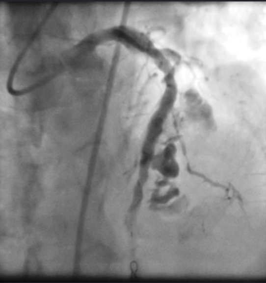
perforation
classifcation system for perforation?

Management of coronary perforation?
- Most can be managed expectantly
- Place covered stent (downside: hard to maneouvre and may block off branch arteries)
- Balloon tamponade
- coil
- gelfoam embolization
o sterile, water insoluble, gelatin based sponge
o thrombosis and inflammation

Causes? Mx?
Causes
improper catheter aspiration and flushing
loose connection in manifold
balloon rupture
Mx
- 100% NRM
- Aspirate air
- Intracoronary adenosine (vasodilator)
Intracoronary adrenaline