BIO 224 LAB PRACTICAL 1 CSN
 Call Kai
 Learn
Learn Practice Test
 Spaced Repetition
 Match
Match Flashcards
 Knowt Play
1/264

There's no tags or description
Looks like no tags are added yet.
Name | Mastery | Learn | Test | Matching | Spaced | Call with Kai | Chat |
|---|
No analytics yet
Send a link to your students to track their progress
265 Terms
The closing of the atrioventricular valves as ventricles contract.
The first sound of a heart beat.
(Sheep Heart) Aorta

The closing of the semilunar valves as the ventricles relax.
The second sound of your heart beat.
(Sheep Heart) Superior Vena Cava

Heart murmurs
Defects in the heart valve are caused by?
(Sheep Heart) Inferior Vena Cava
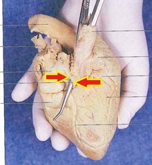
Red electrode
"-" negative, Right wrist above the hand
(Sheep Heart) Pulmonary Trunk

(Sheep Heart) Pulmonary Veins
1 & 2

Black electrode
"+" positive, Left wrist just above the hand
Green electrode
"g" ground, Right wrist two inches above the red electrode
(Sheep Heart) Endocardium

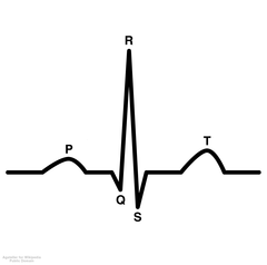
P wave
Depolarization of atrium

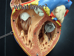
(Sheep Heart) Myocardium
Between red arrows

QRS complex
Depolarization of ventricles
(Sheep Heart) Epicardium

T wave
repolarization of ventricles

(Sheep Heart) Interatrial Septum

repolarization of the atrium
cannot be seen because it occurs during the QRS complex.
(Sheep Heart) Interventricular Septum

Right Atrium
Chamber

(Sheep Heart) Bicuspid Valve (Mitral)
Which valve is this

Left Atrium
Chamber

(Sheep Heart) Tricuspid Valve

Right Ventricle
Chamber

(Sheep Heart) Chordae Tendineae
Chords that pull on the valves to keep close

Left Ventricle
Chamber

(Sheep Heart) Pulmonary Semilunar Valve

Epicardium
superficial layer of the heart wall.
(Sheep Heart) Aortic Semilunar Valve

Myocardium
thick middle layer of the heart wall

(Sheep Heart) Opening to Coronary Sinus

(Sheep Heart) Right Atrium
Chamber

Endocardium
is deep to the myocardium

Aorta

(Sheep Heart) Left Atrium
Chamber
Aortic Arch
"red top"

(Sheep Heart) Right Ventricle
Chamber
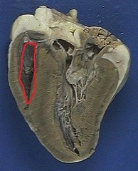
Superior Vena Cava

(Sheep Heart) Left Ventricle
What is B? Chamber

Inferior Vena Cava

Inferior Vena Cava (Posterior)

Pulmonary Trunk

Left pulmonary artery

Right pulmonary artery
#10
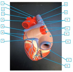
Left pulmonary vein

Right pulmonary vein
#11

Aortic Semilunar Valve

Pulmonary Semilunar Valve

Bicuspid (Mitral) Valve

Tricuspid Valve
right side

Ligamentum Arteriosum
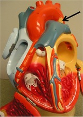
Auricle (right)

Fossa Ovalis

Auricle (left)

Interatrial Septum

Interventricular Septum

Trabeculae Carneae

Papillary Muscle

Chordae Tendineae

Apex of heart

Opening of Coronary Sinus

Coronary Sulcus
groove that marks border between atria and ventricles

Anterior Interventricular Sulcus

Posterior Interventricular Sulcus

Right Coronary Artery

Right Marginal Branch

Posterior Interventricular Artery

Left Coronary Artery

Anterior Interventricular Artery

Circumflex Artery

Small Cardiac Vein

Middle Cardiac Vein

Great Cardiac Vein

coronary sinus (superficial)

Artery
any vessel that carries blood away from the heart
Arteries. They are smaller in diameter and have higher pressure
Blood moves fastest in which vessels? Why?
Agglutinogens
Genetically determined antigens are known as?
Vein
Any vessel that returns blood from a capillary network back to the heart.
Capillaries. They are small, single-cell capacity and have lower pressure
Blood moves slowest in which vessels? Why?
Agglutinins
The presence of these agglutinogens is determined by testing the erythrocytes with antibodies, known as __________________________, that will bind to the antigens.
Agglutination
If the agglutinogen is present on the erythrocytes there will be an antigen-antibody reaction which causes the red blood cells to clump together, called ___________________________.
Arteries, because they need higher pressure; thicker walls
Considering a corresponding artery and vein which has a smaller diameter? Why?
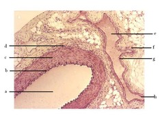
Tunica Interna (artery)
What is B?

Tunica Media (artery)
What is C?

Plethysmograph
An instrument used to measure the amount of blood flowing through an organ.
Hematocrit
The _____________________ is the percentage of the total blood volume that is comprised of the packed red cells.
Tunica Externa (artery)
What is D?

Volume Pulse
The change in blood volume with each cardiac cycle.
40-50%
For men the normal range for hematocrit is?
36-45%
For women the normal range for hematocrit is?
Tunica Externa (vein)
What is F?

Elastic recoil of an artery after the closure of the aortic valve.
Define Dicrotic Notch
Tunica Media (vein)
What is G?

Elastic recoil of the artery after the aortic semilunar valve closes
What occurrence in the body is responsible for the dicrotic notch?
Leukocytes
The _______________________ are responsible for protecting the body against disease.
Tunica Interna (vein)
What is H?
Rising edge corresponds to active contraction and falling edge to relaxation.
Notice that the slope of the rising edge of the volume pulse is much steeper than the slope of the falling edge. Why?
Granulocytes
The _______________________, white blood cells containing cytoplasmic granules, consist of neutrophils, basophils and eosinophils.
Agranulocytes
The _______________________, cells without cytoplasmic granules, consist of monocytes and lymphocytes.