SYS PHYSIO Week 8: Cardiovascular System Continued
 Call Kai
 Learn
 Practice Test
 Spaced Repetition
 Match
 Flashcards
 Knowt Play
1/18

There's no tags or description
Looks like no tags are added yet.
19 Terms
What kinds of junctions connect cardiac autorhythmic and contractile cells? How are these junctions important for normal heart function? In other words, for cardiac contractile cells, from where does the initial electricalexcitation signal come?
Kinds of Junctions Connecting Cardiac Cells
Cardiac muscle cells (cardiomyocytes) are joined end-to-end at specialized regions called intercalated discs. These discs contain three primary types of cell junctions:
Gap Junctions: Communicating channels (made of proteins called connexins) that allow ions and small molecules to pass directly from the cytoplasm of one cell to the next.
Desmosomes (Macula Adherens): Anchoring junctions that physically hold the cardiac muscle fibers tightly together so they do not pull apart when the heart contracts with high force.
Adherens Junctions (Fascia Adherens): Anchoring sites for actin filaments that help link the muscle cells structurally
Importance for Normal Heart Function
Electrical Coupling (Gap Junctions): Gap junctions allow action potentials to pass rapidly from autorhythmic (pacemaker) cells to contractile cells. This enables the heart to contract as a coordinated single unit, or functional syncytium, ensuring the atria and ventricles squeeze in a synchronized, rhythmically timed sequence.
Mechanical Strength (Desmosomes and Adherens Junctions): These mechanical links prevent the cardiomyocytes from tearing apart or separating under the high pressure and constant physical stress of repeated heartbeats.
Origin of the Initial Electrical Excitation Signal
For cardiac contractile cells, the initial electrical excitation signal comes from autorhythmic (pacemaker) cells—specifically, the sinoatrial (SA) node located in the upper wall of the right atrium.
The SA node spontaneously generates the action potential that sets the natural pace of the heart (sinus rhythm).
This electrical signal propagates through the heart's specialized conduction pathway (including the internodal pathways, atrioventricular [AV] node, bundle of His, bundle branches, and Purkinje fibers) before spreading via gap junctions directly into the surrounding cardiac contractile cells, triggering them to contract.
What does an action potential in a cardiac contractile cell look like? What are the ions and permeability changes responsible for depolarization, plateau, and repolarization of the action potential in a cardiac contractile cell? Why is the plateau phase of the action potential so vitally important for proper heart function? In other words, how is this different from skeletal muscle, and why is itthat the heart cannot tetanize?
Phase 4: resting membrane potential. Myocardial contractile cells have a stable resting potential of about -90 mV
Phase 0: depolarization. When a wave of depolarization moves into a contractile cell through gap junctions, the membrane potential becomes more positive. Voltage-gated sodium channels open, allowing Na+ to enter the cell and rapidly depolarize it. The membrane potential reaches about +20 mV before the sodium channels close. These are double-gated channels, similar to the voltage-gated sodium channels of the axon.
Phase 1: initial repolarization. When the sodium channels close, the cell begins to repolarize as potassium leaves through open potassium channels.
Phase 2: the plateau. The initial repolarization is very brief. The action potential then flattens into a plateau as the result of two events: a decrease in potassium permeability and an increase in calcium permeability. Voltage-gated calcium channels activated by depolarization have been slowly opening during phases 0 and 1. When they finally open, calcium enters the cell. At the same time, some “fast” potassium channels close. The combination of calcium influx and decreased potassium efflux causes the action potential to flatten out into a plateau.
Phase 3: rapid repolarization. The plateau ends when caclium channels close and potassium permeability increases once more. The “slow” potassium channels responsible for this phase are similar to those in the neuron: They are activated by depolarization but are slow to open. When the slow potassium channels open, potassium exits rapidly, returning the cell to its resting potential (phase 4)The influx of during phase 2 lengthens the total duration of a myocardial action potential. A typical action potential in a neuron or skeletal muscle fiber lasts between 1 and 5 msec. In a contractile myocardial cell, the action potential typically lasts 200 msec or more.
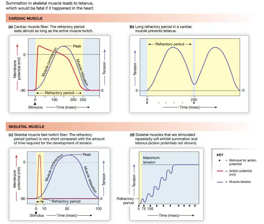
The longer myocardial action potential helps prevent the sustained contraction called tetanus. Prevention of tetanus in the heart is important because cardiac muscles must relax between contractions so the ventricles can fill with blood. To understand how a longer action potential prevents tetanus, let’s compare the relationship between action potentials, refractory periods, and contraction in skeletal and cardiac muscle cells.

Label the cardiac contractile AP

Drawthe different AP’s of cardiac and skeletal muscles. Further explain why tetany cannot happen in cardiac cells.
As you may recall, the refractory period is the time following an action potential during which a normal stimulus cannot trigger a second action potential. In cardiac muscle, the long action potential (red curve) means the refractory period (yellow background) and the contraction (blue curve) end almost simultaneously. By the time a second action potential can take place, the myocardial cell has almost completely relaxed. Consequently, no summation occurs.
In contrast, the skeletal muscle action potential and refractory period are ending just as contraction begins. For this reason, a second action potential fired immediately after the refractory period causes summation of the contractions. If a series of action potentials occurs in rapid succession, the sustained contraction known as tetanus results.
Draw and label the steps of E-C Coupling in a cardiac contractile cell

How does the process of excitation-contraction coupling occur in cardiac contractile cells?
Contraction
An action potential that enters a contractile cell moves across the sarcolemma and into the t-tubules 1 → where it opens voltage-gated L-type calcium channels in the cell membrane 2 → calcium enters the cell through these channels, moving down its electrochemical gradient. Calcium entry opens ryanodine calcium receptor release channels (RyR) in the sarcoplasmic reticulum 3 → This process of EC coupling in cardiac muscle is also called calcium-induced-calcium release (CICR). When the RyR channels open, stored calcium flows out of the sarcoplasmic reticulum and into the cytosol 4 → creating a “spark” that can be seen using special biochemical methods. Multiple sparks from different RyR channels sum to create a calcium signal 5 → Calcium released from the sarcoplasmic reticulum provides about 90% of the calcium needed for muscle contraction, with the remaining 10% entering the cell from the extracellular fluid. Calcium diffuses through the cytosol to the contractile elements, where the ions bind to troponin and initiate the cycle of crossbridge formation and movement 6 → Contraction takes place by the same type of sliding filament movement that occurs in skeletal muscle.
Relaxation
Relaxation in cardiac muscle is generally similar to that in skeletal muscle. As cytoplasmic calcium concentrations decrease, calcium unbinds from troponin, myosin releases actin, and the contractile filaments slide back to their relaxed position 7 → As in skeletal muscle, cacium is transported back into the sarcoplasmic reticulum with the help of a calcium-ATPase 8 → However, in cardiac muscle, calcium is also removed from the cell via the sodium-calcium exchanger (NCX) 9 → One calcium moves out of the cell against its electrochemical gradient in exchange for three sodium entering the cell down their electrochemical gradient. Sodium that enters the cell during this transfer is removed by the sodium-potassium-ATPase 10.
MAP = CO * TPR, which is our guiding light equation for the CV system!
If we want to increase or decrease MAP, how would we change CO and/or TPR?
To Increase MAP
Increase CO: Raise heart rate and/or stroke volume (via sympathetic stimulation or increased venous return).
Increase TPR: Cause vasoconstriction in the arterioles to narrow vessel diameter and raise resistance.
Do both: Simultaneously raise CO and TPR for a larger increase in MAP.
To Decrease MAP
Decrease CO: Lower heart rate and/or stroke volume (via parasympathetic/vagal tone or reduced blood/fluid volume).
Decrease TPR: Cause vasodilation in the arterioles to widen vessel diameter and lower resistance.
Do both: Simultaneously reduce CO and TPR to lower MAP.
What does cardiac output represent, and what are the determinants of cardiac output?
(CO = SV * HR)
What do each of the terms mean? What are the units for each of the terms?
If we want to increase or decrease CO, how would we change SV and/or HR?
Cardiac Output: the volume of blood pumped by one ventricle in a given period of time. (L/min)
Stroke Volume: the amount of blood pumped by one ventricle during a contraction. It is measured in milliliters per beat
Increase CO: Increase SV and/or HR, or both
Decrease CO: Decrease SV and heart rate
Which cardiac cells are responsible for determining HR? Determining SV?
HR: Pacemaker cells in the SA node
SV: Cardiac contractile cells
How does the body regulate CO?
Specifically, how does the body regulate HR and SV to affect changes in CO?
By antagonistic control with the sympathetic speeding up HR and the parasympathetic slowing HR.
SV Regulation:
Contractility is the intrinsic ability of a cardiac muscle fiber to contract at any given fiber length and is a function of calcium interaction with the contractile filaments
Length-tension relationship: as stretch of the ventricular wall increases, so does the SV. If additional blood flows into the ventricles, the muscle fibers stretch, then contract more forcefully, ejecting more blood. The degree of myocardial stretch before contraction begins is called the preload on the heart because this stretch represents the load placed on cardiac muscles before they contract
Frank-Starling Law: SV is proportional to EDV. As additional blood enters the heart, the heart contracts more forcefully and ejects more blood. It means that within physiological limits, the heart pumps all the blood that returns to it.
Venous Return: EDV is normally determined by venous return - the amount of blood that enters the heart from the venous circulation. Determined by three factors: 1) contraction/compression of veins returning blood to the heart (skeletal muscle pump) 2) pressure changes in the abdomen and thorax during breathing (respiratory pump 3) sympathetic innervation of veins.
What are the intrinsic and extrinsic factors regarding regulation?
Intrinsic Regulation: Mechanisms built directly inside the heart (self-governing properties) that adjust performance without outside neural or hormonal signals.
Extrinsic Regulation: Control mechanisms originating outside the heart, primarily from the autonomic nervous system and endocrine (hormonal) system.
How does the body intrinsically regulate HR? How does the body extrinsically regulate HR?
Normally, tonic control of heart rate is dominated by the parasympathetic branch. This control can be shown experimentally by blocking all autonomic input to the heart. When all sympathetic and parasympathetic input is blocked, the spontaneous depolarization rate of the SA node is 90–100 times per minute. To achieve a resting heart rate of 70 beats per minute, tonic parasympathetic activity must slow the intrinsic rate down from 90 bpm.
An increase in heart rate can be achieved in two ways. The simplest method for increasing rate is to decrease parasympathetic activity. As parasympathetic influence is withdrawn from the autorhythmic cells, they resume their intrinsic rate of depolarization, and heart rate increases to 90–100 beats per minute. Sympathetic input is required to increase heart rate above the intrinsic rate. Norepinephrine (or epinephrine) on beta-1 receptors speeds up the depolarization rate of the autorhythmic cells and increases heart rate.
Both autonomic branches also alter the rate of conduction through the AV node. Acetylcholine slows the conduction of action potentials through the AV node, thereby increasing AV node delay. In contrast, the catecholamines epinephrine and norepinephrine enhance conduction of action potentials through the AV node and through the conducting system.
How does the body intrinsically regulate HR? How does the body extrinsically regulate HR?
Cellular mechanisms of autonomic regulation of HR
Parasympathetic: The parasympathetic neurotransmitter acetylcholine (ACh) slows heart rate. Acetylcholine activates muscarinic cholinergic receptors that influence potassium and calcium channels in the pacemaker cell. Potassium permeability increases, hyperpolarizing the cell so that the pacemaker potential begins at a more negative value. At the same time, calcium permeability of the pacemaker decreases. Decreased calcium permeability slows the rate at which the pacemaker potential depolarizes. The combination of the two effects causes the cell to take longer to reach threshold, delaying the onset of the action potential in the pacemaker and slowing the heart rate.
Sympathetic: Sympathetic stimulation of pacemaker cells speeds up heart rate. The catecholamines norepinephrine (from sympathetic neurons) and epinephrine (from the adrenal medulla) increase ion flow through both If and calcium channels. More rapid cation entry speeds up the rate of the pacemaker depolarization, causing the cell to reach threshold faster and increasing the rate of action potential firing. When the pacemaker fires action potentials more rapidly, heart rate increases.
Catecholamines exert their effect by binding to and activating beta-1 adrenergic receptors on the autorhythmic cells. The beta-1 receptors use a cAMP second messenger system to alter the transport properties of the ion channels. In the case of the If channels, which are cyclic nucleotide-gated channels, cAMP itself is the messenger. When cAMP binds to open If channels, they remain open longer. Increased permeability to sodium and calcium during the pacemaker potential phase speeds up depolarization and heart rate.
How does the body intrinsically regulate SV?
Through the Frank-Starling Law
Explain how increased end-diastolic volume (also called preload) causes cardiac muscle cells to contract more forcefully to increase SV
Increased Filling & Stretch:
When more blood returns to the heart during diastole (venous return), the ventricular chamber fills with a larger volume of blood, raising the EDV. This extra volume physically stretches the resting cardiac muscle fibers.Optimal Sarcomere Length:
The stretch increases the length of the sarcomeres (the contractile units of the muscle cells). At resting or lower EDV, cardiac sarcomeres are slightly compressed. Stretching them brings the thick (myosin) and thin (actin) filaments into a more optimal physical overlap.Increased Cross-Bridge Formation:
With ideal actin-myosin overlap, more binding sites are exposed or aligned, allowing a greater number of cross-bridges to form between the filaments when the muscle is activated.Enhanced Calcium Sensitivity:
Stretching the myofilaments also increases their sensitivity to calcium ions (mediated in part by troponin C), making the intracellular contraction machinery more responsive to the calcium released during excitation-contraction coupling.More Forceful Contraction & Higher Stroke Volume:
Because more cross-bridges form and sensitivity to calcium is higher, the muscle cell generates a stronger and more forceful contraction. This extra force ejects a greater volume of blood from the ventricle with each beat, resulting in an increased stroke volume.
What are three ways the body can increase preload? AKA what are the three factors affecting venous return?
Increased Venous Return (Blood Volume): Adding fluid to the circulatory system (such as through intravenous (IV) fluids, blood transfusions, or sodium and water retention triggered by hormones like aldosterone or antidiuretic hormone) raises overall blood volume and pushes more blood back into the heart. Skeletal muscle pump = muscle contractions that squeeze veins (particularly in the legs) and push blood toward the heart. Most active during lower body exercise and not during sitting or motionless standing
Respiratory pump: The respiratory pump is created by movement of the thorax during inspiration (breathing in). As the chest expands and the diaphragm moves toward the abdomen, the thoracic cavity enlarges and develops a subatmospheric pressure. This low pressure decreases pressure in the inferior vena cava as it passes through the thorax, which helps draw more blood into the vena cava from veins in the abdomen. The respiratory pump is aided by the higher pressure placed on the outside of abdominal veins when the abdominal contents are compressed during inspiration. The combination of increased pressure in the abdominal veins and decreased pressure in thoracic veins enhances venous return during inspiration.
Constriction of veins by sympathetic activity is the third factor that affects venous return. When the veins constrict, their volume decreases, squeezing more blood out of them and into the heart. With a larger ventricular volume at the beginning of the next contraction, the ventricle contracts more forcefully, sending the blood out into the arterial side of the circulation. In this manner, sympathetic innervation of veins allows the body to redistribute some venous blood to the arterial side of the circulation.
EXTRA:
Reduced Heart Rate (Slower Heart Rate): A slower heart rate extends the duration of diastole (the filling phase), allowing more time for blood to passively fill the ventricles before the next contraction.
How does the body extrinsically regulate SV?
Through neural and hormonal input. Any chemical that affects contractility is called an inotropic agent {ino, fiber}, and its influence is called an inotropic effect. (Do not confuse this with ionotropic receptors found in the nervous system.) If a chemical increases the force of contraction, it is said to have a positive inotropic effect. For example, the catecholamines epinephrine and norepinephrine and drugs such as digitalis enhance contractility and are therefore considered to have a positive inotropic effect. Chemicals with negative inotropic effects decrease contractility.
Hormonal (Endocrine) Regulation
Catecholamines (Epinephrine and Norepinephrine):
Released from the adrenal medulla into the bloodstream during stress, exercise, or "fight-or-flight" responses.
They circulate to the heart, bind to β₁-adrenergic receptors, and mimic and prolong sympathetic stimulation, enhancing myocardial contractility and stroke volume.
Control of Afterload (Blood Vessels):
The sympathetic nervous system also extrinsically controls smooth muscle tone in the peripheral arteries and arterioles (total peripheral resistance).
Raising or lowering arterial pressure alters afterload—the resistance the heart must pump against. Lowering arterial resistance makes it easier for the heart to eject blood (increasing SV), whereas high afterload makes ejection harder (decreasing SV).
What are the cellular mechanisms of sympathetic regulation of SV (= we call this contractility)?
Note that contractility is distinct from the length-tension relationship. A muscle can remain at one length, but show increased contractility. Contractility increases as the amount of calcium available for contraction increases. Contractility was once considered to be distinct from changes in force resulting from variations in muscle (sarcomere) length. However, it now appears that increasing sarcomere length also makes cardiac muscle more sensitive to calcium thus linking contractility to muscle length.
The signal molecules bind to and activate beta-1 adrenergic receptors on the contractile myocardial cell membrane. Activated beta-1 receptors use a cyclic AMP second messenger system to phosphorylate specific intracellular proteins [p. 173]. Phosphorylation of voltage-gated calcium channels increases the probability that they will open and stay open longer. More open channels allow more calcium to enter the cell.
The catecholamines increase calcium storage through the use of a regulatory protein called phospholamban. Phosphorylation of phospholamban enhances calcium-ATPase activity in the sarcoplasmic reticulum. The ATPase concentrates calcium in the sarcoplasmic reticulum, making more calcium available for calcium-induced calcium release. Because more cytosolic calcium means more active crossbridges, and because the force of contraction is proportional to the number of active crossbridges, the net result of catecholamine stimulation is a stronger contraction.
In addition to increasing the force of cardiac contraction, catecholamines also shorten the duration of contraction. The enhanced calcium-ATPase speeds up removal of calcium from the cytosol. This in turn shortens the time that calcium is bound to troponin and decreases the active time of the myosin crossbridges. The muscle twitch is therefore briefer.
Describe the second mechanism that enchances contractility
A different mechanism that enhances contractility can be triggered by the administration of cardiac glycosides, a class of molecules first discovered in the plant Digitalis purpurea (purple foxglove). Cardiac glycosides include digitoxin and the related compound ouabain, a molecule used to inhibit sodium transport in research studies. Glycosides increase contractility by slowing calcium removal from the cytosol (in contrast to the catecholamines just discussed, which speed up calcium removal). This mechanism is a pharmacological effect and does not occur in the absence of the drug.
Cardiac glycosides have been used since the eighteenth century as a remedy for heart failure, a pathological condition in which the heart is unable to contract forcefully. These highly toxic drugs depress sodium-potassium-ATPase activity in all cells, not just those of the heart. With depressed sodium-potassium-ATPase activity, sodium builds up in the cytosol, and the concentration gradient for sodium across the cell membrane diminishes. This in turn decreases the potential energy available for indirect active transport. In the myocardial cell, cardiac glycosides decrease the cell’s ability to remove calcium by means of the sodium-calcium exchanger. The resultant increase in cytosolic calcium causes more forceful myocardial contractions.