Block 2 - Healthy heart and lungs
 Call Kai
 Learn
 Practice Test
 Spaced Repetition
 Match
 Flashcards
 Knowt Play
1/167

There's no tags or description
Looks like no tags are added yet.
168 Terms
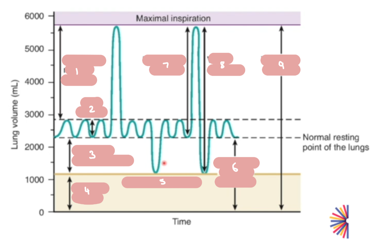

Label the key terms on a spirometry graph.


(Spirometry) Formulas for working out:
total lung capacity (TLC)
vital capacity (VC)
inspiratory capacity (IC)
functional residual capacity (FRC)
TLC = VC + residual volume (RV)
VC = tidal volume (VT) + inspiratory reserve volume (IRV) + expiratory reserve volume (ERV)
IC = VT + IRV
FRC = ERV + RV
What procedure can be used to measure ventilation? What do we use to measure residual volume (RV) and functional residual capacity (FRC)?
Ventilation measured using spirometry
RV and FRC can’t be measured by spirometry, so helium dilution method or plethysmography are used
helium dilution - the drum is filled with helium
plethymsography - patient sits in sealed chamber and breath through pneumotach
Physiological functions of residual volume in the lungs and the meaning of elevated RV and decreased RV.
Residual volume is the amount of air remaining in the lungs after a maximum forceful inhalation
This air is important as it prevents lung collapse - alveoli open at all times, and maintains a continuous reserve of O2 and CO2, preventing sharp, dangerous fluctuations in blood gas levels between breaths
Elevated RV indicates that air is becoming trapped inside chest, preventing normal complete emptying, e.g. in conditions like COPD
Reasons for increased RV include: respiratory muscles have a greater resting length, respiratory movements are less efficient, work required for breathing increases
Low RV indicates lungs cannot expand or hold a normal volume of air, e.g. in restrictive lung diseases like pulmonary fibrosis

Describe and explain changes in spirometry graph between healthy alveoli and COPD alveoli.
residual volume has doubled - dead volume has increased - because patients can’t empty their lungs completely before the next breath begins
total lung capacity is the same
it takes a similar amount of time to inhale, much longer time to exhale - due to lack of elasticity, stiffer lungs
reduced functional range - increased exercise increases the breathing rate - small amounts of air that they can inhale and exhale, less room left in chest cavity to draw in fresh breath
inspiratory capacity has halved - due to dynamic hyperinflation caused by air trapping
Why is alveolar ventilation less than pulmonary ventilation?
Pulmonary ventilation - moving gases into and out of lungs
Alveolar ventilation - volume of air that participates in gas exchange
alveolar ventilation is less than minute ventilation because a portion of every inhaled breath remains in the anatomical dead space - the conducting airways where gas exchange does not occur
Alveolar ventilation formula
Respiratory rate x (tidal volume (VT) - dead space (VD))
Anatomical and alveolar dead space in the respiratory system
dead space refers to the volume of inspired air that doesn’t take part in gas exchange across respiratory membrane
anatomical dead space is the volume of air trapped in the conducting airways that lead to the gas-exchange zones - includes nose, pharynx, trachea, bronchi and terminal bronchioles
alveolar dead space - volume of air that reaches respiratory zone (alveoli) but fails to take part in gas exchange because those alveoli are not perfused with pulmonary capillary blood flow.
this means air is flowing into the alveoli (ventilation), but blood is not flowing through the surrounding capillaries (perfusion)
What is physiological dead space and the clinical impact of it?
physiological dead space - sum of anatomical dead space and alveolar dead space, represents total volume of each breath that is ‘wasted’ and doesn’t eliminate CO2.
When physiological dead space increases, the body must breathe faster or deeper (increasing minute ventilation) just to clear normal amounts of CO2.
Define minute volume and tidal volume (VT) and give equations.
Minute volume - volume of air entering and leaving lungs each minute (pulmonary ventilation)
Minute volume (MV) = Respiratory rate (RR) x Tidal volume (VT)
Tidal volume - air taken in and out with each breath
Tidal volume = dead space (VD) + volume of air entering alveoli (VA)
Define respiratory rate and give normal rates for newborns, young children and adults.
Number of breaths per minute
Newborns: 30-60 breaths/min
Young children: 20-30 breaths/min
Adults: 12-20 breaths/min
What are the two airways of the respiratory system? In which part does gas exchange occur?
Conducting airways
Respiratory airways - where gas exchange occurs
What are some tests of ventilatory function? Measuring airflow
Forced Expiratory Volume in 1 second (FEV1) and Forced Vital Capacity (FVC) and its ratio FEV1/FVC expressed as a % - then measured against predicted values
Peak expiratory flow rate (PEFR) - convenient way of measuring airways obstruction, can be used to monitor asthma or COPD at home


in obstructive condition, e.g. emphysema - similar endpoint FVC, however it takes a lot longer to reach it. FEV1 is reduced by a lot
restrictive condition, e.g. pulmonary fibrosis - maximum is reached fairly quickly but FVC is much lower in the restricted condition
Define Boyle’s Law.
At a constant temperature (T) the volume (V) of a given quantity of gas is 1/ to the pressure (P) it exerts.

basically as volume increases, pressure decreases = they are inversely proportional.
Define Avogadro’s Law. (for gases)
Volume of gas at the same temperature and pressure contain the same number of molecules.
this means that even with different gases, e.g. hydrogen, helium, contain same number of molecules.
they are proportional to each other (moles and volume)
Define Charles’ Law.
At a constant pressure the volume of a gas is directly proportional to its absolute temperature.
this means increasing the temperature increases the volume of a gas at a constant temperature.
What is the Ideal Gas Law?
PV = nRT
pressure x volume = n (moles) x R (gas constant) x T
Define Graham’s Law. (gases)
The rate of diffusion of a gas is inversely proportional to the square root of its molecular weight.
this means a gas with a lower molecular weight diffuses more quickly
Define Henry’s Law.
The quantity of gas that can dissolve in a fluid is equal to the partial pressure of the gas multiplied by the solubility coefficient.
some gases are more soluble than others, e.g. oxygen is less soluble in water than other gases
Dalton’s Law of Partial Pressure
the pressure exerted by a mixture of gases is equal to the sum of the individual partial pressure exerted by each gas in the same volume.
this means if you add up the partial pressure of each of the individual gases making up a mixture of gases, the pressure exerted by the mixture is the same as the pressure exerted by each one individually
e.g. pressure exerted by air = 100, made up of one gas pressure 20, another gas pressure 70 and another gas pressure 10.
Describe the pressure gradients that drive ventilation.
Inspiration: contraction of the diaphragm and external intercostal muscles expands the thoracic cavity. as lung volume increases, intra-alveolar pressure drops below atmospheric pressure. air flows inward down this pressure gradient from the higher pressure outside the body to the lower pressure in alveoli
Expiration: as the diaphragm and external intercostal muscles relax, the thoracic cavity volume reduces. as lung volume decreases via elastic recoil, the intra-alveolar pressure rises above atmospheric pressure and as a result air is pushed out of lungs into the atmosphere down the pressure gradient.
What are the partial pressures of H2O, CO2, O2 and N2 in alveolar air, including units? What is the total pressure of alveolar air?

What does the rate of diffusion of gases from the alveoli to the blood depend on?
partial pressure of the gas
solubility of the gas in the liquid, e.g. CO2 ×20> O2
area available for gas exchange (e.g. dead space)
thickness of alveolar membrane
The ability of a gas to diffuse between alveolar air and blood is measured by what?
its diffusing capacity (or transfer factor) DL
Why is the rate of diffusion of CO2 in blood faster than oxygen?
because CO2 is more soluble, even though it has a higher molecular weight than oxygen
Oxygen:
Solubility in water?
How does it combine with haemoglobin?
Amount of oxygen carried by blood can be expressed as…
dissolves slightly in H2O (greater than N2 but less than CO2)
reversibly combines with haemoglobin (can attach to Hb in places with high O2 levels and detach in places that need O2)
Amount of O2 carried by the blood can be expressed as:
Arterial oxygen content CaO2 (ml/100ml)
Arterial oxygen content (CaO2) is the amount of oxygen bound to Hb plus the amount of oxygen dissolved in arterial blood
% saturation of Hb with O2, SaO2 (%)
the relationship of PaO2 to SaO2 and CaO2 is not linear but forms the oxygen dissociation curve for Hb.
Process of gas exchange in alveoli
O2: Inhaled air in the alveoli has a higher concentration of oxygen than the deoxygenated blood arriving from the pulmonary artery. Oxygen diffuses across the alveolar epithelium and capillary endothelium into the blood, where it binds to haemoglobin in red blood cells
CO2: The blood entering the pulmonary capillaries has a higher concentration of CO2 (produced by cellular respiration) than the air in the alveoli. CO2 diffuses out of the blood, across the capillary and alveolar walls and into the alveolus to be exhaled.
Structure of haemoglobin
composed of four protein globin chains, each surrounding a central heme group
in most adult haemoglobin, there are two alpha chains and two beta chains
each heme group consists of a porphyrin ring with an iron atom in the centre
How does haemoglobin help transport more oxygen in the blood? Oxygen content of plasma and red blood cells?
Oxygen content of plasma normally is 3mL per litre of blood, without haemoglobin, the alveolar pO2 = arterial pO2
Oxygen partial pressure = 100 mmHg
In blood with haemoglobin: oxygen content of red blood cells = 197 mL per litre of blood + 3mL of oxygen per litre in plasma = total oxygen carrying capacity: 200mL oxygen per litre of blood.
Red blood cells with haemoglobin are carrying 98% of their max. load of oxygen
Haemoglobin-oxygen dissociation curve: Explain.

in general, as the dissolved oxygen (pO2) increases, the haemoglobin saturation increases (% of oxygen bound to haemoglobin)
its not a linear, it is sigmoidal (S-shaped) due to the change in shape of haemoglobin (positive cooperativity)
when it binds to the first oxygen, that increases its affinity for the next two oxygen molecules to bind to it
graph plateaus at pO2 > 60mmHg - large changes in pO2 result in very little change in Hb saturation, due to it being harder for fourth oxygen to bind to Hb - there is only one empty site so chances of a free oxygen molecule colliding with and binding to it are significantly lower.
even if pO2 drops a little (e.g. from 100 down to 60mmHg), Hb stays heavily saturated
In the steeper phase of the graph (around 40mmHg or less), even a small drop in pO2 causes Hb to rapidly unload large amounts of oxygen to the cells that need it the most.
Explain shifts in the oxyhaemoglobin dissociation curve.
the curve can shift to the right or the left, which changes how tightly haemoglobin holds onto oxygen (its affinity):
Right shift:
lower affinity for oxygen, easier unloading
due to higher temps, higher pCO2, lower pH (more acidic/acidosis) and higher levels of 2,3-BPG
happens in hard-working muscles or active tissues that need more oxygen
called the Bohr effect
Left shift:
higher affinity for O2
due to lower temp, lower pCO2, higher pH and lower 2,3-BPG
happens in lungs (or fetal haemoglobin- has higher affinity for O2, which needs to pull oxygen from the mother’s blood in the placenta)
What is 2,3-BPG in red blood cells?
2,3-bisphosphoglycerate - organic phosphate molecule found in RBCs
it binds to central cavity of deoxygenated haemoglobin, stabilising it
this binding decreases haemoglobin’s affinity for oxygen, shift the oxygen-haemoglobin dissociation curve to the right
helps haemoglobin unload oxygen more easily into metabolically active tissues
levels increase in response to chronic hypoxia or high altitude to help tissues extract more oxygen when air pressure is low
Carbon monoxide binding to haemoglobin + symptoms
CO combines 200x more readily to Hb than O2
combination is almost irreversible
forms carboxyhaemoglobin
decreasing affinity for Hb for O2
impairing release of O2 at tissues
symptoms of CO poisoning vary depending of COHb level
between 3% and 24% symptoms include acute poisoning, headaches, nausea, blurred vision, dizziness, vomiting
24.3% is the mean level of loss of consciousness
32% is the mean level for fatality
How is carbon dioxide carried in the blood?
carried in the blood in three forms:
when cells excrete CO2, 7% of it dissolves straight into the blood
the other CO2: goes through a series of chemical reactions, forming bicarbonate (HCO3-) which is in the blood (70% of the CO2), and some binds to haemoglobin forming carbaminohaemoglobin (23%)
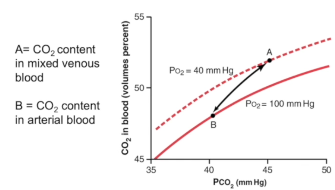

Explain carbon dioxide dissociation curve and Haldane Effect.
more linear and steeper than the oxyhaemoglobin dissociation curve - as there is no cooperativity, doesn’t rely on a saturable 4-site binding mechanism the way oxygen does
more CO2 in venuous blood than arterial blood as veins carry CO2 away from respiring cells
Haldane effect: removing O2 from Hb increases ability of Hb to pick up CO2 and CO2 generated H+ ions.
venuous curve: deoxyhaemoglobin has a higher affinity for CO2 and acts as a better buffer for H+ ions - as blood releases oxygen in tissues it shifts upwards to carry more CO2 at any given pCO2
arterial curve: as blood picks up oxygen in the lungs, the curve shifts downwards, displacing CO2 and forcing it out of the blood to be exhaled
How do the lungs control pH? Normal pH of blood?
CO2 is a metabolic waste product that dissolves in blood and combines with water to form carbonic acid (HCO3-) and hydrogen ions (H+). More CO2 means more H+ ions which lowers blood pH (making it more acidic)
If blood is too acidic (acidosis), brain signals lungs to breathe faster and deeper (hyperventilation), expelling more CO2 into the air - drops H+ levels in the blood and cerebrospinal fluid.
If blood is too alkaline (alkalosis), brain signals lungs to breathe slower and shallower (hypoventilation), retaining CO2 in blood to increase H+ levels and lower pH back to normal
Normal blood pH range: 7.35 and 7.45
Effect of hyperventilation and hypoventilation on CO2 levels in the body
hyperventilation causes low CO2 (hypocapnia) and alkalosis
hypoventilation causes high CO2 (hypercapnia) and acidosis
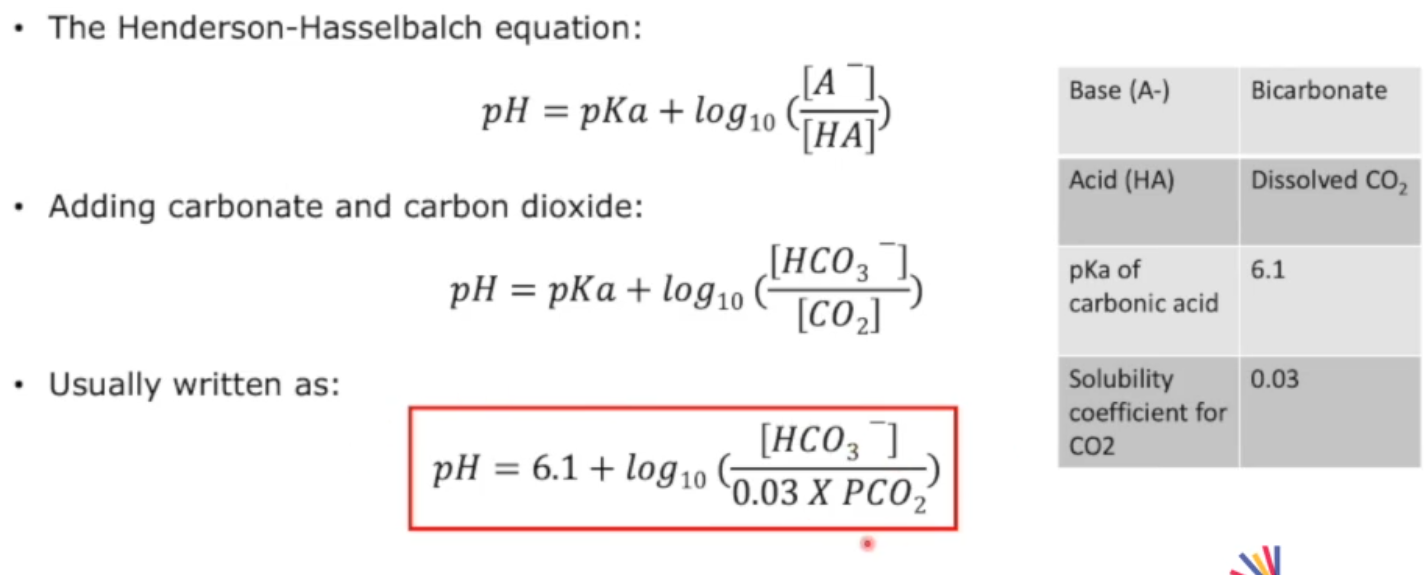
Henderson-Hasselbalch equation to estimate blood pH given bicarbonate ion concentration and partial pressure of arterial carbon dioxide
What enzyme mediates the conversion of carbon dioxide to carbonic acid in red blood cells?
Carbonic anhydrase
When carbon dioxide enters red blood cell, how is the charge of the cell maintained?
carbon dioxide forms HCO3-
bicarbonate ions leave the cell, making it more positive
chloride shift: Cl- ions enter the cell to maintain charge of cell.
Gas exchange at the tissue
some CO2 leaves respiring cells and dissolves into the plasma
majority of CO2 combines with water catalysed by carbonic anhydrase to form carbonic acid (H2CO3) - then dissociates into bicarbonate ions and H+
bicarbonate ions leave the cell and chloride ions enter RBC - chloride shift
some CO2 binds to amino groups on haemoglobin forming carbaminohaemoglobin inside RBCs
lower oxygen concentration at tissues causes haemoglobin to unload its oxygen HbO2 → Hb + O2
Gas Exchange at the lungs
bicarbonate ions move back into red blood cells, combing with H+ to form carbonic acid (H2CO3) and is converted back into CO2 + H2O with enzyme carbonic anhydrase inside RBCs
Carbaminohaemoglobin releases its CO2
free CO2 diffuses out of blood, across the alveolar wall and into the air spaces of the lungs to be exhaled
air inside alveoli has a high partial pressure of oxygen, so oxygen moves down the concentration gradient into red blood cells, binding to haemoglobin to form oxyhaemoglobin
Causes of pH change in the body and state, in general, physiological responses to it?
respiratory, e.g. hyperventilation (alkalosis), hypoventilation (acidosis)
metabolic, e.g. diabetic ketoacidosis (acidosis), vomiting (alkalosis), renal failure (acidosis)
Physiological responses:
buffering by chemicals including HCO3-, Hb and other blood proteins - this is rapid, but limited capacity
changes in ventilation - rapid, but impossible if primary cause is respiratory
changes in renal excretion of H+ and HCO3- - slower response, limited if primary cause is renal
Physiological response to respiratory acidosis
caused by low pH and high pCO2 (lungs retaining CO2)
kidney compensates by increasing plasma HCO3- and excreting H+ in the urine
slow - takes 3-5 days
HCO3- and pH rise
this restores pH towards normal
but HCO3- and pCO2 remain high
Physiological response to respiratory alkalosis
caused by high pH and low pCO2 (lungs lose excessive CO2)
kidney compensates by retaining plasma H+, excreting HCO3- in the urine
slow - takes 3-5 days
HCO3- and pH fall
this restores pH towards normal
but HCO3- and pCO2 remain low
Equation for dissociation of carbon dioxide equilibrium

(Respiratory) Transfer factor measurement - TLCO meaning and how is it measured
transfer factor of the lung for carbon monoxide
measured by breathing a gas containing a small amount of carbon monoxide and helium and looking at the changes between inspired and expired levels. it’s a measure of the quantity of carbon monoxide which is transported across the alveolar-capillary membrane for each kPa of alveolar carbon monoxide (PCO).
the helium is used to assess alveolar volume
(Respiratory) KCO diffusion coefficient meaning and how is it measured?
it is TLCO divided by the alveolar volume to which the gas has access
measured by breathing a gas containing a small amount of carbon monoxide and helium and looking at the changes between inspired and expired levels. it’s a measure of the quantity of carbon monoxide which is transported across the alveolar-capillary membrane for each kPa of alveolar carbon monoxide (PCO).
the helium is used to assess alveolar volume
Davenport diagram - identify the 6 acid-based disorders given pH, pCO2 and bicarbonate concentration.


metabolic acidosis = decrease in arterial pH, usually due to a decrease in plasma bicarbonate concentration (bicarbonate in plasma acts as a base that keeps blood pH normal)
metabolic alkolosis = increase in arterial pH, usually due to an increase in plasma bicarbonate concentration
respiratory acidosis = when your lungs cannot get rid of carbon dioxide causing CO2 to build up in your blood and drop blood pH below normal
respiratory alkalosis = hyperventilation causes body to exhale too much carbon dioxide, driving blood pH above normal
when acidosis/ alkolosis is chronic, the body has time to adapt - renal compensation - kidneys have time to reabsorb/excrete (alkolosis) and generate more bicarbonate (acidosis)
Structural divisions of the respiratory system
Upper respiratory
nose, nasal cavity and paranasal sinuses
pharynx
larynx
Lower respiratory
trachea
bronchi
Describe the external anatomy of the nose (simple)
nasal bone and cartilage covered by facial muscles and skin
apertures (openings) are the nostrils
Functions of the nasal cavity
warms and humidifies inspired air
removes and traps pathogens and particulate matter from inspired air
responsible for sense of smell
drains and clears the paranasal sinuses and lacrimal ducts
resonating chamber
Describe the anatomy of the nasal cavity.
entered anteriorly through the nostrils
opens posteriorly into the nasopharynx through the choanae
mucosa lines the nasal cavity, except for the nasal vestibule which is lined with skin
roof of the nasal cavities is curved and narrow, except at its posterior end where the body of the sphenoid bone forms the roof
floor of nasal cavities is formed by the palatine processes of the maxilla and the horizontal plates of the palatine bone
medial wall of the nasal cavities is formed by the nasal septum
lateral walls of the nasal cavities are made up of the nasal conchae
nasal conchae (superior, middle, inferior) project medially from the lateral wall of the nasal cavity, and meatus (superior, middle, inferior) underlies each concha
sphenoethmoidal recess superior to the superior concha

Function of the conchae (turbinate bones)
scrolled shape increases surface area, warming and moisturising air before it reaches lungs
middle and superior conchae guide air currents toward the olfactory epithelium at top of nasal cavity to help process smells
filter impurities - trap dust, pollen and debris in the mucus lining
Histology of the nasal vestibule and respiratory region of the nasal cavity
Nasal vestibule:
keratinising and non-keratinising stratified squamous epithelium, contains hair follicles that trap large particles
Respiratory region:
ciliated pseudostratified columnar epithelium
goblet cells produce and secrete mucus to humidify air + trap particles
ciliated columnar cells move mucus and trapped debris toward the pharynx
What are the paranasal sinuses?

sinus = hollow space within bone
Where do the paranasal sinuses drain?
frontal sinus drains into middle meatus
maxillary sinus drains into middle meatus
anterior and mid ethmoid air cells drain into middle meatus
posterior ethmoid air cells drain into superior meatus
sphenoid sinus drains into sphenoethmoidal recess
What is sinusitis? symptoms?
inflammation of tissue lining paranasal sinuses
blocked with fluid
symptoms include:
pain, pressure around cheeks, eyes or forehead
blocked or stuffy nose
thick, discoloured mucus from nose or down back of throat
reduced sense of smell/taste
fever, headache, toothache
Function of the nasolacrimal duct
when lacrimal gland secretes tears, eyelids sweep tears towards the lacrimal sac
this drains tears down the nasolacrimal duct to the inferior meatus
nasolacrimal duct drains into inferior meatus
Epistaxis
nosebleed
originate from Kiesselbach’s plexus (or Little’s area) on the lower nasal septum
Kiesselbach’s plexus - lots of blood supply to it as many arteries supply the area, e.g. ethmoidal artery, palatine artery
common causes: local irritation and trauma (nose blowing, picking nose), dry air, medications
Anatomical position of the pharynx + overview
muscular tube
posterior to the nasal cavity, oral cavity and larynx
base of the skull superiorly
cricoid cartilage of larynx inferiorly
3 constrictor muscles: superior, middle, inferior
3 parts: nasopharynx, oropharynx, laryngopharynx
Parts of the pharynx and regions
nasopharynx: from base of the skull to the uvula and soft palate
oropharynx: from soft palate to epiglottis
laryngopharynx: from epiglottis to cricoid cartilage
Structures of nasopharynx
from base of skull to uvula and soft palate
tubal opening (eustachian tube), tubes drain and equalise pressure in middle ear
pharyngeal tonsil (adenoids) and tubal tonsil
tonsils are collections of lymphoid tissue and immune system’s first line of defense
Tonsils in the oropharynx
palatine and lingual tonsils
Anatomical position of the larynx and its functions
superiorly - laryngeal inlet
inferiorly - cricoid cartilage
anteriorly - trachea
posteriorly - oesophagus
functions:
phonation (arytenoid cartilages and vocal ligament)
cough reflex
protection of the lower respiratory tract
What cartillages are within the larynx?
Unpaired: epiglottis, thyroid and cricoid
Paired (one on either side): arytenoids (vocal folds), cuneiform and corniculate


false vocal cords also known as ventricular cords
What is phonation? What produces changes in pitch?
process where airflow from lungs is converted into sound by making the vocal folds vibrate inside the larynx
rima glottidis (aperture between vocal folds) changes shape as vocal folds move
muscles stretch and tense or relax the vocal ligament, and abduct or adduct the vocal folds - these movements permit changes in pitch
Cause of dysphonia
dysphonia - abnormal functioning of voice
impairment of the vocal vibrations
inflammation of the vocal folds - hoarse voice
What is the vertebral level of the arch of cricoid cartilage, superior end of oesophagus and superior end of trachea?
vertebral level C6 (CVI)
Describe the trachea. At what vertebral level does it bifurcate?
main airway
anterior to the oesophagus
has c-shaped cartilage rings bridged by trachealis
it bifurcates (splits into two) into the two primary bronchi at T4/T5 (manubriosternal angle)
Functions of the thoracic cage.
organ protection - encloses and shields vital organs in the chest cavity from external trauma and impact
respiration support - facilitates breathing, rhythmically expanded by the respiratory muscles to create a vacuum that draws air into the lungs
Rib numbers of the true ribs and the false ribs
true ribs: 1-7
false ribs: 8-12
floating ribs: 11, 12
What are the muscles involved in inspiration?
sternocleidomastoid
scalenes
external intercostals
parasternal intercostals
diaphragm
What are the muscles involved in expiration?
internal intercostals
external abdominal oblique
internal abdominal oblique
transversus abdominis
rectus abdominis
Diaphragm
floor of the thoracic cavity and roof of the abdominal cavity - partition between these two regions
dome shaped muscle
when it contracts, the top of the dome flattens down
Process of inspiration
diaphragm contracts and descends
external intercostals contract to raise ribs
volume of thoracic cavity increases
intrathoracic pressure drops below atmospheric pressure
air moves in down the pressure gradient
Process of expiration
expiration can be passive (quiet breathing) or active (forced breathing)
quiet breathing - relies on natural relaxation of diaphragm and external intercostals, combined with the elastic recoil of the lungs
forced expiration - internal intercostals pull the ribs down and in to reduce chest volume, abdominal muscles compress the abdominal organs upward against the diaphragm and pull the lower ribs down, sharply decreasing thoracic volume
What are the blood and nerve supply to the intercostal spaces?
intercostal vein
intercostal artery
intercostal nerve
present in each intercostal space
collateral vein, artery and nerve
Explain the physiological mechanism of a cough
Cough has three phases: inspiratory, compressive and expiratory.
Inspiratory phase - vocal cords (glottis) open wide (abduct). Diaphragm and external intercostal muscles contract to draw a deep breath into lungs, increasing lung volume
Compressive phase - vocal cords snap shut (adduct) to close the glottis, abdominal wall and internal intercostal muscles contract forcefully. Glottis is closed, so air cannot escape, causing pressure (subglottic and thoracic) to build up rapidly in lungs
Expiratory phase - glottis suddenly opens and trapped high-pressure air bursts outwards at extremely high speeds, creating a high-velocity airflow 'spike' that shears mucus and debris off the airway walls and expels them.
Describe the cough reflex arc
Caused by irritation of respiratory tract by gases, smoke and dust
The cough reflex arc:
(afferent pathway) - Chemical or mechanical stimuli activate sensory nerve endings (receptors) in larynx, trachea, bronchi or upper airways. Signals travel via vagus nerve to nucleus tractus solitarius (NTS) in brainstem
(central pathway) - 'cough centre' in pons and medulla processes input.
(efferent pathway) - output signals trave down vagus, phrenic and spinal motor nerves to coordinate respiratory and laryngeal muscles.
in trachea, output is a cough, in lower airways leads to hyperpnoea (deep or fast breathing to match demand for oxygen)
also causes reflex bronchial & laryngeal constrictions
results in expulsion of irritants from respiratory tract
Why is a cough clinically significant?
Indication to diseases such as rhinitis (acute), or from cigarette smoke, pulmonary diseases, and asthma (chronic)
Could also be side effects from medication (ACE inhibitors like ramipril)
Role of opiate receptors in cough and respiration
Opioid receptors are specialised receptors on surface of cells in the brain, spinal cord and digestive tract.
They inhibit pain signals and influence mood, stress, respiration, and digestion.
Opiates suppress the cough reflex primarily by binding to mu-opioid receptors (MOR) and kappa-opioid receptors (KOR) located on presynaptic neurons in the brainstem (medullary cough centres). This inhibits transmission and processing of cough signal.

Label each structure of respiratory system.

What types of epithelium are in the conducting zone of the respiratory system?
pseudostratified with 3 cell types:
ciliated epithelial cells - mucociliary escalator
goblet cells (secretory, non-ciliated)
basal cells (act as stem cells, may contribute to structure)
columnar in nasal cavities, trachea, bronchi
more cuboidal in bronchioles with Clara cells replacing goblet cells
What are the defence mechanisms of the nose and oropharynx?
nasal hairs of the nose filters large particles from inspired air
nose warms and humidifies the air - prevents epithelium drying out
mucosal layer lined with different types of epithelial cells:
respiratory epithelium - bathed in aqueous viscous and sticky mucus to trap particles
ciliated epithelium have a coordinated beat - ‘mucociliary escalator’ push material towards the nasopharynx and oropharynx
irritant particles stimulate irritant receptors and provoke sneezing and coughing to remove material
smell is also a defence mechanism!
Histology of the bronchus
presence of plates of hyaline cartilage
smooth muscle and goblet cells are also present
lined with ciliated pseudostratified columnar epithelium
Histology of terminal bronchiole
terminal bronchioles are smallest and final segments of the conducting portion of the respiratory system, mark end of air transport before respiratory gas-exchange airways begin
no cartilage or glands
epithelium in initial portion is ciliated columnar but becomes cuboid with cilia
absence of goblet cells
final portion un-ciliated smooth muscle - allows autonomic control of airway diameter
contain club cells (clara cells)
What are Club Cells (Clara cells)?
dome-shaped, non-ciliated secretory cells found in bronchioles of the lungs
secrete lipoproteins and glycoproteins to protect the bronchiole epithelium and prevent luminal collapse
act as local stem cells in the bronchiolar epithelium, multiplying and differentiating into ciliated cells to repair and regenerate the airway lining after injury
Cell types present in alveoli
Type I pneumocytes (epithelial cells)
Type II pneumocytes (epithelial cells)
Alveolar macrophages
Neighbouring alveoli interconnected by ‘Pores of Kohn’ - small openings in the walls of adjacent pulmonary alveoli that allow air, fluid and cells to pass between them
What are Type I pneumocytes?
90 - 95% of alveolar surface
highly flattened
simple squamous epithelium
cannot replicate/ divide - if damaged, they’re replaced with Type 2 cells
form occluding junctions with one another
barrier to extracellular fluid into alveolus
type I cells forms thin diffusion barrier - fused with pulmonary capillary endothelium: the blood-gas interface, allows rapid diffusion of oxygen into blood and CO2 out of blood
What are Type II pneumocytes?
more numerous than type I but only about 5-10% of the alveolar surface
cuboidal with apical microvilli
capable of rapid cell division
dispersed among type I pneumocytes and form occluding junctions with them
produce surfactant - reduces surface tension, prevents alveoli from collapsing during exhalation
Sneezing reflex
caused by irritation of nasal mucous membrane due to dust particles, debris and excess fluid accumulation
characterised by deep inspiration followed by forced expiration with open glottis
results in expulsion of irritant from airways
irritation of nasal mucous membrane, olfactory receptors and trigeminal nerve endings in nasal mucosa
afferents from trigeminal and olfactory nerves pass to sneezing centre in medulla
efferent fibres pass from medulla via trigeminal, facial, glossopharyngeal, vagus and intercostal nerves
results in activation of pharyngeal, tracheal and respiratory muscles
Swallowing reflex
initiated by stimuli to dorsum of tongue, soft palate and epiglottis
during swallowing, respiration is inhibited - deglutition reflex
prevents inhalation of food
What is the mucociliary escalator? How does it work? What can damage it?
defence mechanism of the airways
two layers of mucus coating cilia of epithelium - thin sol layer and gel layer on top
sol layer - more fluid and less viscous than gel layer - enables the cilia to beat, if it was more thick, cilia would be inhibited and wouldn’t beat properly
gel layer - thicker layer of mucus, acts as a trap for particulate matter, dust, bacteria
creates semipermeable barrier and allows exchange of nutrients, H2O and gases - it is impermeable to most pathogens
cilia beat in coordinated fashion to removed unwanted particles via ‘escalator’ towards the mouth
mucus from large bronchi to reach pharynx takes about 40 min
from respiratory bronchi it takes several days
escalator can be damaged by inflammation, smoking, pollution, infections
What is the mucus and how does it provide non-specific immunity?
produced by goblet cells
main component of airway mucus are mucins, which are high molecular weight glycoproteins - give it gel-like nature
fluidity and ionic composition of sol phase controlled by epithelial cells
mucus contains:
anti-proteases, e.g. a1-antitrypsin - inhibits proteases released from bacteria and neutrophils, which can be destructive to us
lysozyme - has anti-bacterial and anti-fungal properties (enzyme that targets bacterial cell walls)
anti-microbial properties from lactoferrin (glycoprotein that binds to free iron), peroxidases and neutrophil-derived defensins (disrupt pathogen membranes)
these provide non-specific immunity
What are the main roles and characteristics of pulmonary immune responses?
multifaceted - protective (prevents infections with non-specific immunity) and reactive roles (responds when infection, injury or inflammation occurs)
lungs are immunologically active
healthy lungs - contain relatively few leukocytes
when there are pathological conditions - increased influx and diversity of leukocytes
involves a large number of receptors, cytokines and chemokines
vital in immune homeostasis, infection and inflammation
What are cytokines and chemokines?
cytokines - signalling proteins that activate and regulate immune responses
chemokines - chemoattractant proteins that recruit immune cells to specific locations
both help coordinate immune responses in the lungs (as well as other body parts)
Give examples of innate immune cells and adaptive immune cells.
Innate (general): neutrophils, eosinophils, basophils/mast cells, natural killer cells, dendritic cells, monocytes and macrophages
Adaptive (specialised): T-helper cell, cytotoxic T-lymphocyte, B cell, plasma cell