Features of the PA Chest X-Ray
 Call Kai
 Learn
 Practice Test
 Spaced Repetition
 Match
 Flashcards
 Knowt Play
1/17

There's no tags or description
Looks like no tags are added yet.
18 Terms

In order: trachea, carinus (point where the trachea splits), primary main bronchi/bronchus and secondary bronchi (two on left and three on right).

Left and right lungs.
Lobes can only be seen when there is a pathology present.
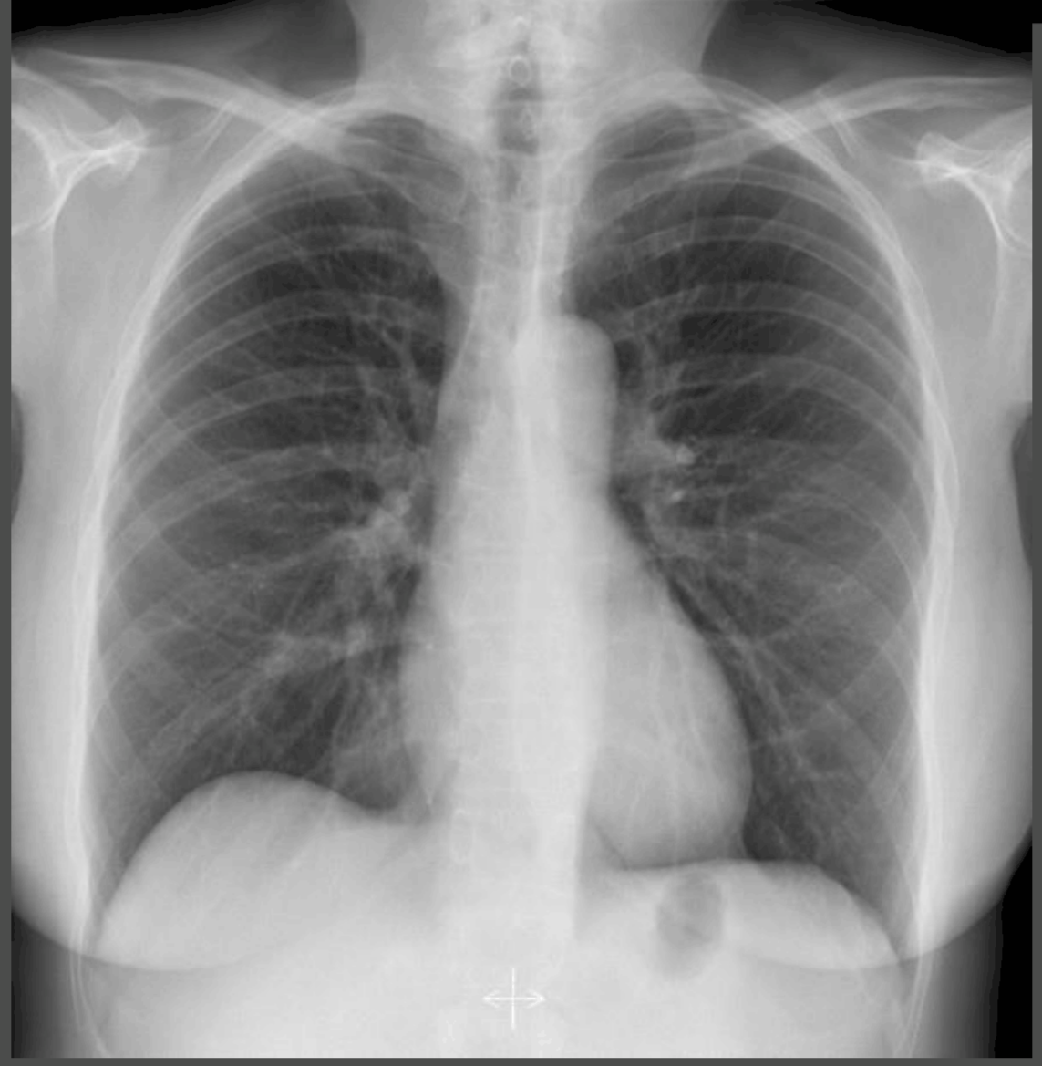
What are the fibrous markings on the radiograph?
Called lung markings
Result from the radiopaque pulmonary (lung) arteries and veins
Normal

Purple: Hilar root or hilar zone.
Hilum: The opening on the lung.
Hilar root: The physical structures which pass through the hilum (i.e., the pulmonary vessels0.
Hilar zone: The area on the X-Ray where the shadow/silhouette of the hilar root is visible.
Orange: Hilar point.
Hilar point: The point where the upper and lower pulmonary vessels
meet/converge at the lateral aspect (lateral face) of the lung.
Left hilar point is usually HIGHER than the right hilar point since the left pulmonary artery ARCHES over the left main bronchus.
Right hilar point is aligned at around T6/T7.

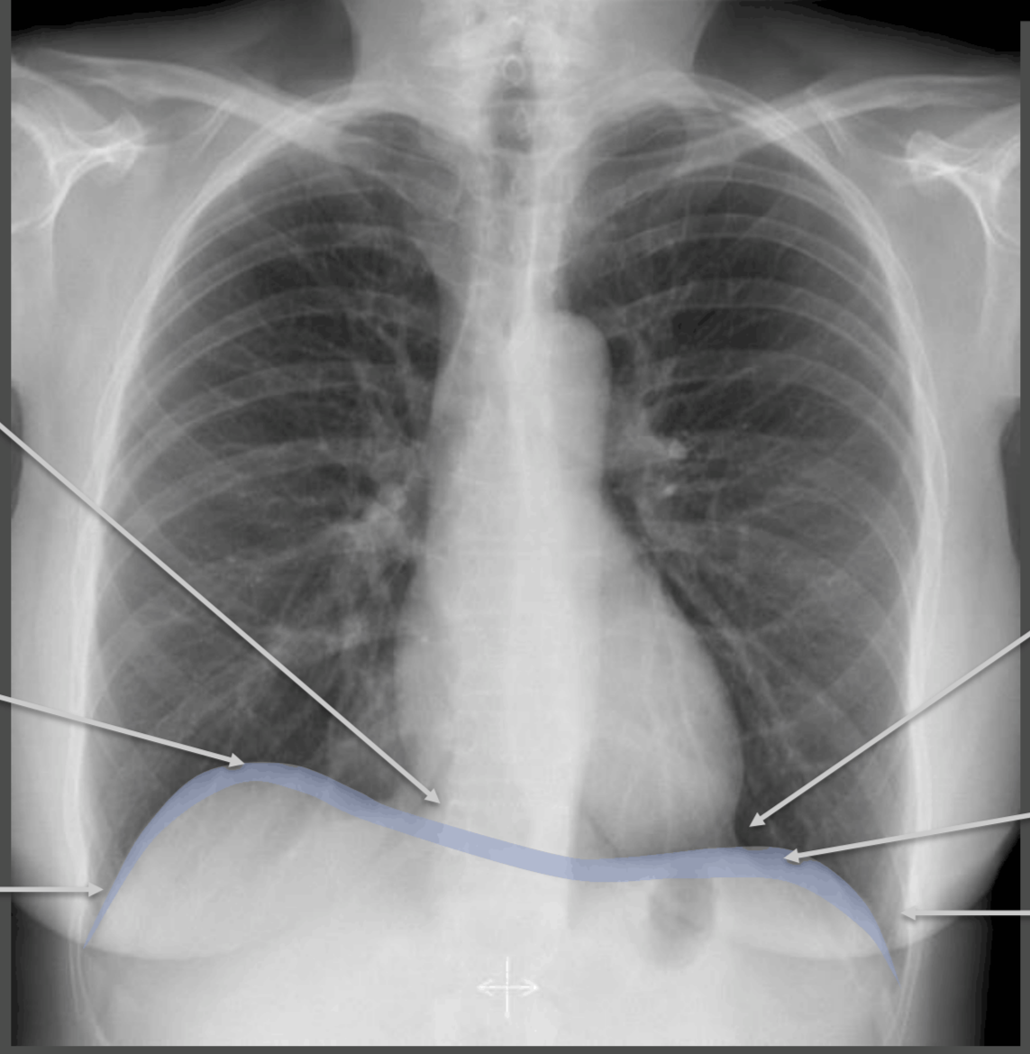
From left to right;
Right costophrenic angle
Right hemidiaphragm
Right cardiophrenic angle
Left cardiophrenic angle
Left hemidiaphragm
Left costophrenic angle

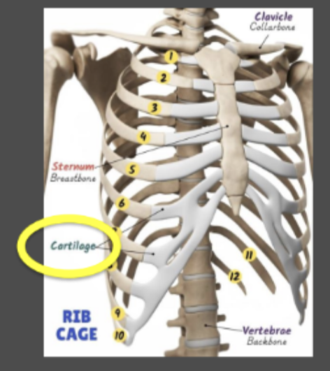
Clavicles

Sternum
Harder to identify since it is superimposed by the vertebral column and the mediastinum.
In order:
Manubrium: Head of the sternum.
Body: Body of the sternum.
Xiphoid process: The lower part of the sternum.

Scapula

Posterior part of the rib
Runs laterally and then curves inward.

One rib entirely
*NB: Costal cartilage is considered SEPARATE from the rib.

Red:
Heart shadow, or the
Cardiac silhouette
*NB: DO NOT call it the heart, SINCE you can not actually see the separate structures of the heart (you just see a radiopaque shadow).

Aortic knuckle (or aortic knob or aortic loop): The rounded upper-part of the aortic silhouette — it is the shadow of the aortic arch.
Parts of the aortic silhouette:
Aortic knob: Rounded upper part of the aortic silhouette.
Ascending aorta contour: The outline of the ascending aorta.
Descending aorta contour: The outline of the descending aorta.


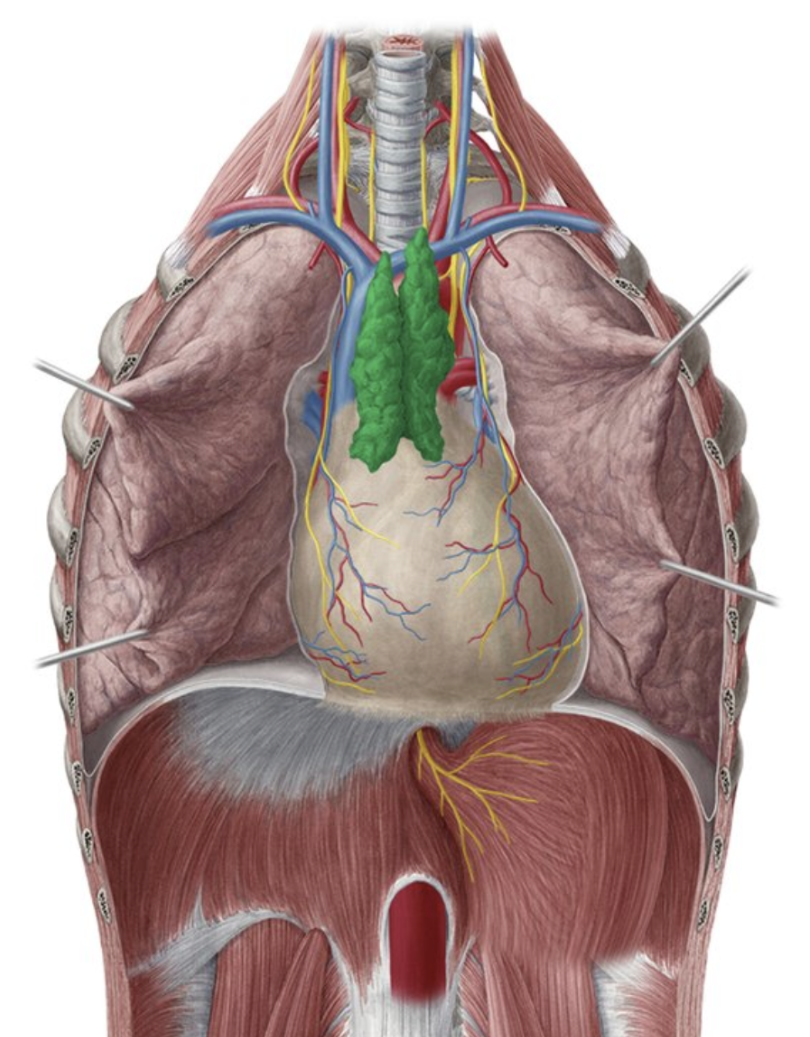
Thymus
Harder to see since it is superimposed by other structures such as the vertebrae and the trachea.
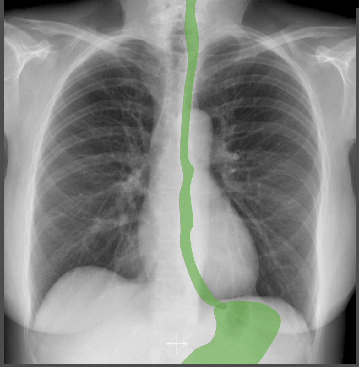
Oesophagus and stomach (cardia, fundus and pylorus).

Spleen
Slightly radiolucent structure medial and slightly superior to the spleen is called the gastric bubble.
Gastric bubble: A pocket of gas that is formed when air that is swallowed when you eat; normally found in the fundus.

Liver
Reason why the hemidiaphragm sits more superior compared to the left hemidiaphragm since it pushes the right hemidiaphragm more up.


Breast tissue.

Anterior part of the rib.
Image is slightly wrong since the rib does not connect all the way to the sternum; the costalcartilage exists between the sternum and the rib.