Exam 3 - Tumors
 Call Kai
 Learn
 Practice Test
 Spaced Repetition
 Match
 Flashcards
 Knowt Play
1/127

There's no tags or description
Looks like no tags are added yet.
128 Terms
What is the second MC primary malignant tumor of bone
Osteosarcoma
What is the third MC primary malignant tumor of bone?
Chondrosarcoma
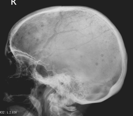
What is the finding/sign and what is the most likely diagnosis and what would be the appropriate follow-up testing to accomplish?
Rain drop skull
Multiple myeloma
Protein electrophoresis (PEP)
What is a plasmacytoma?
A solitary site of multiple myeloma (typically lytic & expansile)
What is the MC primary malignant tumor of the bone that metastases to bone?
Ewing’s sarcoma (also mets to lungs)

How would you describe the periosteal rxn?
Onion skinned (laminated or multilamellated)

What is the diagnosis?
Lytic mets

What is the likely diagnosis in this 15 year old patient?
Ewing’s sarcoma
What are the top three MC cancers and what are the top three cancers that are responsible for bone mets?
Breast
Prostate
Lung
What is the second MC primary malignant tumor of bone in a child?
Ewing’s sarcoma
A 14 year old female patient w/ pain in the mid-shaft region of the left femur has a permeative lytic destructive change with a multi-layered periosteal rxn, cortical destruction and a soft tissue mass. What is the most likely diagnosis?
Ewing’s sarcoma
What is the likely diagnosis in a 15 year old patient with a painful permeative lytic destructive region with areas of sclerosis noted in the metaphyseal region of the proximal tibia with a spiculated periosteal rxn and a soft tissue mass?
Osteosarcoma

What is the most likely diagnosis in this 45 year old male patient with pain for the last few months?
Chondrosarcoma
Bence Jones proteinuria is most likely associated with ___
Multiple myeloma
What is the typical age range associated with an osteosarcoma?
10-25 & > 60
Bimodal age distribution

What is the most likely diagnosis?
Osteosarcoma
What is the typical age range associated with a chondrosarcoma?
40-60 yo
What is the typical age range for a Ewing’s sarcoma?
10-25 yo
Cortical saucerization and an onion skinned periosteal rxn are often associated with a/an ___
Ewing’s sarcoma
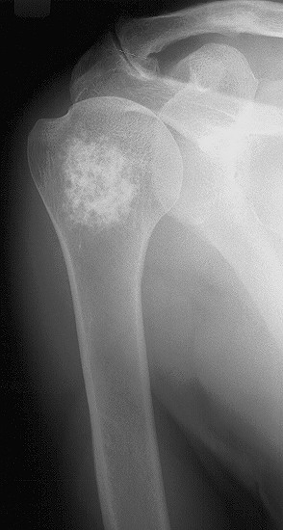
What is the ddx for this patient?
Chondrosarcoma
Enchondroma
Calcified medullary infarction
What is the classic location for an osteosarcoma?
Metaphyseal → knee and proximal humerus
What is the classic location for Ewing’s
Diaphyseal
What is the fourth MC primary malignant tumor of bone?
Ewing’s sarcoma
__ is a malignant proliferation of plasma cells.
Multiple myeloma
What is the third MC blood cancer?
Multiple myeloma

What is the finding and what is the most likely diagnosis and what is the appropriate follow up?
“Raindrop skull”
Multiple myeloma
Protein electrophoresis

What is the most likely diagnosis?
Lytic mets
Moth eaten lytic destructive changes
Male → from lung
Female → from breast
What age is the most likely for an osteosarcoma?
10-25
What is the most likely etiology of a pathological spine fx in a child?
Eosinophilic granuloma
What is the most likely etiology for a “winking owl” sign?
Lytic mets
Lytic mets involving the spine in a 56 yo female would likely have come from the ___
Breast
What is the MC malignant tumor of bone?
Mets
What is the MC primary malignant tumor of bone?
Multiple meyloma
Which type of mets is MC?
Lytic
Would a periosteal rxn be more likely associated with a primary or secondary bone tumor?
Primary
__ is a type of periosteal rxn and is considered the most aggressive type of a periosteal rxn
Codman’s triangle
Which type of bone destruction is considered the most aggressive?
Permeative

Describe the zone of transition
Describe the lytic change
Describe the location
Does this appear benign or aggressive?
Short/narrow b/c thick sclerotic border
Geographic
Central and subtrochanteric
Very benign

What are the arrows pointing at?
Does this appear benign or aggressive?
Perpendicular periosteal rxn (sunburst/hair on end)
Aggressive

What is the arrow pointing at?
Layered periosteal rxn
Onionskin periosteal rxn
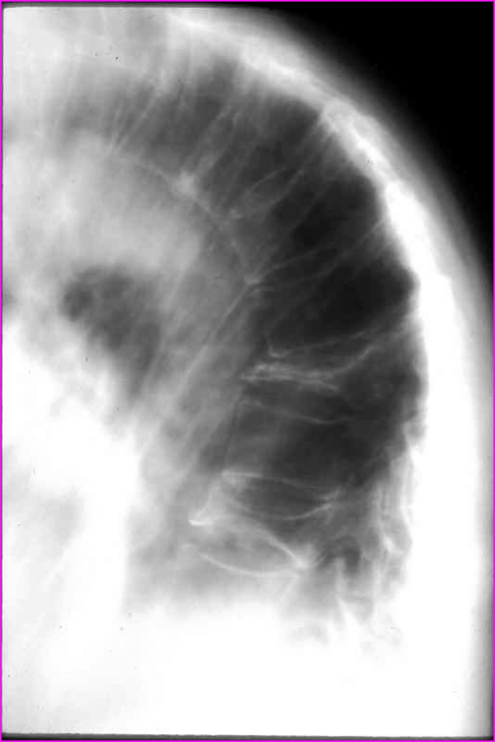
What is the finding, what is the differential, and what is the most likely diagnosis in this patient?
Multiple pathological fractures
Osteoporosis, lytic mets, and multiple myeloma
Osteoporosis
What is the most likely etiology for a one eyed pedicle sign?
Lytic mets

What is the finding and what is the DDX and what is the most likely cause in this patient?
One eyed pedicle sign at L1 on the left
Congenital absence, destructive changes from a primary/secondary bone tumor, infection, or soft tissue tumor
Lytic mets (female patient → breast cancer)
What is the MC malignant tumor of bone?
Metastasis

What is the diagnosis and what is the most likely primary cancer site?
Blastic mets → prostate primary
What is the general rule of thumb concerning bone marrow signal intensity on a T1 weighted MRI?
Bright is right
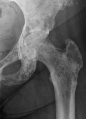
What is the most likely diagnosis?
Mixed mets (lytic and blastic changes)

What is the differential diagnosis in this 53 yo patient?
Bone island or blastic mets
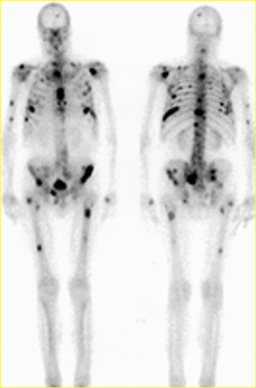
What type of imaging is this?
Bone scan/radionuclide scintigraphy

Describe the lesion, including the zone of transition, and does it look benign or aggressive?
Central geographic lytic change w/ thick sclerotic border and a hazy internal matrix (ground glass appearance) w/ short zone of transition in subtrochanteric region of femur
Benign

What are the arrows pointing at?
Does this appear benign or agressive?
Perpendicular periosteal rxn (sunburst)
Aggressive

What is the arrow pointing at?
Onion-skin periosteal rxn

What is the finding and what is the differential and what is the most likely diagnosis in this patient?
Multiple pathological fxs
Osteoporosis
What is the most likely etiology for a one-eyed pedicle sign?
Lytic mets

What is the finding, what is the DDx, and what is the most likely cause in this patient?
One-eyed pedicle sign and L1 on the left
Congenital absence, destructive changes from primary/secondary bone tumor, or infection
Lytic mets
What is the MC malignant tumor of bone?
Metastasis
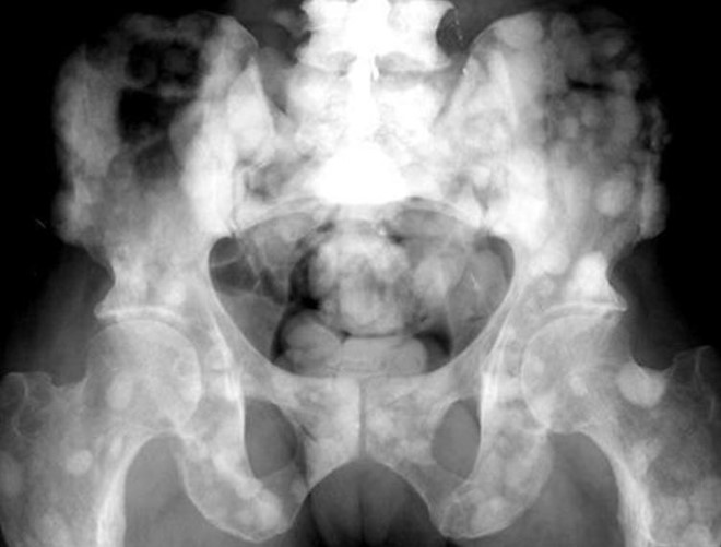
What is the diagnosis and what is the most likely primary cancer site?
Blastic mets
Prostate
What is the general rule of thumb concerning bone marrow signal intensity on a T1 weighted MRI?
Bright is right
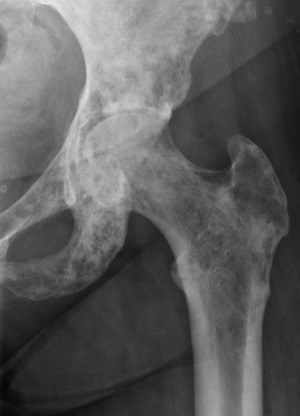
What is the most likely diagnosis?
Mixed mets

What is the differential in this 53 year old pt?
Bone island and blastic pathology
Tumor matrix calcification described as rings and arc, stippled, flocculation or popcorn like would suggest which type of tumor?
Cartilaginous based tumor
A “ground glass” appearance of tumor matrix would suggest a/an __
Fibrous type of tumor
What are the top three MC cancers?
Breast, prostate, and lung
What is the MC pathway for metastasis?
Hematogenous dissemination

What description would you use to describe the tumor matrix calcification and what would it suggest or represent?
Stippled, punctate, or flucculent = Cartilage based tumor
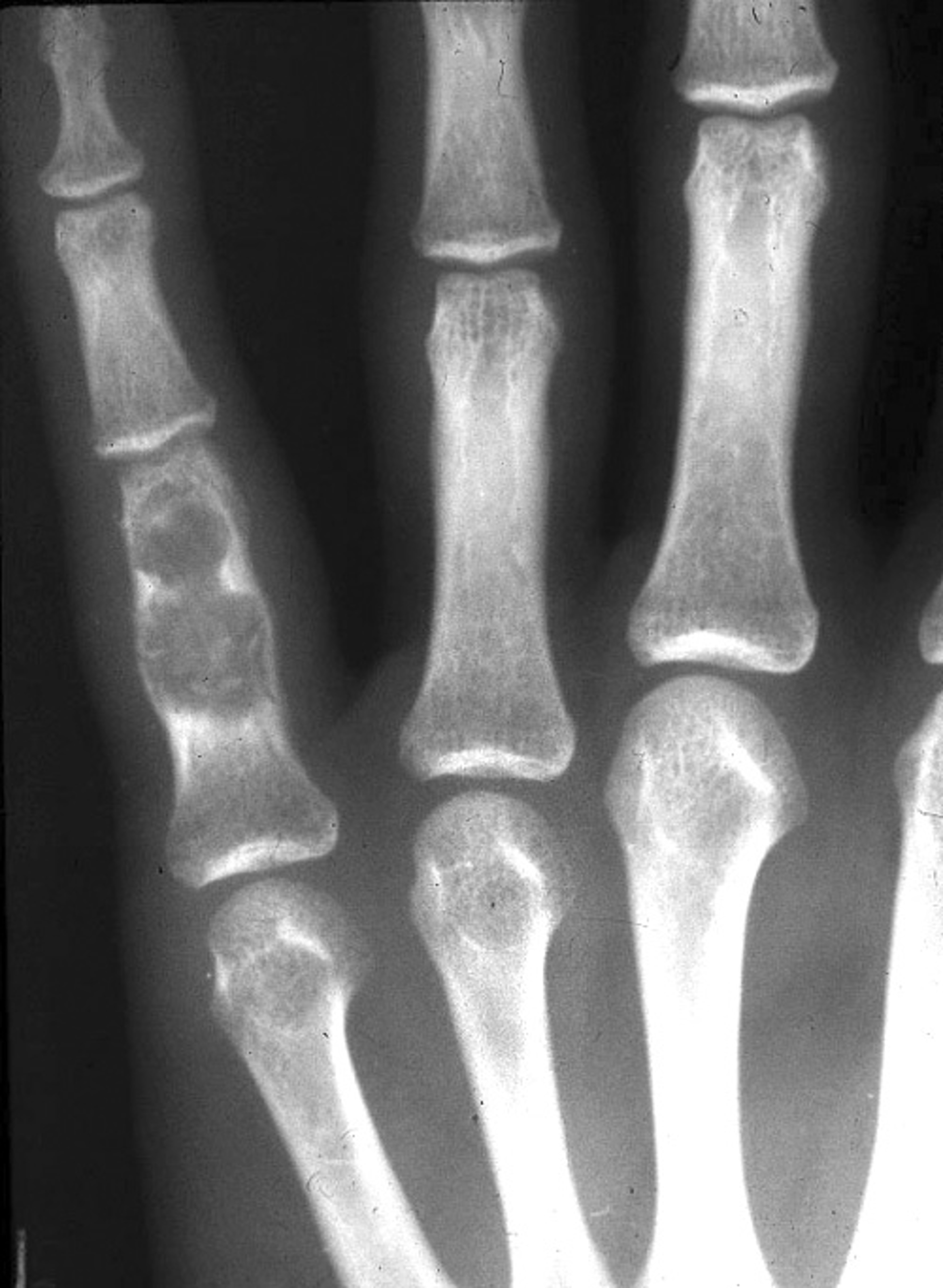
Describe the lesion and what is the zone of transition? Does it look benign or aggressive?
Central geographic lytic change w/ slight expansion & cortical thinning in the proximal phalanx of digit #5
Short zone of transition
Benign
Which type of periosteal reaction is considered to be most aggressive?
Codman’s triangle

How would this area of destruction be described?
Geographic lytic w/ thin sclerotic border & short zone of transition
What is the differential diagnosis for a pathological spine fx?
Osteoporosis, lytic mets, & multiple myeloma
What are the three MC cancers?
Breast
Prostate
Lung
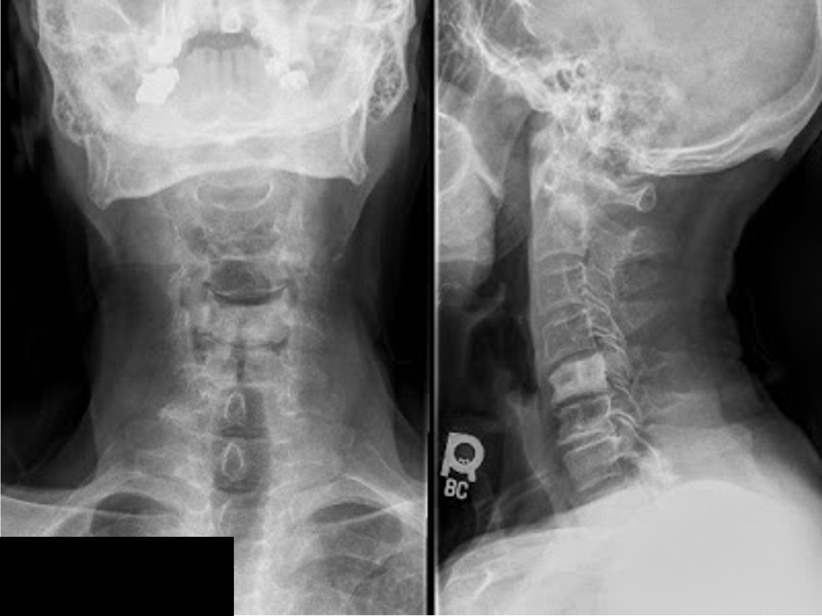
What is the most significant finding, DDx, and what is the most likely Dx?
Ivory vertebra at C5
Blastic mets, Paget’s, and lymphoma
Blastic mets
What is the MC primary malignant tumor of bone? (TQ)
Multiple myeloma
A 17 yo patient w/ hip pain for months, x-rays reveal cortical thickening, reactive sclerosis and a 0.5 cm rounded radiolucency in the medial aspect of the intertrochanteric region on the left. ___ is the most likely diagnosis.
Osteoid osteoma
What is the usual age range associated with a giant cell tumor?
20-40

This is an 18 yo patient w/ pain for months. What is the most likely diagnosis?
ABC or osteoblastoma

What is the finding and what is the most likely diagnosis?
One eyed pedicle sign
Lytic mets

What is the finding and likely diagnosis?
Eccentric oval shaped radiolucency in the epiphysis of distal tibia
Chondroblastoma
An expansile neural arch tumor in a young patient is most likely a/an ___
ABC
Fluid-fluid levels on an MRI associated with a geographic, eccentric metaphyseal lytic lesion would likely be associated with a/an ___
ABC
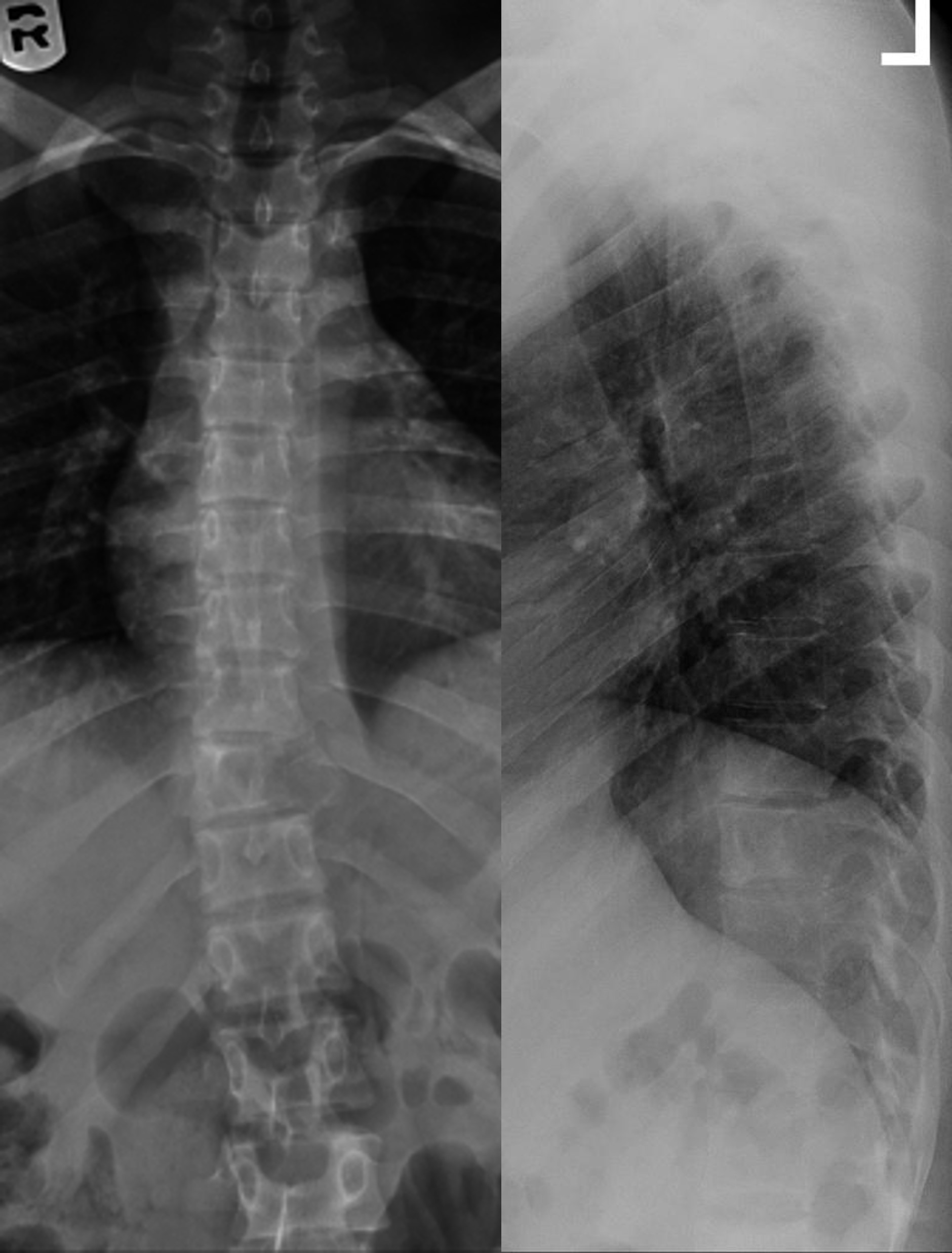
What are the findings and DDx in this 16 yo patient with pain?
One eyed pedicle sign at T10 on left
ABC or osteoblastoma
Your 16 yo patient has a central, geographic, lytic lesion that is somewhat truncated in appearance measuring 5cm in length in the metaphyseal region of the proximal humerus. What is the most likely diagnosis?
SBC
Your 16 yo patient has a painful, lytic, eccentric geographic lesion with a short zone of transition in the metaphyseal region of the proximal tibia. There is no associated soft tissue mass or periosteal rxn. The likely diagnosis is a/an ____
ABC

This lesion is painful. What is the most likely diagnosis in this patient?
Chondroblastoma
What age range is typically seen with an osteoid osteoma?
10-25

12 yo male patient with pain. Describe the lesion and what is the likely diagnosis?
Small rounded radiolucency with slight cortical thickening
Osteoid osteoma
What is the MC benign tumor of the hand?
Enchondroma
Your 17 yo patient has a painful expansile lytic lesion in the neural arch region of L4. What is the most likely diagnosis?
ABC
__ is the MC reason for a one eyed pedicle sign
Lytic mets
A fibrous cortical defect or a non-ossifying fibroma could be called a/an __
Fibrous xanthoma
Your 30 yo male patient has a painful 7cm expansile, eccentric, geographic lytic lesion in the distal metaphyseal and epiphyseal region of his left femur. What is the most likely Dx?
GCT
What is osteoporosis circumscripta?
Lytic change of the calvarium during the lytic phase of Paget’s
Banana fxs are associated with ___
Paget’s disease
Are most hemangiomas symptomatic or asymptomatic? What is the follow-up for an hemangioma?
Asymptomatic (MC) → no follow-up
Symptomatic → MRI
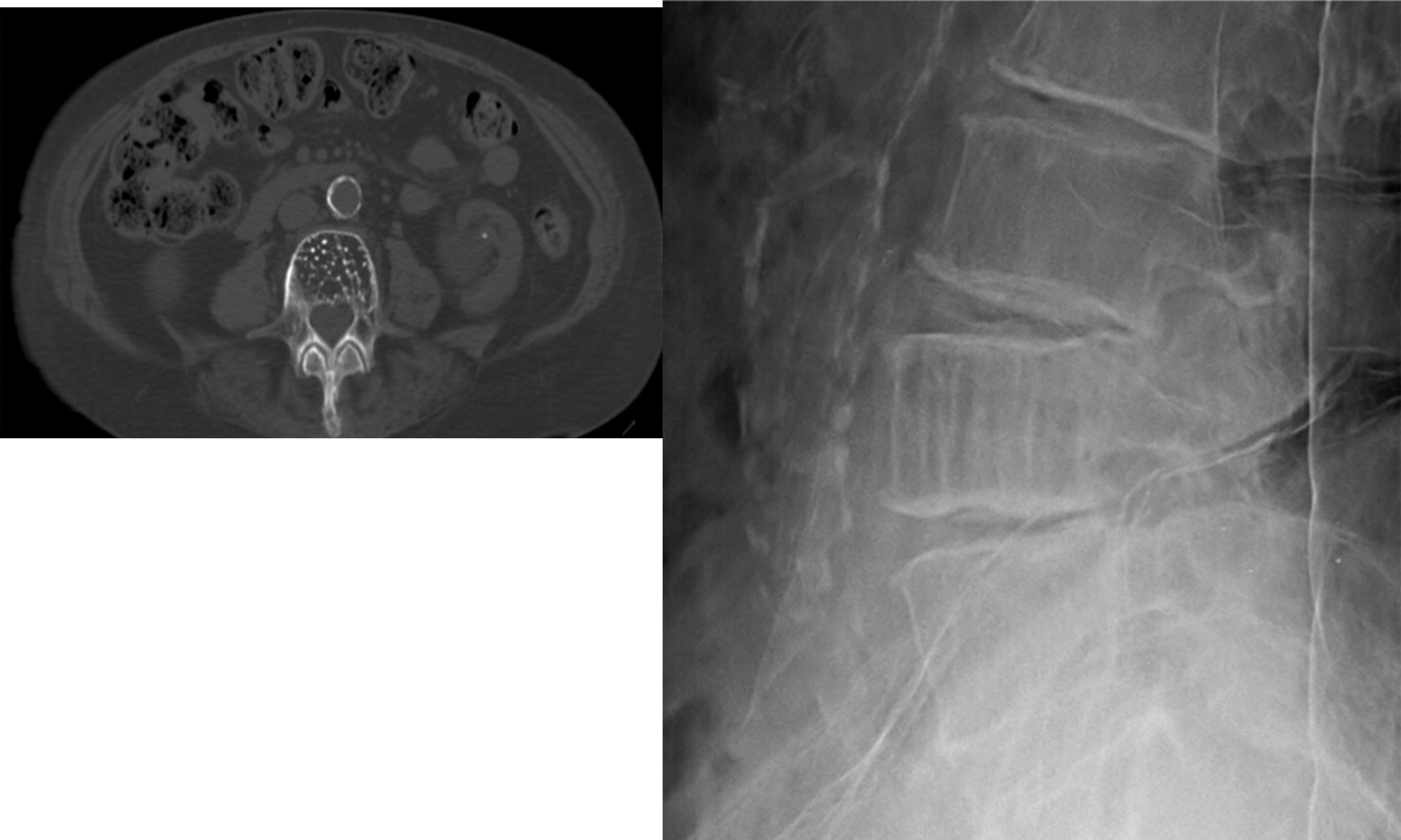
Other than the x-ray, what type of imaging is noted? What is the appearance/sign called on the specialized imaging and what is the likely diagnosis?
CT
Polka-dot appearance
Hemangioma
A “picture frame” vertebra is consistent with ___
Paget’s
What is Maffucci’s syndrome?
Multiple enchondromas and multiple soft tissue hemangiomas

What is the diagnosis?
Ollier’s disease

What is the diagnosis?
Fibrous dysplasia
What is the differential for an ivory vertebra?
Blastic mets, Paget’s, and lymphoma

What is the diagnosis?
Pedunculated osteochondroma