L6 - Erythrocyte Structure and Function II
 Call Kai
 Learn
 Practice Test
 Spaced Repetition
 Match
 Flashcards
 Knowt Play
1/39

There's no tags or description
Looks like no tags are added yet.
40 Terms
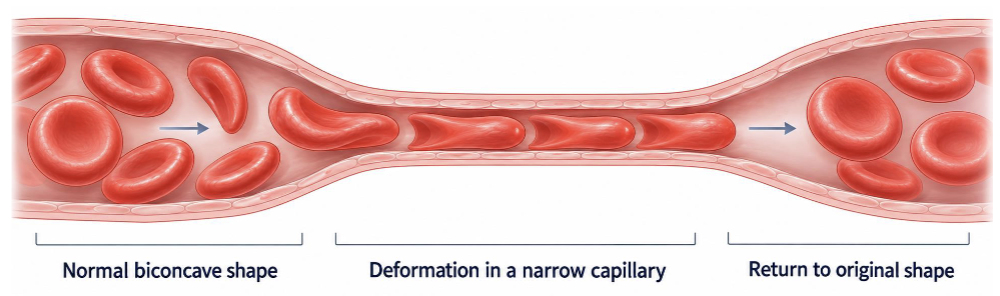
Why is RBC Deformability Essential for Normal Function?
RBCs must bend to pass through narrow capillaries and return to their biconcave shape in larger vessels
Deformation may also facilitate oxygen unloading through connections between the cytoskeleton and hemoglobin
What are the Main Components of the RBC Membrane and Their Functions?
Cytoskeleton: Provides shape, elasticity, rigidity, and deformability
Lipid bilayer: Acts as a permeability barrier, retains hemoglobin, and is relatively noncompressible
Membrane anchor proteins: Connect the lipid bilayer to the cytoskeleton
Cholesterol: Contributes to membrane stiffness and stability

What is the Role of Spectrin in RBC Structure?
Spectrin forms an elastic cytoskeletal lattice beneath the lipid bilayer
Membrane anchor points connect this lattice to the bilayer, allowing RBCs to deform and recover their normal shape

What is the Difference between Physiologic and Pathologic RBC Deformation?
Physiologic: Elastic, reversible deformation supported by the cytoskeletal lattice
Pathologic: Persistent or irreversible shape changes caused by membrane or cytoskeletal injury, reducing flexibility
How does ATP Maintain RBC Membrane Function?
ATP generated through Embden–Meyerhof glycolysis supports:
Membrane deformation
Cation pumps
Ionic gradients
Cell volume regulation
ATP depletion decreases RBC deformability.
How do Ionic Gradients Differ between Most Mammalian RBCs and Dog/Cat RBCs?
Most mammals: RBC interiors are relatively potassium-rich
Dogs and cats: Relatively sodium-rich RBC pattern
ATP-dependent pumps help maintain these ionic gradients
Delayed blood sample processing can alter measured electrolyte concentrations

How can Plasma Lipid Abnormalities Affect RBC Morphology?
RBC membrane lipids are in equilibrium with plasma lipids
Abnormal cholesterol or lipid concentrations can change membrane composition and produce abnormal RBC shapes, particularly acanthocytes
What are the Major Causes and Consequences of RBC Membrane Abnormalities?
Causes:
Structural defects involving membrane proteins or lipid
Metabolic defects involving ATP or calcium regulation
Oxidative injury, intracellular organisms, or abnormal hemoglobin aggregates
Consequences:
Decreased deformability
Abnormal RBC shapes (poikilocytosis)
Capillary obstruction or fragmentation
Premature RBC destruction (hemolysis)
Why is Intravascular Hemolysis Potentially Harmful?
Intravascular hemolysis releases free hemoglobin into circulation
Free hemoglobin can cause secondary tissue damage, particularly to the kidneys
What are the Normal RBC Shapes across Different Species?
Most mammals: Biconcave, discoid RBCs
Dogs: Prominent central pallor
Cats and horses: Less obvious central pallor on routine smears
Camelids: Normally oval or elliptical RBCs
Birds and reptiles: Oval, nucleated RBCs.

What is Poikilocytosis, and Why is It Diagnostically Important?
Poikilocytosis refers to abnormal RBC shapes
Specific morphologies can suggest underlying membrane damage, metabolic abnormalities, mechanical injury, or disease
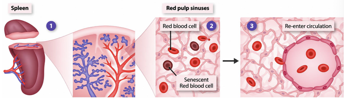
What is Splenic Conditioning, and How does It Remove Abnormal RBCs?
Healthy, deformable RBCs pass through narrow splenic red-pulp passages and return to circulation
Rigid or damaged RBCs become trapped and are phagocytosed by splenic macrophage
The spleen can also remove RBC inclusions, including nuclear remnants
Excessive abnormal RBC production may overwhelm splenic filtration, allowing abnormal cells to appear on blood smears

Name the Abnormal RBC Morphology and What Causes Their Formation?
ANSWER → Echinocytes
Morphology: RBCs with multiple short, pointed surface projections
Causes:
Artifacts: Underfilled EDTA tubes or rapid smear drying (crenation)
Pathologic: Electrolyte abnormalities, uremia, and rattlesnake envenomation
Mechanism: Osmotic disturbances or membrane damage alter RBC shape

Name the Abnormal RBC Morphology and What Causes Their Formation?
ANSWER → Acanthocytes
Morphology: RBCs with irregularly distributed projections of varying lengths, often rounded or blunt
Causes:
Hepatic disease and lipid abnormalities
RBC fragmentation
Neoplasia, including hemangiosarcoma, lymphoma, and osteosarcoma
Mechanism: Altered membrane lipid composition, particularly excess cholesterol in the outer bilayer leaflet, produces irregular projections
Acanthocytes may be physiologic in pigs, calves, and rabbits

What is the Difference between Echinocytes and Acanthocytes?
Echinocytes:
Short, pointed, more uniformly distributed projections
Associated with electrolyte disturbances, uremia, and artifacts
Acanthocytes:
Irregular, often blunt, unevenly distributed projections
Associated with lipid abnormalities, liver disease, and neoplasia


Name the Abnormal RBC Morphology and What Causes Their Formation?
ANSWER → Target Cells (Codocytes)
Morphology: RBCs with a target-like central appearance
Associations:
Regenerative anemia
Iron deficiency anemia
Hepatobiliary disease
Mechanism: Changes in the relationship between membrane surface area and cell volume, including reduced hemoglobin content in iron deficiency

Name the Abnormal RBC Morphology and What Causes Their Formation?
ANSWER → Schistocytes
Morphology: Mechanically fragmented RBCs
Associations:
Thrombi
Disseminated intravascular coagulation (DIC)
Some vascular tumors
Mechanism: RBCs are torn by fibrin strands or abnormal blood flow within vessels


Name the Abnormal RBC Morphology and What Causes Their Formation?
ANSWER → Keratocytes
Morphology: RBCs with horn-like projections or blister-type changes
Associations:
Thrombi
Disseminated intravascular coagulation (DIC)
Vascular tumors
Mechanism: Mechanical injury damages part of the RBC membrane without completely dividing the cell, producing a horned appearance

What is the Difference between Schistocytes and Keratocytes?
Schistocytes: RBC fragments resulting from mechanical shearing
Keratocytes: RBCs with horn-like projections resulting from partial membrane damage
Both suggest mechanical RBC injury, often associated with fibrin strands, thrombi, or abnormal vascular flow


Name the Abnormal RBC Morphology and What Causes Their Formation?
ANSWER → Spherocytes
Morphology: Small, rounded, dense RBCs with diminished central pallor
Mechanism:
1. Antibodies bind the RBC membrane
2. Macrophages partially phagocytose antibody-coated membrane
3. The remaining membrane reseals around the cytosol, producing a spherical RBC

What is the Clinical Significance of Spherocytes?
Numerous spherocytes, particularly in dogs, strongly suggest immune-mediated hemolytic anemia (IMHA)
A few spherocytes alongside schistocytes and acanthocytes may indicate RBC fragmentation
Spherocytes must be interpreted with additional clinical and laboratory evidence

Name the Abnormal RBC Morphology and What Causes Their Formation?
ANSWER → Heinz bodies
Morphology: Aggregates of oxidized globin attached to the inner RBC membrane
Mechanism:They form when oxidative damage causes hemoglobin proteins to denature and aggregate
These aggregates reduce RBC deformability and may contribute to hemolysis
Why are Cats Particularly Susceptible to Heinz Body Formation?
Feline hemoglobin contains numerous sulfhydryl (SH) groups that are sensitive to oxidation
Up to approximately 5% of feline RBCs may contain Heinz bodies under normal conditions
What are Common Causes of Heinz Body Formation, and How are They Identified?
Causes:
Oxidant exposures such as acetaminophen, mothballs, and skunk musk
Endogenous oxidants from accumulated metabolic products
Identification:
Routine stain: Pale red RBC protrusions
New methylene blue: Makes Heinz bodies easier to identify


Name the Abnormal RBC Morphology and What Causes Their Formation?
ANSWER → Eccentrocytes
Morphology: RBCs with hemoglobin displaced toward one side
Causes: Eccentrocytes indicate oxidative injury and may occur with or without Heinz bodies
Mechanism: Oxidative membrane damage causes portions of the membrane to adhere to each other, displacing hemoglobin
What are the Main RBC Antioxidant Pathways and Their Functions?
Embden–Meyerhof glycolysis: Produces ATP and NADH
Methemoglobin reductase system: Restores oxidized heme iron (Fe³⁺) to functional Fe²⁺
Pentose phosphate pathway (PPP): Produces NADPH to maintain reduced glutathione
Glutathione system: Protects hemoglobin and membrane proteins from oxidative damage
How does the Glutathione Antioxidant System Protect RBCs?
Reduced glutathione (GSH) helps detoxify hydrogen peroxide (H₂O₂) into water
GSH becomes oxidized glutathione (GSSG)
Glutathione reductase converts GSSG back into GSH
This regeneration requires NADPH from the Pentose phosphate pathway
The lecture also emphasizes selenium-dependent antioxidant protection.


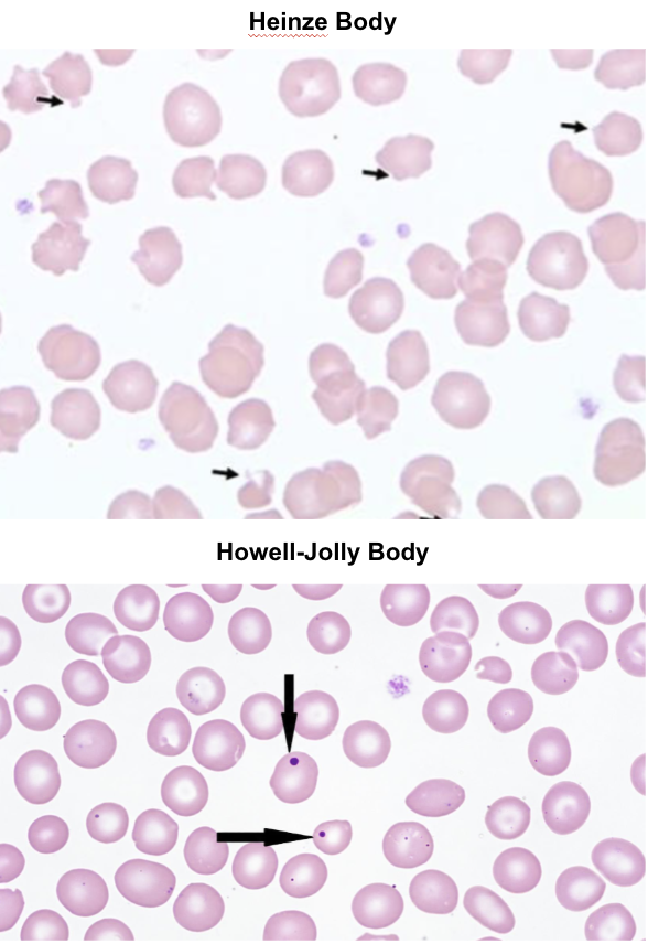
What is the Difference between Heinz Bodies and Howell–Jolly Bodies?
Heinz bodies: Oxidized hemoglobin aggregates, typically pale red on routine smears and better visualized with new methylene blue
Howell–Jolly bodies: Small nuclear remnants that stain blue-purple on routine smears
Howell–Jolly bodies may increase with regenerative anemia, splenic dysfunction, or splenectomy
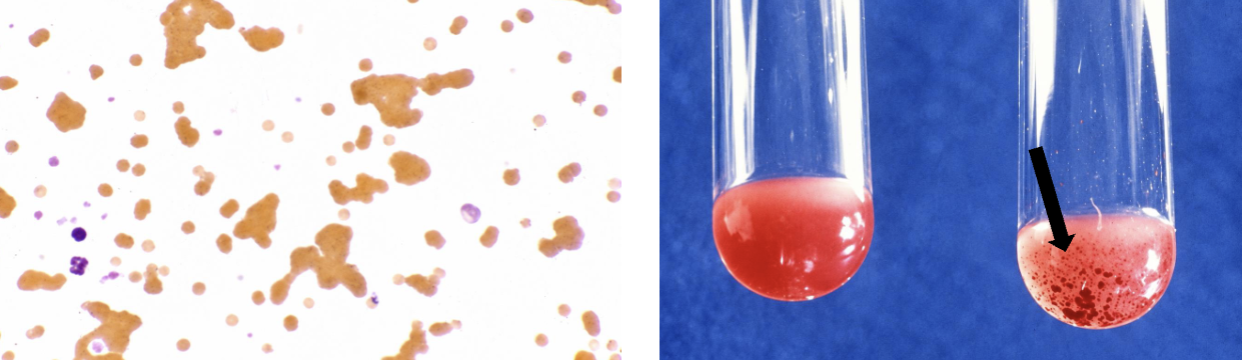
What is RBC Agglutination, and What is Its Clinical Significance?
Agglutination occurs when antibodies cross-link RBC surfaces, causing cells to clump together
It is an important hallmark of IMHA and may also occur following incompatible blood transfusions
What is RBC Senescence, and What Causes Aging RBCs to Be Removed?
Senescence is the progressive aging of RBCs as they approach the end of their species-specific lifespan
Changes include:
ATP depletion and metabolic failure
Reduced antioxidant protection and gradual hemoglobin oxidation
Alterations in membrane proteins recognized by senescence antibodies
Decreased deformability
Aging RBCs become trapped in the spleen and are removed by macrophages
What happens to Hemoglobin after Senescent RBCs are Phagocytosed?
Hemoglobin is separated into:
Globin: Degraded into reusable amino acids
Heme: Broken down to recover iron and degrade the potentially oxidative heme ring
Iron may be stored as ferritin or hemosiderin for future use.
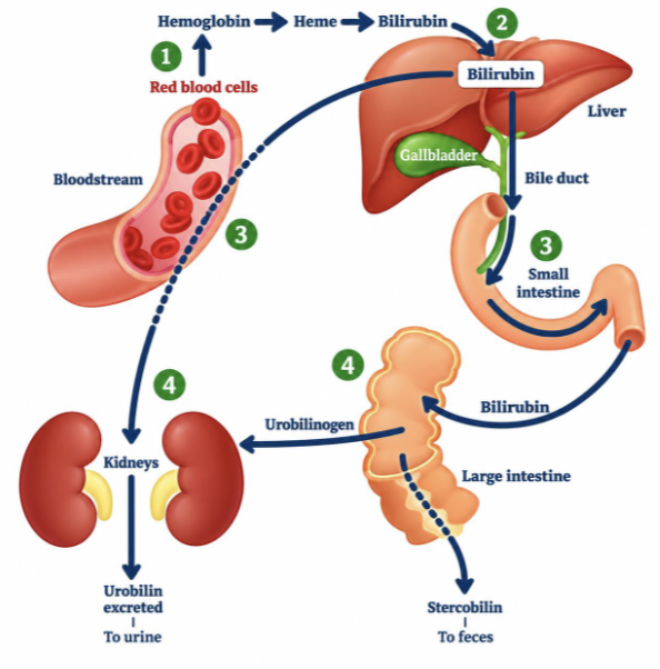
What is the Pathway of Heme Degradation to Bilirubin?
Heme oxygenase converts heme into biliverdin
Biliverdin reductase converts biliverdin into bilirubin
Unconjugated (indirect) bilirubin binds albumin and travels through plasma to the liver
Hepatocytes conjugate bilirubin with glucuronide
Conjugated (direct) bilirubin enters bile and travels to the intestine

What is the Difference between Unconjugated and Conjugated Bilirubin?
Unconjugated (indirect): Produced from heme degradation, poorly water-soluble, and transported bound to albumin
Conjugated (direct): Processed by hepatocytes through glucuronidation, becoming more water-soluble for biliary excretion
What Happens to Bilirubin after It Enters the Intestine?
Intestinal bacteria process bilirubin into downstream products, including urobilinogen
These products contribute to:
Stercobilin: Fecal pigment
Urobilin: Urinary pigment
How can Hemolysis Affect Bilirubin Metabolism?
Increased RBC destruction produces more heme for degradation, increasing bilirubin production
If bilirubin production exceeds the body's ability to process and eliminate it, bilirubin may accumulate and contribute to icterus
What are the Major Stages Where Bilirubin Metabolism can Be Disrupted?
These disturbances can cause abnormal bilirubin accumulation or excretion
Prehepatic: Increased RBC destruction and bilirubin production
Hepatic: Impaired bilirubin uptake, conjugation, or handling by the liver
Posthepatic: Impaired biliary excretion or downstream elimination

Table 1. RBC Membrane Components & Functions
Component | Main Function |
Cytoskeleton (spectrin) | Maintains RBC shape, elasticity, and deformability |
Lipid bilayer | Permeability barrier; retains hemoglobin |
Membrane anchor proteins | Connect lipid bilayer to cytoskeleton |
Cholesterol | Contributes to membrane stiffness and stability |
ATP (glycolysis) | Supports deformation, ion pumps, and cell volume regulation |
Cation pumps | Maintain ionic gradients and osmotic balance |
Key concept: RBC deformability depends on the interaction between the cytoskeleton, lipid bilayer, and ATP-dependent processes.
2. Poikilocytes — Morphology, Mechanism & Clinical Associations
RBC Type | Morphology | Mechanism / Associations |
Echinocyte | Multiple short, pointed projections | Osmotic/electrolyte disturbances, uremia, envenomation, or EDTA artifact |
Acanthocyte | Irregular, blunt projections | Altered membrane lipids, liver disease, fragmentation, or neoplasia |
Codocyte (target cell) | Target-like central appearance | Altered membrane-to-volume relationship; iron deficiency, regenerative anemia, liver disease |
Schistocyte | Fragmented RBC | Mechanical shearing from fibrin, thrombi, or DIC |
Keratocyte | Horn-like projections | Partial membrane damage from mechanical fragmentation |
Spherocyte | Round, dense, reduced central pallor | Membrane loss following partial macrophage phagocytosis; IMHA |
Eccentrocyte | Hemoglobin displaced to one side | Oxidative injury causes membrane adhesion |
Important distinctions:
Echinocytes vs. Acanthocytes: Uniform pointed projections vs. irregular blunt projections.
Schistocytes vs. Keratocytes: Complete fragmentation vs. partial membrane damage.
Spherocytes: Strongly associated with IMHA when numerous, especially in dogs.
Table 3. RBC Oxidative Damage & Antioxidant Pathways
Pathway / System | Main Function | Key Molecule |
Embden–Meyerhof glycolysis | Supplies energy and reducing equivalents | ATP, NADH |
Methemoglobin reductase | Restores oxidized heme iron (Fe³⁺ → Fe²⁺) | NADH |
Pentose phosphate pathway | Supplies reducing power for antioxidant defense | NADPH |
Glutathione system | Detoxifies peroxides and protects RBC membrane/globin | GSH |
Glutathione reductase | Regenerates reduced glutathione (GSSG → GSH) | NADPH |
Key distinction: Methemoglobin reductase protects hemoglobin's functional iron state, whereas the glutathione system protects membrane and globin proteins against oxidative damage.
Table 4. RBC & Hemoglobin Degradation
Step | Process | Product / Fate |
1 | Splenic macrophages phagocytose senescent RBCs | Hemoglobin released for degradation |
2 | Globin breakdown | Amino acids recycled |
3 | Heme breakdown | Iron recovered and heme ring degraded |
4 | Heme oxygenase | Heme → biliverdin |
5 | Biliverdin reductase | Biliverdin → unconjugated bilirubin |
6 | Albumin transport | Unconjugated bilirubin → liver |
7 | Hepatic conjugation | Conjugated bilirubin |
8 | Biliary excretion | Conjugated bilirubin → bile → intestine |
9 | Intestinal bacterial metabolism | Urobilinogen and downstream pigments |
10 | Final elimination | Stercobilin (feces), Urobilin (urine) |
Remember: Iron and globin are recycled, while the potentially oxidative heme ring must be degraded.