Protein Binding Interactions
 Call Kai
 Learn
Learn Practice Test
 Spaced Repetition
 Match
Match Flashcards
 Knowt Play
1/71

There's no tags or description
Looks like no tags are added yet.
Name | Mastery | Learn | Test | Matching | Spaced | Call with Kai | Chat |
|---|
No analytics yet
Send a link to your students to track their progress
72 Terms
Identify the five (5) types of protein binding partners.
Proteins and peptides
Nucleic acids
Small molecules and ligands
Plasma/carrier proteins
Membranes
Identify and describe a homomeric complex.
Homomeric proteins, i.e., homo-oligomers, are functional quaternary complexes comprised of two or more copies of the tertiary polypeptide unit.
Identify and describe a heteromeric complex.
Heteromeric proteins, i.e., hetero-oligomers, are functional quaternary complexes comprised of two or more different tertiary subunits.
What is the role of peptides in relation to being a protein binding partner?
Protein-protein binding interactions arise when multiple tertiary protein subunits are bound together by non-covalent and/or covalent interactions.

What is HIV-1 protease?
HIV-1 protease is a homo-oligomer of two (2) identical 99-amino-acid subunits. Each subunit possesses a catalytic aspartic acid Asp25 that dimerize to form the active site of the homo-oligomer.
HIV-1 protease plays an essential role in cleaving large Gag-pol polypeptide precursors into specialized proteins during budding and maturation.

What is HIV-1 reverse transcriptase?
HIV-1 reverse transcriptase is a hetero-oligomer complex of p51 and p66 monomers.
HIV-1 reverse transcriptase plays an essential role in the reverse transcription of viral RNA into complementary DNA (cDNA).
What is GLP-1R?
GLP-1R is a class-B GCPR that carries out the effects of GLP-1 (ligand) via phosphorylating signaling pathways involved in insulin production and secretion.

What is hemoglobin?
Hemoglobin is a heterotetramer of two (2) identical, tightly-bound αβ heterodimer, i.e., a dimer of dimers. Hemoglobin plays an essential role in nutrient, gas, and waste transport.

What is the role of nucleic acids, i.e., RNA and DNA, in relation to being a protein binding partner?
Most eukaryotic organisms store their genetic information in the form of nucleosomes, i.e., a DNA-histone complex where a 177-base-pair DNA strand is wound 1.65 times around a histone octamer.
Histones and transcription factors bind to DNA in order to control gene expression.
Poly(A)-binding proteins bind RNA to regulate stability and protein translation.
What are the roles of small molecules and ligands in relation to being a protein binding partner?
Small molecules commonly function as ligands that bind to the catalytic active site (receptor) of large proteins.
This mechanism is governed by protein-ligand interactions.
Proteins and enzymes possess active sites (binding domains) that small molecules—i.e., neurotransmitters, hormones, or drug molecules—can bind to, which induces conformational and functional changes in the protein.
Structure is function.

What is Imatinib?
Imatinib, commonly referred to as Gleevec, is a tyrosine kinase inhibitor (ligand) that inhibits the BCR-Able kinase (receptor) in chronic myeloid leukemia.
What is the role of plasma/carrier proteins in relation to being a protein binding partner?
Broadly speaking, proteins can function as molecular carriers that reversibly bind to fragile ligands, playing an essential role in:
Transporting substances
Regulating substrate plasma concentrations
Describe albumin.
Albumin is a blood protein that plays an essential role in fluid balance, nutrient transport, and overall metabolic function. Albumin is exclusively produced in the liver, and is the most abundant protein in the human blood plasma.
Structurally, albumin is a single polypeptide chain comprised of 585 amino acids with 17 disulfide bridges and one free cysteine (Cys34). Albumin has three (3) structurally similar α-helical domains with two subdomains, A and B, respectively, that form an asymmetrical heart.
Domains: I, II, III
Subdomains: IA, IB, IIA, IIB, IIIA, IIIB
Identify the major endogenous binding partners of albumin.
Fatty acids (primary physiological cargo)
Bilirubin
Hormones
Metal ions, e.g., calcium, zinc, and copper
Amino acids and metabolites
Water
What is the role of lipid membranes in relation to being a protein binding partner?
The cell membrane is a semipermeable lipid bilayer. A cell’s semi-permeability is largely attributed to the surface proteins anchored in the membrane.
Identify the two (2) broad categories of membrane-bound proteins.
(1) Integral proteins
α-helix protein → receptors
Helical bundle protein → enzymes, transporters, receptors
β-barrel protein → channel proteins
(2) Peripheral proteins
α-helix protein → enzymes, anchors, carrier proteins
Identify the two (2) broad types of protein binding.
Irreversible and reversible binding
Define reversible binding mechanisms.
Reversible binding mechanisms are governed by weak, transient non-covalent bond interactions between protein structures.
Define irreversible binding mechanisms.
Irreversible binding mechanisms are governed by strong, covalent bonds that permanently attach a ligand or toxin.
Identify the major non-covalent bonds involved in reversible protein binding interactions.
Protein binding interactions are driven by the following non-covalent interactions:
(1) Hydrophobic interactions
Hydrophobic interactions are largely driven by what is entropically favorable for water molecules.
This results in the molecular propensity of “like dissolves like”.
(2) Hydrogen bond formation
Attraction between H+ and halogens (N, O, or F)
Governed by partial charges
(3) Electrostatic forces (ionic bonds/salt bridges)
Non-covalent interactions consistent with Coulomb’s law.
(4) Van der Waals interactions
Arise from spontaneous dipole formations between proximal molecules as a consequence of constant electron movement.
Describe the function and significance of protein-protein interactions.
Protein-protein interactions are essential to biochemical processes, e.g., cell proliferation, growth, signal transduction, etc.
Aberrant protein-protein interactions are largely associated with disease:
Cancer
Infectious pathogens
Neurodegenerative disorders
What is the main issue that arises with targeting protein-protein interaction (PPI) interfaces?
Small-molecule drugs are typically characterized by an MOA that involves some type of binding-specificity to a particular binding domain (active site/receptor) via non-covalent interactions.
These drugs are intended to target protein-ligand binding interactions.
Conversely, protein-protein interfaces have no such pockets, frequently being large, flat, and hydrophobic. This makes PPI interfaces traditionally difficult to target.
What are protein domains?
A protein domain is an element of a protein’s overall structure that is stable and folds independently from the rest of the polypeptide.
What are protein interfaces?
Protein interfaces are the contact region of adjacent protein subunits.
What are protein motifs?
Protein motifs are small, distinct regions of an amino-acid sequence that is shared among different proteins.

What characteristics determine if a protein interaction is transient or stable?
The PPI’s intrinsic interface properties determine whether a protein interaction will be transient or stable:
(1) Transient interactions
Characterized by domain-domain and domain-motif interactions.
Weak transient interactions bind quickly, and dissociate quickly.
Strong transient reactions change when an affinity switch occurs.
Governed by non-covalent interactions
Low binding affinity (high Kd)
Loose binding
(2) Stable interactions
Characterized by a large interface between two folded proteins
Governed by covalent interactions
High binding affinity for native structure (low Kd)
Tight binding
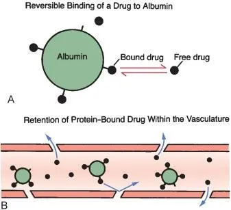
Describe drug-protein binding interactions.
Drug-protein binding interactions are typically reversible, noncovalent, nonspecific interactions between drug molecules and nonpharmacological binding partners, i.e., plasma proteins.
Drug molecules can function as competitive and/or allosteric inhibitors that can displace endogenous ligands with weaker binding affinity.
Bound drug-protein complexes can not target the main pharmacological entity.
Only unbound drug molecules can cross membrane barriers and bind to pharmacological targets to produce a therapeutic effect.
Identify the two (2) ways in which binding reactions can be described.
Binding reactions can be described via:
Equilibrium constants (Ka and Kd)
Kinetic constants (kon and koff)
Describe equilibrium constants.
Equilibrium constants describe the position of substrates at equilibrium, i.e., determines which side of the reaction is more thermodynamically favorable.
Mathematically defined as Ka = Keq = [products]/[reactants] = [PL]/([P][L]), where:
P = protein
L = ligand
PL = product
Equilibrium constants can demonstrate the binding affinity of a reaction:
(a) Association constant (Ka = M-1 = 1/M)
Ka > 1 → more products than reactants at equilibrium
Tight binding (strong binding affinity)
Ka = 1 → equal concentration of products and reactants at equilibrium
Ka < 1 → more reactants than products at equilibrium
Weak binding affinity
(b) Dissociation constant (Kd = M)
Kd > 1 → more reactants than products at equilibrium
Weak binding affinity
Kd = 1 → equal concentration of products and reactants at equilibrium
Kd < 1 → more products than reactants at equilibrium
Tight binding (strong binding affinity)
![<p>Equilibrium constants describe the position of substrates at equilibrium, i.e., determines which side of the reaction is more thermodynamically favorable.</p><ul><li><p>Mathematically defined as K<sub>a </sub>= K<sub>eq </sub>= [products]/[reactants] = [PL]/([P][L]), where:</p><ul><li><p>P = protein</p></li><li><p>L = ligand</p></li><li><p>PL = product</p></li></ul></li></ul><p></p><p>Equilibrium constants can demonstrate the binding affinity of a reaction:</p><p><strong>(a) Association constant (K<sub>a</sub> = M<sup>-1</sup> = 1/M)</strong></p><ul><li><p>K<sub>a</sub> > 1 → more products than reactants at equilibrium</p><ul><li><p>Tight binding (strong binding affinity)</p></li></ul></li><li><p>K<sub>a</sub> = 1 → equal concentration of products and reactants at equilibrium</p></li><li><p>K<sub>a</sub> < 1 → more reactants than products at equilibrium</p><ul><li><p>Weak binding affinity</p></li></ul></li></ul><p></p><p><strong>(b) Dissociation constant (K<sub>d </sub>= M)</strong></p><ul><li><p>K<sub>d</sub> > 1 → more reactants than products at equilibrium</p><ul><li><p>Weak binding affinity</p></li></ul></li><li><p>K<sub>d</sub> = 1 → equal concentration of products and reactants at equilibrium</p></li><li><p>K<sub>d</sub> < 1 → more products than reactants at equilibrium</p><ul><li><p>Tight binding (strong binding affinity)</p></li></ul></li></ul><p></p>](https://assets.knowt.com/user-attachments/80373b61-f8c7-4a7d-be67-6e60678532cf.png)
Describe kinetic constants.
Kinetic constants describe how fast a reaction is progressing in relation to the concentration of reactants.
Kinetic constants can be demonstrated in two (2) ways:
Reaction velocity
Rate constants

Describe reaction velocity.
Reaction velocity is the speed at which complexes are being formed and/or broken apart.
There are two (2) reaction velocities in a reaction:
(1) Forward reaction velocity (vforward)
The rate at which molecular complexes are forming per unit of time (M/s).
vforward = kon[P][L]
Dependent on [free reactants]
Dependent on the on-rate
(2) Reverse reaction velocity (vreverse)
The rate at which molecular complexes are breaking apart per unit of time (M/s).
vreverse = koff[PL]
Dependent on [bound complexes]
Dependent on the off-rate
At equilibrium, kon[P][L] = koff[PL] = kon/koff = Keq
Describe rate constants.
Rate constants determine the intrinsic probability of molecules bonding or breaking in a reaction.
There are two (2) rate constants in a reaction:
(1) Association rate constant (kon = 1/Ms)
The intrinsic probability of two molecules binding upon collision.
Typical range = 106-108 1/Ms
Maximum range = 108-109 1/Ms
Also known as the diffusion limit
Governed by translational/rotational diffusion
Minimum range = 103 1/Ms
Attributed to major conformational gating, restricted pocket access, or multi-step induced-fit rearrangements.
kon is influenced by three (3) variables:
[a] Diffusion
Theoretically, if every single collision resulted in a binding interaction, kon would only depend on how fast the ligand finds the active site.
This is the principle of Smoluchowski’s limit.
Also called the diffusion limit
[b] Electrostatics
The general range assumes that collisions occur out of random chance.
However, Coulomb interactions between charged molecules create favorable conditions for binding interactions.
This is what raises the kon to 109 1/Ms.
[c] Conformational gating
Put simply: if the binding domain is blocked or restricted, a collision interaction with a ligand will not result in binding.
If a conformational change to a binding-incompetent structure occurs before a ligand can bind to an active site, a bottleneck is added.
(2) Dissociation rate constant (koff = 1/s)
The intrinsic probability/frequency of bound complexes breaking apart in a reaction.
Rate constants are independent of concentration, and only vary with temperature, pH, and buffer conditions.
Explain the difference between reaction velocity and rate constants.
Reaction velocities determine the speed of bond formation and breakage, while rate constants determine the intrinsic probability of bond formation and breakage occurring in a reaction.
Additionally, rate constants are concentration-independent variables, while reaction velocities are concentration-dependent variables.
Explain the difference between equilibrium constants and rate constants.
Equilibrium constants demonstrate where a reaction settles at equilibrium, while the rate constant demonstrates how fast a direction is going in one direction.
Define fractional occupancy.
Fractional occupancy is the proportion of available binding sites, receptors, or molecular locations currently occupied by a ligand, molecule, or atom.
In other words, “what fraction of active sites currently have a ligand bound to them?”
Fractional occupancy is mathematically defined as θ = [L]/{[L]+Kd}, where:
[L] = ligand concentration
Kd = dissociation constant
When [L] = Kd, the fractional occupancy is 0.50 (50%). This is governed by the law of mass action, which states that the speed at which reactants turn into products depends directly on how much reactant is present.
Identify the three (3) types of protein-ligand binding models.
Lock-and-key
Induced fit
Conformational selection
Define the lock-and-key model, and describe its accuracy.
The lock-and-key model is a theoretical model that assumes a fixed, rigid complementary relationship between a substrate and its corresponding active site.
Mathematically represented as P + L ⇌ PL
This model is oversimplistic, and does not consider conformational changes that may be induced by the noncovalent interactions.
Define the induced fit model, and describe its accuracy.
The induced fit model posits that non-covalent ligand-protein interactions induce a conformational change in the active site that tightly binds the substrate.
Mathematically represented as P + L ⇌ PL ⇌ P’L
This model takes binding specificity into consideration. However, it is still insufficient in predicting binding affinities.
Define the conformational selection model, and describe its accuracy.
The conformational selection model assumes that a protein takes on several conformational variations. This model postulates that eventually, the protein will sample a binding-competent conformation that a ligand will bind to.
Mathematically represented as P ⇌ P’ ⇌ P’ + L ⇌ P’L
Are the protein-ligand binding models sufficient for explaining binding affinities?
No. There are several limitations to all three models, of which the most pressing are: (1) the models’ propensity to oversimplify and idealize reactions, and (2) the model’s failure to explain dissociation reactions.
Additionally, it is important to note that conformational selection is difficult to prove from binding kinetics alone.

Identify the different components of a free-energy diagram.
(A) Axes
Y-axis → Gibbs free energy (G)
X-axis → reaction coordinate
(B) Substrates
Reactants
Intermediates
Products
(C) Phases
Ground state
Transition state
(D) Gibb’s free energy
ΔG → free energy difference
ΔGon → barrier for association
ΔGoff → barrier for dissociation
Define desolvation.
Desolvation is the mechanism by which a solvation ring around a molecule is broken.
Describe the four-step landscape of a free-energy diagram.
(1) Approach
The ligand encounters an an active site.
This forms an ensemble of loose, diffusion-controlled complex configurations governed by:
Electrostatics (long-range interactions)
Partial desolvation
Determines kon
(2) Barrier
Characterized by:
Partial desolvation
Short-range interactions, i.e., hydrogen-bonding and Van der Waals forces
Determines koff
(3) Transition State
The transition state is a transient saddle point on the energy landscape that represents the highest energy configuration of partial desolvation.
(4) Partitioning
From here, the protein complex either falls apart or forms a more stable, lower energy intermediate.
Describe the role of the transition state in the induced fit model.
In the induced fit model, the transition state involves concerted or sequential energy barrier for local refolding and contact.
Describe the role of the transition state in the conformational selection model.
In the conformational selection model, the transition state mirrors the intrinsic conformational switching barrier (protein crosses barrier independent of the ligand) and binding engagement (ligand binds to binding-competent protein conformation).
What is the Arrhenius equation?
The Arrhenius equation mathematically links a reaction’s Ea to its rate constant and temperature.
This equation is mathematically defined as k = Ae-Ea/RT, where:
k = rate constant
A = frequency factor (collision frequency)
Ea = activation energy
R = gas constant = 8.314 J/kmol
T = temperature (Kelvins)
Describe endergonic reactions.
Endergonic reactions are non-spontaneous chemical reactions that require net input of energy in order to be driven, represented by ΔG > 0.
Endergonic reactions produce energy-storing products that sit at a higher, more unstable energy state.
Describe exergonic reactions.
Exergonic reactions are spontaneous reactions that release a net output of energy, represented by ΔG < 0.
Exergonic reactions produce more stable products that sit at a power energy state than the reactants.
What is Gibb’s free energy?
Gibb’s free energy (G) demonstrates the maximum amount of free energy available to do useful work at a constant time and pressure.
ΔG < 0 → favorable, spontaneous binding
ΔG = 0 → equilibrium
ΔG > 0 → unfavorable, non-spontaneous binding
Gibb’s free energy helps determine if a reaction is favorable or not.
Identify the three (3) formulas associated with the Gibb’s free energy.
ΔG = ΔH - TΔS
ΔG° = -RT ln(Ka) = RT ln(Kd)
ΔG = ΔG° + RT ln(Q)
Define ΔG = ΔH - TΔS, and describe describe the benefits and limitations of this formula.
This formula posits that Gibb’s free energy is defined by the change in enthalpy and the change in entropy.
This formula is a sufficient for determining the spontaneity of a reaction. However, the change enthalpy and entropy, respectively, are difficult values to experimentally obtain.
Define ΔG° = -RT ln{[PL]eq/([P]eq[L]eq)} = -RT ln(Ka) = RT ln(Kd), and describe the benefits and limitations of this formula.
This formula posits that, at equilibrium (ΔG = 0), the standard Gibb’s free energy is derived from the equilibrium constant K, vice versa. This formula is an application of ΔG = ΔG° + RT ln(Q) at equilibrium.
ΔG° < 0 → reactant-favoring equilibrium, where K > 1
Strong binding affinity
Spontaneous in the forward direction
Thermodynamically favors the forward reaction
ΔG° = 0 → equilibrium, where K = 1
ΔG° > 0 → product-favoring equilibrium, where K < 1
Weak binding affinity
Spontaneous in the reverse direction
Thermodynamically favors the reverse reaction
A huge limitation of this formula is that it demonstrates Gibbs free energy under standard conditions, which is not useful for predicting what happens at experimental concentrations (which tend not to be under standard conditions).
Define ΔG = ΔG° + RT ln(Q), and describe the benefits and limitations of this formula.
This equation represents Gibbs free energy under non-equilibrium, non-standard conditions. This formula is optimal for calculating the change in free energy under experimental conditions.
Does the spontaneity of a reaction determine its kinetics?
No. Just because a reaction is spontaneous, does not necessarily mean that the reaction is inherently “faster”.
Describe the significance of enzymes.
Enzymes play a critical role in metabolism, diagnosis, and therapeutics.
Enzyme catalysis is necessary in metabolic reactions because most uncatalyzed reactions are too slow on their own.
Enzyme levels can be diagnostic (e.g., myocardial infarction, liver disease).
Enzymes can be used therapeutically (e.g., digestive enzymes, DNAase for cystic fibrosis)

Describe the function of enzymes.
Enzymes are (generally) catalytic proteins that accelerate the reaction rate by stabilizing the transition state, consequently lowering the energy of activation.
Enzymes can lower activation energy by:
Binding (possessing strong binding affinity for) the transition state, not substrate (induced fit model).
Since the transition state can not stably exist, the enzyme will imperfectly bind to the substrate via non-covalent interactions, which drive conformational changes and that force the complex towards the transition state.
Enabling enzyme-substrate interactions to optimize the proximity and orientation of reactive chemical groups
Increasing functional group reactivity
Contorting substrates in such a way that facilitates bond-breaking, helping to reach the transition state
Providing an alternative reaction pathway to products.
Enzymes catalyze nearly every biochemical reaction in the body.
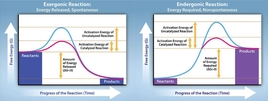
Do enzymes alter the equilibrium?
No. Enzymes do influence the kinetics (speed) of a reaction by proportionally lowering the activation energy (Ea) for both the forward and reverse reactions so that equilibrium can be reached faster.
Enzymes do not impact the thermodynamics of a reaction. This is because the equilibrium is determined by the overall Gibb’s free energy of a reaction; the reactants and products remain unchanged and independent of enzyme catalysis.
This means that enzymes do not influence the spontaneity of a reaction.

Describe the mechanism of enzymes in an exergonic reaction.
Enzymes are (generally) catalytic proteins that increase the reaction rate by stabilizing the transition state, consequently lowering the energy of activation.

Describe the mechanism in endergonic reactions.
Strictly speaking, an enzyme does not influence the thermodynamics—i.e., the total free energy—of a reaction. This means that a stand-alone endergonic reaction is still nonspontaneous and unfavorable even with the presence of an enzyme; energy is still required to bring the reaction to completion.
To make nonspontaneous reactions occur in biology, cells can employ three (3) tricks:
(1) Reaction coupling (general)
Cells can use ATP by coupling exergonic reactions (e.g., ATP hydrolysis) with endergonic reactions to produce a ΔG < 0.
(2) Product removal
This idea is governed by Le Chatelier’s principles: if product is consumed and consequently removed, the reaction quotient decreases (Q < K), shifting the equilibrium to the forward direction.
(3) Substrate modification (specific)
This is a specific subtype of reaction coupling.
A cell can phosphorylate a substrate into a higher energy configuration that results in a less-stable intermediate.
Then, this high-energy configuration can function as the driving force that can bring an endergonic reaction to completion by releasing the free energy an endergonic reaction would need to use.
This sequence can be demonstrated via the following:
Reaction I (phosphorylation): A + ATP ⇄ A-P + ADP
Reaction II (displacement): A-P + B ⇄ C + Pi (leaving group)
Most common way of driving endergonic reactions.

Identify and describe an example of covalent coupling.
Glutamine synthesis is a prime example of covalent coupling:
Glutamate reacts with ATP (i.e., is phosphorylated) to form glutamyl phosphate, a high-energy phosphate intermediate.
Glutamyl phosphate then reacts with ammonia in a displacement reaction to form glutamine.
This reaction is largely driven forward by a 2-step mechanism involving a phosphorylation and a displacement reaction.
Another type of covalent coupling, which I don’t feel like explaining here, is aminoacyl-tRNA and polypeptide bond formation.
Identify the two (2) broad types of enzymes.
Processive enzymes
Distributive enzymes
Describe the structure and function of processive enzymes.
Processive enzymes are enzymes characterized by their ability to catalyze multiple consecutive reactions without releasing their substrates.
Processive enzymes are ring-shaped tethered proteins with an enclosed active site that can tightly bind substrates.
Describe the structure and function of distributive enzymes.
Distributive enzymes are enzymes that dissociate after a single catalytic reaction.
How can you increase the processivity of an enzyme?
You can increase the processivity of an enzyme by:
Increasing its binding affinity by fusing a non-specific substrate binding domain.
Transplanting functional regions to form a more processive homolog.
Recruiting an accessory processivity factor.
Identify the three (3) types of work performed by enzymes.
Chemical work
Transport/osmotic work
Mechanical work
Describe the mechanism of chemical work, and provide an example.
Chemical work is primarily characterized by the formation and destruction of bonds.
An enzyme that performs chemical work is amylase:
Amylase is an enzyme secreted by the pancreas and salivary glands.
Amylase plays an essential role in the catabolism of starch into maltase (disaccharide).
Gene copies of amylase increased after the agricultural revolution (development of sedentary societies) 12,000 years ago when it conferred an evolutionary advantage to break down starch (introduction of corn diet due to maize production).
Modern humans carry 2-20 copies of these structural genes.
α-amylase is an endogenous enzyme that catalyzes a 2-step catabolic mechanism that hydrolyzes (break) the internal α-1,4-glycosidic bonds in polysaccharide chains to form maltose disaccharides.
Amylase has three (3) catalytic active sites:
Asp197
Glu233
Asp300
Asp197 attacks the sugar’s anomeric center, while Glu233 donates a proton to the glycosidic bond. This forms a glycosylated covalent intermediate.
Asp300 is said to play a role in stabilizing the intermediate.
This intermediate then undergoes deglycosylation and hydrolyzes the Asp197-polysaccharide bond, cleaving the starch chain into short maltose disaccharides.
These chains are later further broken down into glucose monomers via maltase.
Do cats produce amylase?
No, cats do not produce amylase. Your cats are fucking fat because you keep on a diet with a bunch of filler products, i.e., corn.
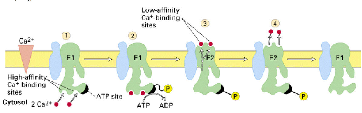
Describe the mechanism of transport work, and provide an example.
Transport work is primarily characterized by pumping substances against a concentration gradient.
One important example of osmotic work can be found in Ca2+-ATPase:
Ca2+-ATPase is an alternating-access pump that pumps two (2) Ca2+ ions out of the cytosol per one (1) ATP molecule.
Low intracellular [Ca2+] is necessary for signal transduction pathways to take place.
Ca2+-ATPase cycles between two (2) conformations:
(1) High-affinity conformation (E1)
As aforementioned, low intracellular [Ca2+] is an intrinsic, necessary cytosolic property.
Therefore, since cytosolic Ca2+ is already limited, the cytosol-facing pump conformation must have a high-affinity for Ca2+ in order to bind any ions at all.
(2) Low-affinity conformation (E2)
Converse to intracellular [Ca2+], extracellular [Ca2+] is incredibly high.
Therefore, the lumen-facing pump conformation must have low-affinity for Ca2+ in order to ensure that the Ca2+ ions leave the pump and enter the extracellular environment.
Describe the mechanism of mechanical work, and provide an example.
Mechanical movement, as implied by the name, is governed by directional movement.
A prime example of a mechanism governed by mechanical work is helicase:
Helicases are motor enzymes that convert chemical energy (i.e., ATP) into two (2) things:
Directional movement along nucleic acids
Double-helix unwinding
The general mechanism of helicase is as follows:
(1) ATP binds at the interface between two neighboring subunits in a hexameric helicase.
Helicase is a ring of six (6) subunits, meaning that it also possesses six interfaces.
Helicase, therefore, has six ATP binding domains.
(2) ATP binding stabilizes the protein interfaces, strengthening the helicase’s binding affinity to DNA.
(3) ATP binding then induces conformational changes in the helicase that complete the active site.
(4) These conformational shifts enable the helicase to translocate unidirectionally across one strand of the nucleic acid in a 5’-3’ or 3’-5’ direction.
Step sizes range from 1-2 nucleotides per one hydrolyzed ATP.
(5) As the helicase translocates, it actively destabilizes the hydrogen bonds holding complementary base pairs together.
Helicase binding to newly exposed single strands thermodynamically traps the open conformation.
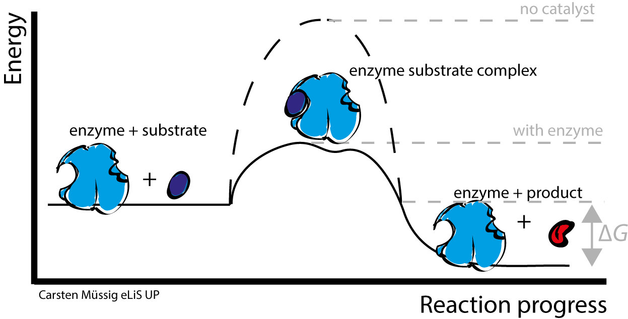
What is wrong with this diagram?
This is a graphical demonstration of a free-energy profile of an enzyme-substrate reaction.
The fundamental issue with this diagram is that it attempts to represent the unmodified enzyme-substrate intermediate as the transition state.
Intermediates are species at an energy minimum between steps.
Transition states demonstrate the highest free-energy structure reached during a reaction. This state is rare, transient, and unusable.
Transition states cannot be isolated from a reaction.

Why can an enzyme bind the transition state more tightly than the substrate, and still leave the overall free energy unchanged?
Enzymes have the highest binding affinity for the transition state.
This is because an enzyme’s catalytic efficiency is attributed to an intramolecular electric field that permanently favors the transition states over the ground states.
Since the transition state can not stably exist, the enzyme will imperfectly bind to the substrate via non-covalent interactions, which will drive conformational changes that force the complex towards the transition state.
Electrostatic stabilization of the transition state is the predominant reason for tighter binding. However, as aforementioned, enzymes cannot alter the equilibrium of a reaction. Therefore, while the activation energy (Ea) is lowered, the overall free energy remains unchanged.