Lesson 66: Structure and Function of the Pharynx and Esophagus
 Call Kai
 Learn
 Practice Test
 Spaced Repetition
 Match
 Flashcards
 Knowt Play
1/59

There's no tags or description
Looks like no tags are added yet.
60 Terms
The pharynx
passage and that connects the nasal and oral cavities to the esophagus and larynx respectively.
role in both respiration and digestion
Anatomy of the pharynx

Oropharynx
Caudal to the oral cavity
From Palatoglossal folds and arches, to the hyoid apparatus.
The soft palate
separates the nasopharynx from oropharynx
Oral and oropharyngeal surface, nasopharyngeal surface
Pharyngeal constrictors
Sequentially contract to push food into the esophagus
Hyopharyngeus
Thyropharyngeus
Cricopharyngeus
Pharyngeal Dilators
Enlarges the pharynx during swallowing(the bolus will assist this act)
Stylopharyngeus
Pharyngeal shortners
Pterygopharyngeus,
Palatopharyngeus
Muscles of the pharynx

Motor innervation to the pharynx muscles
Pharyngeal plexus
Vagus nerve (CN X) –
Glossopharyngeal Nerve (CN IX)
Cranial root of the Accessory nerve (CN XI)
Sensory innervation to the lumen of the pharynx
Glossopharyngeal nerve (CN IX)
Vagus nerve (CN X)
Trigeminal nerve CN V (Maxillary branch)
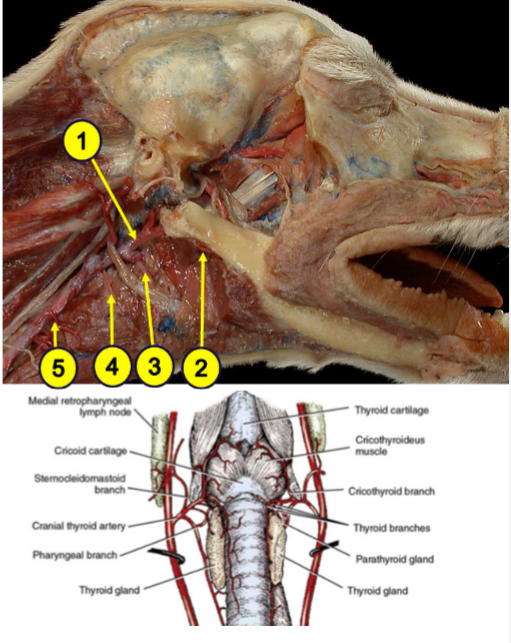
Arterial: Branches of the external carotid a.(1)
Ascending pharyngeal a: Major blood supply
Branches from the lingual(3)and facial(2)aa.
Cranial laryngeal a.(4)
Cranial thyroid a. (5) – Pharyngeal branch
Venous drainage of the pharynx
Similar pattern with satellite veins
Pharyngeal plexuses and satellite veins finally drain into internal and external jugular veins
Horse: pharynx species differences
Long (capacious), but narrow pharynx, adapted for obligate nasal breathing.
Laryngeal opening is directed more towards the nasopharynx
longer soft palate that lies beneath the epiglottic cartilage, preventing oral breathing,
Horse guttural pouch
modified eustachian/auditory tubes connected to the nasopharynx
Ruminants species differences
adapted for regurgitation, remastication (Cud chewing), and re-swallowing of food, and to prevent Aspiration during regurgitation.
Well developed pharyngeal muscles, wider pharynx to allow swallowing of larger boluses, limited oral breathing in ruminants
Pigs species difference
Larger pharynx relative to head size
Pharyngeal diverticulum
The nasopharynx histology
(involved in respiration) has ciliated epithelium to trap and clear debris
The oropharynx and laryngopharynx histology
protective stratified squamous epithelium to withstand mechanical stress.
Tonsils - Lymphoid tissue of the pharynx
Either embedded within the mucosa(Mucosa-associated lymphoid tissue - MALT) or distinctly visible
Pharyngeal tonsils
Associated with nasopharynx,
Tubal (around opening of the auditory tube)
Palatine tonsils
Associated with oropharyngeal surface
Not clearly visible in dogs, cats, bovine. Pigs do not have palatine tonsils. Horse and Small ruminant – radially visible

Lingual tonsils
embedded in the mucosa of the tongue (root)
well developed in The horse, bovine and small ruminants
Palatine Tonsil, Horse.
5. Diffuse lymphatic tissue,
3. tonsillar crypts,
7. Lymphatic nodule,
11. Stratified squamous epithelium,

Horse palatine tonsils
Horses have scattered lymphoid tissue rather than a well-defined tonsillar structure.
However, they are visible on the ventro-lateral aspect of the oropharynx
Ruminants palatine tonsils
large palatine tonsils with deep crypts for antigen trapping, but they are embedded in the latero-dorsal wall (submucosa) of the oropharynx.
Not readily visible, but the opening leading to the tonsillar (lymphoid) tissue can be seen. swelling of the pharyngeal wall can be seen upon enlargement.
Palatine tonsils: dogs and cats
well defined tonsils with deep crypts in the palatine tonsils, making them more prone to tonsillitis
The tonsils in the dog and cat are covered by a mucosal fold(Semilunar fold) forming a tonsillar fossa/pouch.
not readily visible unless inflamed and enlarged.
Palatine tonsils: pigs
The palatine tonsils are absent.
However, they have numerous tonsils all along the oral surface of the soft palate called tonsils of the soft palate. Numerous openings are seen leading to the lymphoid tissue in the soft palate.
Transverse section through
oropharynx
a Oropharynx ;
b Nasopharynx;
1 Lingual tonsil ;
2 Palatine tonsil ;
J Tonsil of the soft palate;
4 Pharyngeal tonsil ;
5 Tubal tonsil

Structure of the esophagus
tubular and muscular hollow organ that connects the pharynx to the stomach, allowing the passage of food and liquids.
It Extends from Cricoid cartilage to the cardia of the stomach.
The cervical part: esophagus
Extends from the pharynx (Cricoid cartilage of the larynx) to the cardia of the stomach (just past the diaphragm).
Related to the trachea, hypaxial muscles, carotid sheath, recurrent laryngeal nerves etc.
Cranial/Upper Esophageal Sphincter(C/UES)– Cricopharyngeus m. and Thyropharyngeus m.
● Pharyngoesophageal limen (Red Arrow)
Lower Esophageal Sphincter (LES)/Cardiac sphincter

Esophagus: The Thoracic part
from thoracic inlet to the esophageal hiatus of the diaphragm; Within the mediastinum
Thymus, cranial vena cava, brachiocephalic trunk, trachea, lungs, base of the heart, aorta, and Vagus nn.
Slightly on the left side (more left in the caudal mediastinum)
Esophagus: motor innervation
Somatic (Voluntary) Control – For striated muscle portions → pharyngeal branches of the Vagus nerve (CN X).
recurrent laryngeal nerve, a branch of the vagus, controls most of the esophageal musculature in species with extensive skeletal muscle (e.g., ruminants, dogs)
Esophagus: sensory innervation
Afferent (sensory) fibers from the Vagus nerve and sympathetic fibers detect stretch, pain, and temperature changes, sending signals to the brainstem.
Esophagus: Autonomic (Involuntary) Control – For smooth muscle portions
Parasympathetic innervation via the Vagus nerve (CN X) enhances peristalsis and mucus secretion.
Sympathetic innervation from the cervicothoracic ganglion and middle cervical ganglion regulates esophageal tone and sphincter function
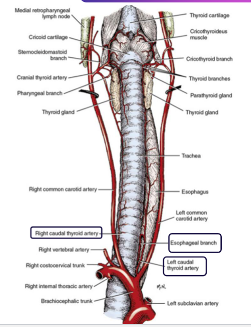
Arterial blood supply: Cervical Esophagus (Neck Region)
Supplied by the esophageal branches of the thyroid arteries (e.g., cranial and caudal thyroid arteries)
Arterial blood supply: Thoracic Esophagus (Chest Region)
Receives blood from the bronchoesophageal artery, a branch of the thoracic aorta or intercostal arteries
Arterial blood supply: Abdominal Esophagus (Near the Stomach)
Supplied by branches of the left gastric artery, which arises from the celiac artery
Venous drainage of the esophagus
The cervical and thoracic esophagus drains into the jugular veins, bronchoesophageal veins, and the azygos vein
The abdominal esophagus drains into the portal vein, linking it to the hepatic circulation.
Anatomy of esophageal blood supply
Esophagus Mucosa
The innermost layer, lined with stratified squamous epithelium to resist mechanical damage from food
Esophagus Submucosa
Contains glands that secrete mucus to lubricate food passage
Lamina propria submucosa, Muscularis mucosae
Esophagus Muscularis
Composed of skeletal and/or smooth muscle, depending on the species
esophagus Adventitia
The outermost connective tissue layer that provides support and attaches the esophagus to surrounding structures.
Serosa (abdominal part only)
The esophageal wall layers

Esophagus, Mid-region, x.s., Dog. The glands of the dog’s esophagus are predominantly mucous. They are located in the submucosa throughout the length of the esophagus of the dog. Stratified squamous epithelium
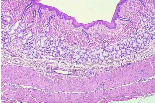
Esophagus, Cranial, l.s., Cat. few mixed glands are present in the submucosa. In the cat, horse, and ruminant, esophageal glands occur only near the junction of the esophagus and pharynx. Stratified squamous epithelium

Esophagus, Cranial, x.s., Horse. The papillated stratified squamous epithelium shows a distinct keratinized layer

Esophagus, Cranial, l.s., Pig. The stratified squamous Epithelium is keratinized in pig, horses and ruminants. Note the abundance of mucous glands in the submucosa.

Esophagus: Muscular coat; composed of skeletal and/or smooth muscle, depending on the species
Ruminants and dogs: Entirely skeletal muscle, allowing voluntary control.
Horses, pigs, and cats: The upper part is skeletal muscle, transitioning to smooth muscle towards the stomach
Esophageal obstructions: Most probable locations
Thoracic inlet, Base of the heart and just cranial to the Esophageal hiatus
Megaesophagus
Carnivores in general are prone
Usually occurs just before the diaphragm
Benign Esophageal stricture
May occur during healing process, adhesion of the mucous membrane.
Mechanism of swallowing: Oral Phase (Voluntary Phase)
conscious control and involves food prehension, ingestion, mastication, and the formation of a food bolus.
tongue pushes the bolus towards the oropharynx → soft palate elevates to close off the nasopharynx, preventing food from entering the nasal cavity
Mechanism of Swallowing: Pharyngeal Phase (Involuntary Reflex Phase)
rapid and involuntary, controlled by the swallowing reflex in the brainstem
The bolus stimulates sensory receptors in the oropharynx, triggering the swallowing reflex.
hyoid apparatus and larynx move forward and upward to help close the airway → epiglottis moves/folds backward to cover the laryngeal opening → pharyngeal muscles (constrictors) contract sequentially to propel the bolus into the esophagus
Mechanism of Swallowing: Esophageal Phase (Involuntary Phase)
upper esophageal sphincter relaxes, allowing the bolus to enter the esophagus → Peristalsis (wave-like contractions) moves the bolus down the esophagus → lower esophageal sphincter relaxes, allowing the bolus to enter the stomach
Neural control of swallowing
Afferent (Sensory) fibers in the glossopharyngeal nerve, the pharyngeal and esophageal branches of the Vagus nerve and the cranial laryngeal nerve /activate the solitary tract nucleus (NS)
Brain Components (Integration) solitary tract nucleus (NS), a sensory nucleus → central pattern generator (CPG) for swallowing (reticular formation – Brainstem) → nucleus ambiguus (NA) a motor nucleus

Motor Component (Execution) of swallowing
Via the “CPG for swallowing”, the neurons in the nucleus ambiguus (NA), the motor neurons of the swallowing muscles via Vagus nerve, generate sequential swallowing activity.
Interconnections between the left and right CPGs coordinate their activity.
Nucleus ambiguous (Motor), Solitary tract nucleus (Sensory): Cranial nerves involved
Trigeminal nerve (CN V) – mastication
Facial nerve (CN VII) – lip closure
Glossopharyngeal nerve (CN IX) – pharyngeal sensation and contraction
Vagus nerve (CN X) – pharyngeal and esophageal control
Hypoglossal nerve (CN XII) – tongue movement