Sep 22 Neuroanatomy
 Call Kai
 Learn
Learn Practice Test
 Spaced Repetition
 Match
Match Flashcards
 Knowt Play
1/89

There's no tags or description
Looks like no tags are added yet.
Name | Mastery | Learn | Test | Matching | Spaced | Call with Kai | Chat |
|---|
No analytics yet
Send a link to your students to track their progress
90 Terms
![<p>What are the three parts of the Diencephalon [Image]?</p>](https://assets.knowt.com/user-attachments/1029a153-f539-4209-8593-629a048c040e.png)
What are the three parts of the Diencephalon [Image]?
Thalamus, Hypothalamus, epithalamus - Pineal gland [Image]
![<p>Thalamus, Hypothalamus, epithalamus - Pineal gland [Image]</p>](https://assets.knowt.com/user-attachments/01de087a-66fd-4675-90df-25632cb775e1.png)
What is the general function of the thalamus?
Gatekeeper for sensory information - “selective attention”
What is the general function of the hypothalamus?
maintains homeostasis (ANS). Pituitary gland (posterior) which secretes hormones.
What is the general function of the epithalamus - pineal gland?
Secretes melatonin (sleep/wake cycle regulation)
What are the three main functions of the thalamus?
Motor - balance Basal Ganglia + Cerebellum. Cortical input: Vision/Hearing, Touch/Pressure/Proprioception, Temperature + Pain. Limbic system.
What are the areas of the thalamus and what functions do they correspond with?
Anterior - motor. Posterior - vision/hearing. Lateral - Touch/Pressure/Proprioception/Temperature + Pain. Medial - limbic system.
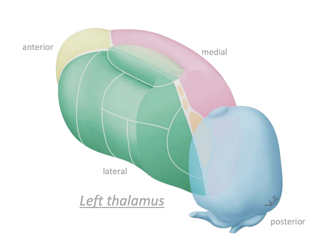
What does the thalamus do with sensory information?
Key for selective attention, filtering most sensory information.
How large is the cerebellum?
10% of the brain's volume, but 50% of the total neurons.
![<p>How is the cerebellum connected to the brain [Image]?</p>](https://assets.knowt.com/user-attachments/f57ff07d-9a04-4e99-bbe1-46dd9c88d319.png)
How is the cerebellum connected to the brain [Image]?
Via peduncles [Image].
![<p>Via peduncles [Image].</p>](https://assets.knowt.com/user-attachments/92e7f9d0-6444-4891-87c1-bce9d8057ea4.png)
What is the structure of the cerebellum?
A cortex with grooves (fissures) and ridges (folia). Deep nuclei. 2 hemispheres w/ lobes. Central “vermis”.
![<p>What are the lobes and hemispheres of the cerebellum [Image]?</p>](https://assets.knowt.com/user-attachments/e19cf9fd-a4ef-49ef-8379-7ca249750d8e.png)
What are the lobes and hemispheres of the cerebellum [Image]?
[Image] Flocculonodular lobe, anterior lobe, posterior lobe. Vermis. Lateral hemispheres (left hemisphere, right hemisphere.
![<p>[Image] Flocculonodular lobe, anterior lobe, posterior lobe. Vermis. Lateral hemispheres (left hemisphere, right hemisphere.</p>](https://assets.knowt.com/user-attachments/d6bea354-f752-48f1-ac2a-5e3e9240f61c.png)
What is the purpose of the flocculonodular lobe?
Eye movements.
What is the purpose of the vermis?
Axial control.
What is the purpose of the lateral hemispheres of the cerebellum?
Appendicular control.
What is the main function of the cerebellum?
Motor coordination + balance. Integrates sensory information to modulate motor output. Important for complex movement.
![<p>What are the three parts of the brainstem [Image]?</p>](https://assets.knowt.com/user-attachments/b861fb95-ff87-4662-9783-40ae18e39782.png)
What are the three parts of the brainstem [Image]?
Midbrain, Pons, Medulla [Image].
![<p>Midbrain, Pons, Medulla [Image].</p>](https://assets.knowt.com/user-attachments/b9127289-e7eb-48df-9911-b7d2fb7683eb.png)
What does the brainstem connect?
Connect cortex to peripheral nervous system + cerebellum.
How many cranial nerves are in the brainstem?
Most cranial nerves (10).
What does the midbrain connect?
Connects pons, medulla, and cerebellum with cortex.
What are the key structures of the midbrain and their functions?
Superior colliculus - visual reflexes (tracking). Inferior colliculus (auditory integration). Substantia nigra (dopamine production (basal nuclei)).
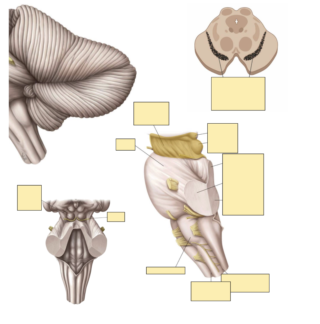
Diagrams of the midbrain?
[Image]
![<p>[Image]</p>](https://assets.knowt.com/user-attachments/fceb4706-25de-4aac-ac49-38a76093d89f.png)
Which cranial nerves does the midbrain have?
Cranial nerves III, IV
What is the function of the Pons?
Transmit information from cerebrum to cerebellum and medulla. Transmit sensory information from periphery to brain.
![<p>Where is the Pons located [Image]?</p>](https://assets.knowt.com/user-attachments/6fb272c4-3d32-404b-9dec-360afbf206c1.png)
Where is the Pons located [Image]?
Anterior to 4th ventricle [Image].
![<p>Anterior to 4th ventricle [Image].</p>](https://assets.knowt.com/user-attachments/0269a4f0-f3ac-412f-ad94-06f8dd4d2a87.png)
Which cranial nerves does the Pons have?
Cranial nerves V - VIII.
What is the Medulla continuous with?
Continuous with spinal cord.
![<p>What are the parts of the medulla [Image]?</p>](https://assets.knowt.com/user-attachments/dd50b963-d109-4435-ae81-2d59886586a3.png)
What are the parts of the medulla [Image]?
[Image] Pyramid (anterior - motor). Olive (lateral). Dorsal columns (posterior - sensory).
![<p>[Image] Pyramid (anterior - motor). Olive (lateral). Dorsal columns (posterior - sensory).</p>](https://assets.knowt.com/user-attachments/35cc154d-409f-4e20-adea-76d1af9a1649.png)
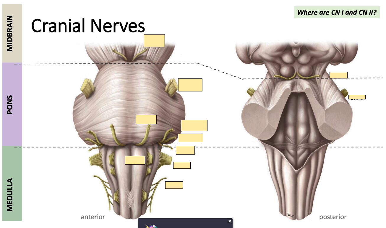
![<p>Identify parts of the midbrain, pons, and medulla from anterior and posterior views [Image]</p>](https://assets.knowt.com/user-attachments/b5e9c7ad-9e99-4b7f-8d8e-0f7a57ff4e15.png)
Identify parts of the midbrain, pons, and medulla from anterior and posterior views [Image]
Slide 21

Which carnival nerves does the Medulla have?
Cranial nerves IX - XII.
How many cranial nerves are there?
12 pairs in total.
What type of information do cranial nerves carry?
Sensory, motor, or both.
What is the mnemonic for cranial nerves?
Oh Oh Oh To Touch And Feel Very Green Velvet A H.
What are the names of the cranial nerves?
Olfactory, optic, oculomotor, trochlear, trigeminal, abducens, facial, vestibulocochlear, glossopharyngeal, vagus, accessory, hypoglossal
What is the mnemonic for sensory, motor, or both for cranial nerves?
Some say marry money but my brother says big brains matter more.
What is the function of the olfactory nerve?
olfaction
What is the function of the optic nerve?
Vision
What is the function of the oculomotor nerve?
Eye muscles
What is the function of the trochlear nerve?
Superior oblique motor
What is the function of the trigeminal nerve?
Face sensation
What is the function of the abducens nerve?
Lateral rectus motor
What is the function of the facial nerve?
Muscles of facial expression + taste
What is the function of the vestibulocochlear nerve?
Balance + hearing
What is the function of the glossopharyngeal nerve?
Taste + swallowing
What is the function of the vagus nerve?
Visceral autonomic
What is the function of the accessory nerve?
Sternocleidomastoid + Trapezius motor
What is the function of the hypoglossal nerve?
Tongue movements
What are the 4 origins of cranial nerves?
Cortex, Midbrain, Pons, Medulla
Which cranial nerves originate at the cortex?
Olfactory, optic
Which cranial nerves originate at the midbrain?
Oculomotor, trochlear
Which cranial nerves originate at the Pons?
Trigeminal, abducens, facial, vestibulocochlear.
Which cranial nerves originate at the Medulla?
Glossopharyngeal, vagus, accessory, hypoglossal
What does the size of the nerve determine?
How much information it needs to carry.
What are the 3 branches of the trigeminal nerve?
Ophthalmic (V1), Maxillary (V2), Mandibular (V3)
![<p>Locations of the branches of the trigeminal nerve [Image]</p>](https://assets.knowt.com/user-attachments/b8e6e462-b96e-41e7-87dc-49b7503e0271.png)
Locations of the branches of the trigeminal nerve [Image]
[Image]
![<p>[Image]</p>](https://assets.knowt.com/user-attachments/8aef20c7-d08c-4390-af83-81b275f7343b.png)
What are the trigeminal nerve’s motor and sensory functions?
Sensory to the face, proprioception of the mouth. Motor (V3) - muscles of mastication.
What are the facial nerve’s motor and sensory functions?
Motor: Muscles of facial expression. Sensory: Taste buds (ant ⅔ of the tongue)
Location of where the facial nerve leads [Image]
Image

What are the glossopharyngeal nerve’s sensory and motor functions?
Sensory: Oropharynx, pharynx + taste buds (posterior ⅓ of the tongue); tympanic membrane. Motor: Swallowing; Parotid gland secretion.
Location of where the glossopharyngeal nerve leads?
[Image]
![<p>[Image]</p>](https://assets.knowt.com/user-attachments/14a93459-bd76-401e-9202-e449c467118d.png)
What is the motor and sensory function of the vagus nerve?
Largest source of ANS (Parasympathetic) control. Visceral sensory from + motor to organs of thoracic and abdominal cavities. Motor to pharynx, larynx, and soft palate (swallowing, vocalization).
Locations of where the vagus nerve leads?
[Nerve]
What is the motor and sensory function of the accessory nerve?
Motor to sternocleidomastoid and trapezius.
Locations of where the accessory nerve leads to?
[Image].
![<p>[Image].</p>](https://assets.knowt.com/user-attachments/bbcd3056-c954-4cb4-82e8-682a231772a5.png)
What is the function of the spinal cord in situ?
Motor output, sensory input, reflexes and interface with the PNS
Where does the spinal cord end?
L1 - L2. At conus medullaris and Cauda equina.
What is the Cauda equina?
Bundle of nerve roots located at the base of the spinal cord, nerves branch out to periphery.
What is the location and function of white matter within the spinal cord?
Periphery. Longitudinal tracts of axons. Sensory (to brain). Motor (from brain).
What is the location and function of grey matter within the spinal cord?
Central “H”. Contains: neuron cell bodies, glial cell bodies. Dorsal horn = sensory, ventral horn = motor.
![<p>Where is the dorsal horn and ventral horn [Image]</p>](https://assets.knowt.com/user-attachments/c2f32ea3-d217-4d48-8406-98088331f570.png)
Where is the dorsal horn and ventral horn [Image]
[Image]
![<p>[Image]</p>](https://assets.knowt.com/user-attachments/30ba68b3-c5fc-41c8-9a6e-c9c7d302de6a.png)
What are the 2 parts of the skull?
Neurocranium (surrounding the brain). Visceracranium (surrounding the face).
What is a part of the neurocranium?
Frontal bone, parietal bone, temporal bone, occipital bone, sphenoid bone.
For which parts of the neurocranium are multiple parts?
We have 2 of each bone. For the frontal bone, it starts separate but fuses together during childhood.
What is a part of the viscerocranium?
Zygomatic, maxilla, mandible
What are sutures?
Fibrous joints connecting bones of the neurocranium. Very tight + limited movement = protection.
![<p>What are the 3 sutures [Image]?</p>](https://assets.knowt.com/user-attachments/ae1cbca0-e6f3-4b8d-b941-5a3b83fa5bf4.png)
What are the 3 sutures [Image]?
[Image] Coronal suture, pterion, sagittal suture
![<p>[Image] Coronal suture, pterion, sagittal suture</p>](https://assets.knowt.com/user-attachments/f4f4f3b1-9d3b-4624-846c-a3e3a72638f6.png)
What is the pterion?
Fusion point for 4 bones in the cranium. Location of the temple. Fragile.
What do sutures exist as at birth?
Sutures have not closed, exist as fontanelles - membranous connective tissue that overlies the brain.
When does fontanelle closure occur by?
Age 2
Which fontanelles become the coronal and pterion suture respectively?
Anterior fontanel. Anterolateral fontanel.
What are the parts of the spine [Image]?
24 vertebrae (7 cervical, 12 lateral, 5 lumbar). 1 sacrum (5 fused vertebrae). 1 coccyx. [Image]
What is between each vertebra?
An intervertebral disc that absorbs shock.
What are the parts of the vertebra by physically making yourself one?
[Image].
![<p>[Image].</p>](https://assets.knowt.com/user-attachments/bfb166d6-b150-4348-9ed5-24eba7461a10.png)
![<p>Diagrams of vertebra [Image]?</p>](https://assets.knowt.com/user-attachments/5ebcddc2-7b29-48e7-980a-eea09944374d.png)
Diagrams of vertebra [Image]?
[Image]
![<p>[Image]</p>](https://assets.knowt.com/user-attachments/62e6a877-1e4d-4826-8854-740aa89610eb.png)
Where do nerves come out from the vertebral column?
Intervertebral foramen.

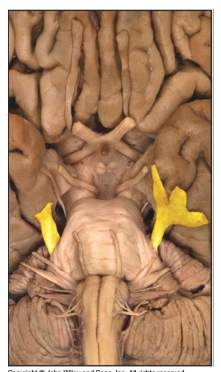
Trigeminal nerve

Facial nerve

Glossopharyngeal nerve

Vagus nerve

Accessory nerve