The Nervous System: Written
 Call Kai
 Learn
 Practice Test
 Spaced Repetition
 Match
 Flashcards
 Knowt Play
1/144

Earn XP
Description and Tags
Chapter 8
145 Terms
Three main functions of the nervous system
Monitors the body's internal and external environments, integrates sensory information, and coordinates voluntary and involuntary responses of other organ systems.
Central nervous system (CNS)
The brain and spinal cord. It processes sensory data, transmits motor commands, and supports higher functions such as intelligence, memory, and emotion.

Peripheral nervous system (PNS)
All nervous tissue outside the CNS. It carries communication between the CNS and the rest of the body.
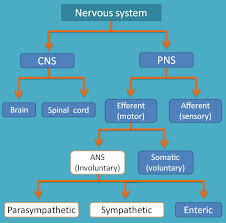
Afferent division of the PNS
Carries sensory information from receptors in body tissues and organs to the CNS.
Receptors
Sensory structures that detect changes in the internal or external environment or respond to specific stimuli.
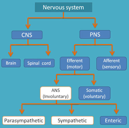
Efferent division of the PNS
Carries motor commands from the CNS to muscles and glands.
Effectors
Target organs and tissues, such as muscles and glands, that respond to motor commands.

Somatic nervous system (SNS)
Controls skeletal muscle contractions, including voluntary movements and subconscious reflexes.

Autonomic nervous system (ANS) / visceral motor system
Automatically regulates smooth muscle, cardiac muscle, glandular secretions, and adipose tissue.
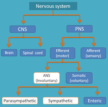
Sympathetic vs. parasympathetic divisions
Sympathetic activity speeds heart rate, while parasympathetic activity slows it.

Enteric nervous system (ENS)
A network of nervous tissue in the walls of the digestive tract that helps control digestive functions independently of the CNS.

Neurons
The basic functional units of the nervous system. They communicate with one another and with other cells.
Neuroglia (glial cells)
Support neural tissue, regulate the environment around neurons, and act as phagocytes; they far outnumber neurons and can divide.

Four main parts of a typical multipolar neuron
Cell body, dendrites, a single axon, and one or more axon terminals.
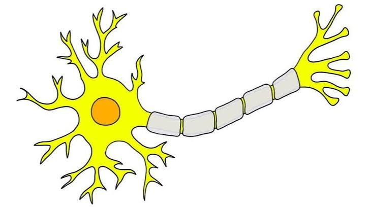
Dendrites
Branching, sensitive extensions that receive incoming signals.
Axon
A single long extension that carries outgoing signals away from the cell body toward axon terminals.
Axon hillock
A thickened region of the cell body where action potentials begin.
Axon collaterals
Branches that extend from an axon.
Axon terminals (synaptic terminals / synaptic knobs)
Found at the tips of axon branches. They form part of a synapse.
Synapse
A site where a neuron communicates with another cell.

Nissl bodies
Clusters of rough endoplasmic reticulum and free ribosomes in neuron cell bodies. They contribute to the gray appearance of areas containing cell bodies.

Why are typical CNS neurons not replaced after injury?
Most neurons lack centrioles and cannot undergo mitosis.
Adult neural stem cell exceptions
Neural stem cell activity occurs in the nose, where olfactory receptors regenerate, and in the hippocampus, which is involved in memory storage.
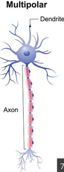
Multipolar neurons
Have two or more dendrites and one axon. They are the most common CNS neurons, and all motor neurons controlling skeletal muscles are multipolar.

Unipolar neurons
Have continuous dendrites and axon with the cell body off to one side. Most sensory neurons of the PNS are unipolar.

Bipolar neurons
Have one dendrite and one axon with the cell body between them; small and rare, found in special sense organs for sight, smell, or hearing.
Sensory (afferent) neurons
Receive information from sensory receptors and relay it to neurons in the CNS.
External receptors
Detect the external environment.
Proprioceptors
Monitor the position and movement of skeletal muscles and joints.
Visceral (internal) receptors
Monitor digestive, respiratory, cardiovascular, urinary, and reproductive systems. They provide sensations of swelling, deep pressure, and pain.
Motor (efferent) neurons
Carry instructions from the CNS to effectors.
Somatic motor neurons vs. visceral motor neurons
Somatic motor neurons of the SNS innervate skeletal muscles while visceral motor neurons of the ANS innervate cardiac muscle, smooth muscle, and glands.
Interneurons (association neurons)
Located in the brain and spinal cord, it interconnect neurons, distribute sensory information, coordinate motor activity, and support memory, planning, and learning.
Astrocytes
Largest and most numerous CNS neuroglia. They help maintain the blood-brain barrier, provide a structural framework, and help repair damaged neural tissue.
Oligodendrocytes
CNS glial cells that form myelin around portions of several axons. Myelin electrically insulates axons and increases action-potential speed.
Myelin, internodes, and nodes of Ranvier
Myelin is the insulating membrane wrapping an axon, myelin-covered regions are internodes, and gaps between them are nodes.
White matter vs. gray matter in the CNS
White matter is dominated by myelinated axons while gray matter contains neuron cell bodies, dendrites, and unmyelinated axons.
Microglia
Smallest and least numerous CNS neuroglia. They are phagocytic cells that engulf cellular waste and pathogens.
Ependymal cells
Simple cuboidal epithelial cells lining CSF-filled CNS cavities, including brain ventricles and the spinal cord's central canal. Some produce CSF and their cilia help circulate it.
Satellite cells
PNS neuroglia that surround and support neuron cell bodies.
Schwann cells (neurolemmocytes)
PNS glial cells that cover every axon outside the CNS. Each can myelinate one segment of one axon.
Oligodendrocytes vs. Schwann cells
Oligodendrocytes can myelinate portions of several CNS axons while a Schwann cell myelinates one segment of one PNS axon, but can enclose portions of several unmyelinated axons.
Ganglia
Collections of neuron cell bodies in the PNS. These cell bodies are surrounded by satellite cells.
Nerves
Bundles of axons in the PNS. They may contain both sensory and motor axons.
Center vs. nucleus (CNS)
A center is a collection of neuron cell bodies with a common function while a center with a discrete boundary is a nucleus.
Neural cortex
A thick layer of gray matter covering portions of the brain surface.
Tracts and columns
Tracts are bundles of CNS axons sharing origins, destinations, and functions while spinal cord tracts form larger groups called columns.
Pathways
Include both gray and white matter and link brain centers with the rest of the body. Sensory pathways ascend and motor pathways descend.
Demyelination
Progressive destruction of myelin sheaths in the CNS or PNS, causing gradual loss of sensation and motor control.
Multiple sclerosis (MS)
A demyelination disorder affecting axons in the optic nerve, brain, and/or spinal cord. The signs can include partial vision loss and problems with speech, balance, and motor coordination.
Membrane potential (transmembrane potential)
A potential difference across the plasma membrane caused by separation of electrical charges.
Resting membrane potential of a neuron
-70 mV; the cytoplasmic surface has an excess of negative charge compared with the extracellular surface.
Major ion distribution at rest
Extracellular fluid has high Na+ and Cl−; intracellular fluid has high K+ and negatively charged proteins.
Leak channels vs. gated channels
Leak channels are always open while gated channels open or close in response to specific chemicals, ions, or changes in membrane voltage.
Sodium-potassium exchange pump
Exchanges three intracellular Na+ ions for two extracellular K+ ions, helping maintain the resting membrane potential.
Depolarization
A shift in membrane potential toward 0 mV. Which is caused by Na+ entering the cell.
Hyperpolarization
A shift in membrane potential away from 0 mV, such as when additional K+ leaves the cell.
Graded (local) potentials
Changes in membrane potential that diminish with distance from the stimulation site.
Action potential
A propagated change in membrane potential in an excitable cell. In a neuron it is also called a nerve impulse.
Excitable membranes
Membranes containing voltage-gated ion channels that can propagate action potentials. They are found in skeletal muscle fibers, cardiac muscle cells, and neuron axons.
Threshold
The critical membrane potential that must be reached for an action potential to occur.
All-or-none principle
A stimulus that brings an excitable membrane to threshold triggers a typical action potential. A stimulus that does not reach threshold triggers none.
Initial segment of an axon
The first portion of an excitable axon membrane. An action potential begins here when it depolarizes to threshold.
Refractory period
The period from opening of voltage-gated Na+ channels at threshold until repolarization is complete. This limits how rapidly action potentials can be generated.
Continuous propagation
Action-potential transmission along unmyelinated axons as adjacent membrane segments are activated in sequence. About 1 m/s.

Saltatory propagation
Action-potential transmission in myelinated axons, where the impulse jumps from node to node. About 18–140 m/s.
Why does an action potential move forward rather than backward?
The previous axon segment is still in its refractory period and cannot immediately generate another action potential.

What does myelin do to action-potential propagation?
It blocks ion flow across internodes, so the local current depolarizes the next node and produces faster saltatory propagation.
Motor unit
A single motor neuron and all the muscle fibers it innervates.
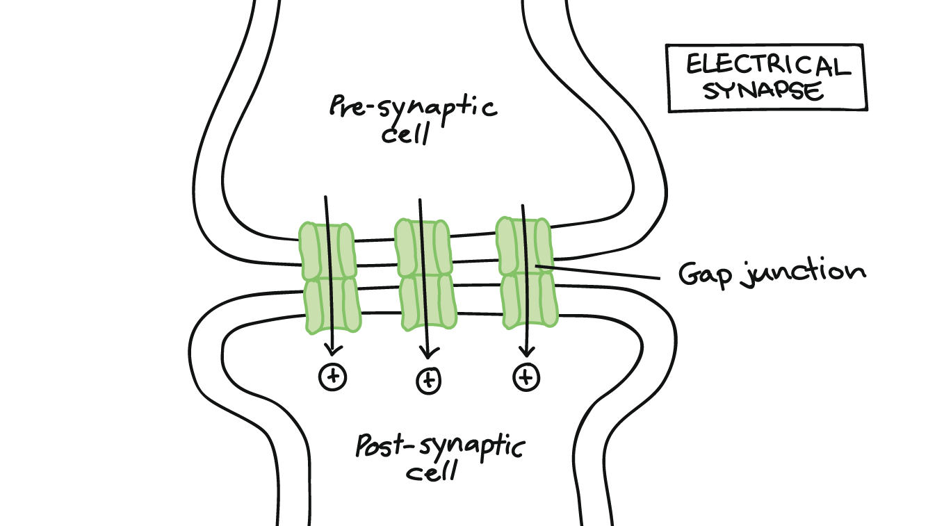
Synapse communication direction
Information passes in one direction. From the presynaptic (sending) neuron to the postsynaptic (receiving) cell.
Presynaptic neuron
The sending neuron. Its axon terminal releases neurotransmitter.
Postsynaptic neuron
The receiving neuron. Neurotransmitter binds to receptors on its membrane.
Synaptic cleft
The narrow space between opposing plasma membranes at a synapse.

Synaptic vesicles
Vesicles in axon terminals that contain molecules of a specific neurotransmitter.
Neuroeffector junction
A synapse between a neuron and another type of cell.
Neuromuscular junction vs. neuroglandular junction
At a neuromuscular junction, a neuron communicates with a muscle cell. At a neuroglandular junction, it regulates a secretory cell.
Cholinergic synapse
A synapse that releases acetylcholine (ACh).
Four main events at a cholinergic synapse
1. An action potential arrives and depolarizes the axon terminal.
2. Ca2+ enters and triggers vesicle exocytosis and ACh release.
3. ACh binds postsynaptic receptors, opening chemically gated
Na+ channels; threshold may trigger an action potential. 4.
Acetylcholinesterase removes ACh.
Role of calcium at a cholinergic synapse
Depolarization opens voltage-gated Ca2+ channels. Ca2+ entry triggers synaptic vesicle exocytosis and ACh release.
Acetylcholinesterase (AChE)
An enzyme in the synaptic cleft and postsynaptic membrane that breaks ACh into acetate and choline.
Norepinephrine (NE)
Also called noradrenaline. It’s important in the brain and portions of the ANS. Synapses that release it are adrenergic.
Other neurotransmitters named in the chapter
Dopamine, GABA, serotonin, nitric oxide (NO), and carbon monoxide (CO).
Excitatory vs. inhibitory neurotransmitter effects
Excitatory effects depolarize the postsynaptic membrane. Inhibitory effects hyperpolarizes it.
Typical effects of ACh and NE
Usually excitatory and depolarizing on postsynaptic neurons. NE is broken down by monoamine oxidase.
Typical effects of dopamine, GABA, and serotonin
Usually inhibitory because they tend to hyperpolarize postsynaptic neurons.
What determines whether a postsynaptic neuron fires?
The balance between depolarizing and hyperpolarizing stimuli arriving at that moment.

Neuronal pool
A group of interconnected interneurons with specific functions and a limited number of input sources and output destinations.
Divergence
A neural circuit in which information spreads from one neuron to several neurons or from one neuronal pool to multiple pools.
Convergence
A neural circuit in which several neurons synapse on a single postsynaptic neuron; allows multiple sources to influence the same motor neurons.
Meninges
Three specialized membrane layers surrounding the brain and spinal cord; provide physical stability and shock absorption, while blood vessels deliver oxygen and nutrients.
Three meningeal layers, superficial to deep
Dura mater, arachnoid, pia mater.
Dura mater
The tough, fibrous outermost meningeal layer.
Dural folds and dural sinuses
Dural folds are inward extensions of the dura that help hold the brain in position; large collecting veins called dural sinuses lie between the layers of a fold.
Spinal epidural space
Space between spinal dura mater and the walls of the vertebral canal; contains areolar tissue, blood vessels, and adipose tissue.
Arachnoid and subarachnoid space
The arachnoid is the middle meningeal layer; the subarachnoid space beneath it contains CSF and a web of collagen and elastic fibers.
Pia mater
The innermost meningeal layer, firmly bound to underlying neural tissue; highly vascular and carries blood vessels along the CNS surface.
Epidural vs. subdural hemorrhage
Epidural hemorrhage is bleeding between the dura mater and skull; subdural hemorrhage is bleeding between the dura mater and arachnoid.
Major functions of the spinal cord
Carries sensory impulses to the brain and motor impulses from the brain; also integrates information and controls spinal reflexes.
Cervical enlargement
Spinal cord enlargement supplying nerves to the shoulder girdles and upper limbs.
Lumbosacral enlargement
Spinal cord enlargement providing innervation to the pelvis and lower limbs.