Test 2 MS2
 Call Kai
 Learn
 Practice Test
 Spaced Repetition
 Match
 Flashcards
 Knowt Play
1/79

There's no tags or description
Looks like no tags are added yet.
80 Terms
Acute Respiratory Distress Syndrome (ARDS)
Requires mechanical ventilation
Results from another condition (i.e. trauma)
Life-threatening condition
What differs ARDS from respiratory failure?
ARDS has an acute onset
ARDS shows severe hypoxemia — despite O2
Refractory hypoxemia
in ARDS there are Bilateral infiltrates on chest x-ray
ARDS common causes
Sepsis
Pneumonia
Aspiration
ARDS risk factors
60 y/o or older
Trauma
Traumatic brain injury
Drowning
Drug overdose
Recent cardiac-related surgery
Smoking
Excessive alcohol use
ARDS- clinical cues
Tachypnea
Respiratory distress
Tachycardia
Crackles in lungs
ARDS potential complications
Infection
Respiratory complications
GI complications
Cardiac complications
Renal failure
MODS
ARDS testing
Arterial blood gas
Chest x-ray
Complete blood count (CBC)
Find the cause
Principles of ARDS management

ABCDEF Bundle - ARDS
A- Assess, prevent, and manage pain
B- Both spontaneous awakening trials (SAT) and spontaneous breathing trials (SBT)
C- choice of analgesia and sedation
D- Delirium: assess, prevent, and manage
E- Early mobility and exercise
F- Family engagement and empowerment
ARDS- Prone positioning
Improves oxygenation
Reduces hypoxemia
Enhances ventilation-perfusion matching
Promotes more uniform distribution of air in lungs
Decreases atelectasis
Reopens collapsed alveoli
Improves lung compliance
Reduces ventilator-induced lung injury
Facilitates drainage of respiratory secretions
Aids in clearing airways
Reduces risk of ventilator-associated pneumonia
Respiratory Failure
A general term for when the respiratory system fails
There is an altered gas exchange
There are 2 main concerns:
Oxygenation
Carbon dioxide elimination
Causes for Respiratory Failure- Pulmonary causes
Tumor
Pneumonia
COPD
Asthma
Cystic fibrosis
Causes for Respiratory Failure- Non-pulmonary causes
Muscle weakness
Neuromuscular disease
Medications
Narcotic/sedative overdose
Chest wall trauma
Respiratory failure risk factors
Pollution, air irritants
Smoke and second-hand smoke
65 y/o or older
Alcohol and drug use (overdose)
Opioid use
Sedative use
Noncompliance with respiratory medications
Pulmonary embolism (PE)
A blockage that causes the pulmonary artery blood flow to be altered
Emboli can be made of:
Blood
Air
Fat
Cancer
Tumors
Foreign body

PE- Virchow’s Triad
Virchow's triad is a medical model that outlines the three primary categories of factors that lead to blood clot formation (thrombosis) inside a blood vessel
Increased coagulability
Venous stasis
Endothelial tissue injury
PE Risk factors
T- Trauma, travel
H- Hypercoagulable state, hormone replacement therapy
R- Recreational drugs (IV)
O- Older age (>40)
M- Malignancy
B- Birth control pills or patches
O- Obesity, obstetrical (pregnancy, 1st 6 weeks postpartum)
S- Surgery
I- Immobilization
S- Serious illness
Use THROMBOSIS to remember
PE clinical cues
Common:
Dyspnea
Chest pain
Cough
More severe
Arrhythmias
Shock
Syncope
PE complications
Dec. cardiac output
Dec. perfusion/oxygenation
Lung damage
Heart strain/damage
Pleural effusion
Testing for PE
Chest X-ray
EKG (nonspecific ST/R wave abnormalities)
VQ Scan
CT angiography (standard)
D-dimer (elevated)
ABG (hypoxemia/hypocapnia)
PE Treatment
Anticoagulation
Immediate
Long term
Thrombolytic therapy
TPA (Tissue plasminogen activator)
Hypotension, no bleeding risk
For massive PE
Surgical
Embolectomy
Rare
Other options failed or are not possible
Hemodynamically unstable
Bleeding precautions
R- razor (electric only)
A- aspirin (NO!)
N- Needles (small gauge)
D- Decreased needle sticks
I- Injury (protect from)
Intracranial Pressure (ICP)
Normal pressure ranges between 5-15 mmHG
Increased ICP decreases cerebral perfusion and causes ischemia, cell death, and further edema (herniation)
The earliest sign of increasing ICP is change in LOC
Signs of Increased ICP (IICP)
Headache
Increased BP
Confusion
Shallow breathing
Nausea

Causes of IICP
Head injury (most common)
Brain tumor
Subarachnoid hemorrhage
Toxic/viral encephalopathy
Cerebral edema
ICP Fluctuations
Persons body temperature
Oxygenation status, especially CO2 and O2 levels
Body position
Arterial and venous pressure
Anything that increase intra-abdominal or thoracic pressure (vomiting, coughing, bearing down, etc.)
Cerebral Perfusion Pressure (CPP) Formula
CPP = MAP - ICP
Adequate CPP is 60-100 mmHG
CPP <50 mmHG results in permanent neurologic damage
A CPP <30 mmHG results in ischemia and is incompatible with life
Maintaining CPP
CPP > 70 is the goal
Monitor respiratory status; even slight hypoxia or hypercapnia can impact cerebral perfusion
Assess VS and neurologic status every 15 minutes to every hour
Strategies to reduce cerebral edema
Cerebral edema peaks 24-36 hours
Strategies to control factors that increase ICP
Avoid extreme head rotation
CPP complications
Cerebral edema and herniation
Decreased cerebral perfusion pressure
Seizure
Fluid and electrolyte abnormalities
SIADH
DI
Hyperthermia
Nutrition problems
Brain death
Cerebral edema
Fluid buildup around the brain
Can be fatal if untreated
Cerebral edema common causes
Blood-brain barrier interruptions
Brain injury
Excess sodium then water
Meningitis
Subarachnoid Hemorrhage
Blood between arachnoid and pia mater
Subarachnoid Hemorrhage Risk factors
Older then 50
Brain injury
Hypertension
Smoking
Subarachnoid Hemorrhage Clinical Cues
Early signs
Sudden “thunderclap” headache
“Worst headache of my life”
Double vision
Altered LOC
Nausea/vomiting
Neck pain
Stiffness
Late signs:
Vasospasm
Cerebral ischemia
Coma
Seizures
Rebleeding
Hydrocephalus
Head Injury: Primary injury
Due to the initial damage
Contusions
Lacerations
Damage to blood vessels
Acceleration/deceleration injury
Due to foreign object penetration
Head injury: Secondary injury
Damage evolves after the initial insult
Cerebral edema
Ischemia
Chemical changes associated with the trauma
Head injury manifestations
Depends on the severity and location of the injury
Scalp wounds
Tend to bleed heavily, portals for infection
Skull fractures
Usually have localized, persistent pain
Fractures of the base of the skill
Raccoon’s eyes
Battle’s signs
ecchymosis behind the ears
CSF leak
Halo sign
Ring of fluid around the blood stain from drainage
Glasgow coma scale
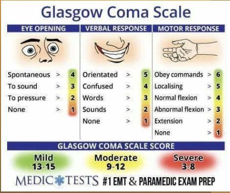
Decorticate vs decerebrate posturing

Head injury safety
Fall risk
Seizure precautions
Up with assist
Increased rounding
Education
wear a seatbelt
wear helmets/protective gear for sports
adequate lighting
limit alcohol and illicit substances
don’t drive impaired
avoid driving if taking medications that cause drowsiness or dizziness
Get help if feeling angry/frustrated when caring for a child
Spinal cord functions
Reflexes
Motor control
Sensory information
Autonomic function
Spinal cord injury causes
Motor vehicle accidents
Falls
Violence
Sports
Surgical injuries
Birth defects
Infections
Cancer
Arthritis
Osteoporosis
Spinal cord injury risk factors
Spinal fractures
Ages 16-30 and older then 65
Not wearing protective gear
No seatbelt use
Chemical impairment
Areas of violence
Medical conditions
Spinal cord injury clinical cues
C1-4: Ventilator dependence
C1-7: Limited proprioception
T1-8 Affects trunk movement, lack of abdominal control
T9-12 Limited abdominal control
L1-S5: Loss of bowel and bladder functioning, affects sexual function
Spinal cord injury treatment
Stabilization
Fix cause if possible
Limit complications
Spinal cord injury prevention and education
wear seatbelt
car with airbags
don’t drive impaired
Fall prevention
Protective gear for sports
Bike helmets
Ground level trampolines, single use
Responsible gun use
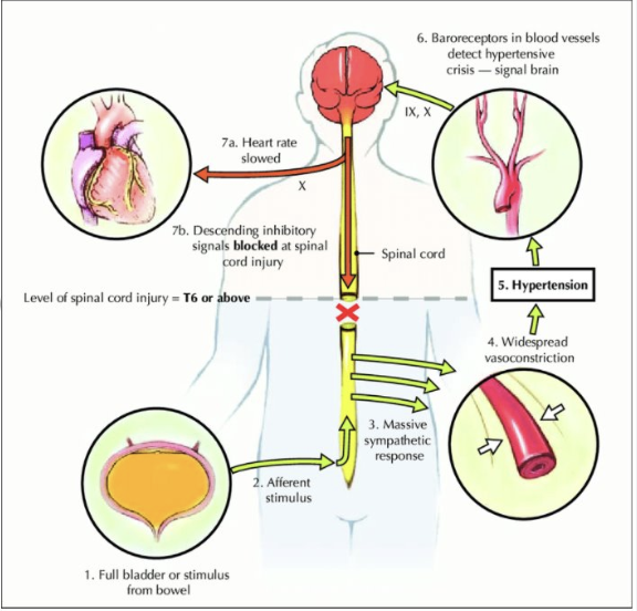
Autonomic Dysreflexia
Massively uncompensated
Cardiovascular reaction
Sympathetic nervous system
Sudden high BP
Sudden low HR
T6 or above
Most common immediately after spinal cord injury
Spinal cord injury resilience

What is wound healing altered by
Poor perfusion
Poorly controlled diabetes
Vasoconstriction
Immunosuppression
Venous insufficiency
Types of tissue injuries
Moisture
Friction
Shearing
Pressure
Burns
Trauma
Infection STONEES assessment
S-Size becoming larger
T-Temperature increasing
O-Osseous, bone exposed
N-New breakdown
E-Erythema
E-Exudate
S-Smell
Healing factors for skin
Perfusion
Nutrition
Protein
Vitamins A, B, C, D, E
The fire triangle
Oxygen, heat, and fuel
Burn Prevention
No smoking in bed
No smoking with oxygen
Keep matches/lighters away with children
Avoid toxic gases
No methamphetamine cooking
Avoid cyanide
Avoid carbon monoxide
Have a fire extinguisher and know how to use it
Max water temperature setting 120F/48.9C
Avoid electrical wires when digging outside
No flammable liquids to start a fire
Watch loose clothes while cooking
Working smoke detectors and carbon monoxide detectors
First degree burn
Damage to the epidermis, such as sunburn
second degree burn
Damage to epidermis and dermis
third degree burn
Damage to epidermis and dermis that will require skin grafting
fourth degree burn
same as third degree but extending fat layer
fifth degree burn
same as 3rd degree burn but extending into muscle layer
sixth degree burn
same as 3rd degree but extending to the bone
Burn ABCDE Assesement
A: Airway, assess for patency and inhalation injury
B: Breathing, assess quality and rate of respirations
C: Circulation, assess blood pressure, pulse, skin color
D: Disability, assess for neurologic impairment or other trauma
E: Exposure and environmental control, remove all clothing, jewelry, and contact lenses. Observe for any remaining burning substances on the skin and remove them. View entire body, including the client’s back. Assess temperature to avoid making client hypothermic
Burn Surface area
Total body surface area
Is an assessment of injury to the skin
Rule of nines
most common, easy to use
The lung and browder method
More precise, includes age of pt
Palmer method
Small burns, for scattered burns
Rule of nines burns
Head 9%
Right arm 9%
Left arm 9%
Front 18%
Back 18%
Right leg 18%
Left leg 18%
Genital 1%
Parkland formula for burns
2-4ml x weight kg x TBSA = 24 hour replacement
unless stated, assume 4ml
½ in 8 hours
half of total volume divided by 8
½ in 16 hours
half of total volume divided by 16
Complications for burns
Wound infection
Pneumonia
Bacteremia
Dehydration
Kidney injury
Acute compartment syndrome (ACS)
Sepsis
Acute respiratory distress syndrome (ARDS)
Compartment syndrome
painful
Decreased blood flow
Acute or chronic

Burns Grafting
Decrease infection risk
Prevent further loss of:
Fluid
Protein
Electrolytes
Heat loss
Autograft:
Patients own skin
Allograft:
Biologic from donors
alive or decreased
Xenograft:
From animals
usually pigs
Do for burns
Cool the burn with water
apply aloe vera
bandage the burn
take pain reliever
Don’t for burns
Don’t use oils
Don’t use egg
Don’t pop blisters
Don’t use ice
Med math
order/given * mL
Drip Rate (gtt/min)=Time (minutes) / Volume (mL)×Drop Factor (gtt/mL)
lbs/2.2= kg
Safe dosage → parameter * weight = range
Cardiac Output Formula
CO = Heart rate× Stroke volume
Mean Arterial Pressure (MAP) formula
MAP= SSP+ 2(DBP) / 3
The average pressure in the arteries during one cardiac cycle
Better indicator of perfusion to vital organs than SBP
70-100 is normal
65-70 is adequate
64 and below is concern for poor perfusion
Ataxia
Inability to coordinate muscle movement
Rigidity
Characteristic features are stiffness, inflexibility, and the inability to bend, twist, or stretch
Halo sign
Contains blood and cerebrospinal fluid (CSF)
Delerium
Disorientation, can progress to irreversible brain damage
Flaccidity
Lack of muscle tone
Battle’s sign
Ecchymosis behind the ear
Spasticity
Is a type of hypertonia that causes continuous contraction, tightness, and stiffness in muscles
Monro Kellie hypothesis
the skull is a fixed, rigid space containing three main components:
Brain tissue
Blood
Cerebrospinal fluid (CSF)
Because the skull cannot expand, the total volume of these three components must remain relatively constant.
If one component increases in volume, the other components must decrease to compensate.
If compensation fails, intracranial pressure (ICP) increases