Sectional Sonography: Liver Exam
 Call Kai
 Learn
Learn Practice Test
 Spaced Repetition
 Match
Match Flashcards
 Knowt Play
1/136

There's no tags or description
Looks like no tags are added yet.
Name | Mastery | Learn | Test | Matching | Spaced | Call with Kai | Chat |
|---|
No analytics yet
Send a link to your students to track their progress
137 Terms
Liver
• INTRAPERITONEAL ORGAN: (RUQ)
• LOCATION: Rt hypochondrium, epigastrium, part of lt hypochondrium
Lies beneath the RIGHT COSTAL MARGIN
Large size displaces digestive gases, “window”
• BORDERS:
SUPERIOR / ANTERIOR / SOME POSTERIOR SURFACES - are in contact with the diaphragm
INFERIOR- rests on the upper abdominal organs
Capsule of liver is known as
Glisons Capsule

Glison’s Capsule
double fold of peritoneum
Indications For Liver For Exam
• Abnormal liver function tests (LFTs)
• Jaundice
• Hepatomegaly
• Findings on other imaging modalities
• Surveillance / Screening for chronic hepatitis/cirrhosis or hepatocellular carcinoma
• Suspicion for stenosis, thrombosis of the hepatic vasculature
Liver Functions
• Formation of bile, antibodies, blood clotting factors, urea, and uric acid
• Activity of reticuloendothelial tissues, RBC destruction
• Synthesis and deamination of amnio acids
• Converts lipids into cholesterol, and protein to carbs and fats
What does the liver store
glycogen, amino acids, fats, vitamins A, D, and B complex, Iron and copper
Liver responsible in the formation of
Serum albumin
Globulin
Antibodies
Heparin*
Fibrinogen*
Prothrombin* *: used for blood clotting
Liver Sonography
• Homogeneous, only interrupted by blood vessels
• Has mid level echoes
• Smooth contour / echotexture
• Hyperechoic to renal parenchyma (isoechoic occasionally)
• Should not be more than 15-17 cm in length.
In order from HYPERechoic to HYPOechoic:
Pancreas → liver → renal cortex
Sinusoids
blood from small branches of the hepatic arteries and the hepatic portal vein → hepatic sinusoids → blood from the hepatic arteries and hepatic portal vein mix → liver sinusoids are partially lined with phagocytic Kupffer’s cells, which remove bacteria, foreign matter, and weakened blood cells → hepatic veins → heart

Four lobes of liver
Right
Left
Quadrate
Caudate
How many segments does the liver have ?
8
The left lobe is divided into
left medial and left lateral

The right lobe is divided into
right anterior and right posterior


1
falciform ligament: divides left lateral from left medial lobes as well as the LHV

2
ligament teres
Left Lobe
• LHV separates left lateral from left
• In contact with the undersurface of diaphragm
• To the left of the Main Lobar Fissure and MHV
• Receives blood from the LPV & LHA branches
• Venous drainage through LHV
RIGHT LOBE OF LIVER
In contact with the right lateral undersurface of the diaphragm
• Larger than left 6:1
• Divided into anterior and posterior segments by the RHV.
• Receives blood from RPV & RHA
• Venous drain through RHV
• Posterior surface is the porta hepatis, GB and IVC
• Contains bulk of hepatic tissue
Quadrate Lobe
• Is the medial segment of the left lobe
• Between the GB fossa and falciform ligament
• Anterior to the porta hepatis
CAUDATE LOBE
• Smallest lobe of liver
• Separated from left lobe by ligamentum venosum
• It receives blood from Rt & Lt portal venous & hepatic artery branches
• Anterior border is ligamentum venosum posterior is IVC
BARE AREA
• Superior and posterior aspect of liver.
• Lies directly on the diaphragm, IVC courses through.
• No peritoneum so IVC can enter into the chest.

Hepatic Nomenclature Couinauds / Liver Divisions
• Longitudinally divided into four sections, divided through the hepatic veins
• Transversely divided by an invisible plane through the right and left portal veins

Porta Hepatis
where structure enter and leave the liver. A “gate”.
Portal Triad is located at porta hepatis
• The portal vein enters
• The hepatic artery (proper) enters
• The Common Bile Duct exits
CBD, portal vein, and hepatic artery form …
the Portal Tria AKA: Mickeys Sign

Portal Triad
contained within a connective tissue sheath which gives the portal vein an echogenic wall
Hepatic Vessels
• Hepatic arteries
• Hepatic veins
• Portal Veins
Sinusoids:
Vascular channels
receive blood arterial and portal flow
exit through central vein
Liver Vasculature
• Hepatic artery (enter at porta hepatis)
Common, Proper & Right, Left
• Hepatic veins (superior and posterior)
Left, Middle, Right
• Portal veins (enter at porta hepatis)
Main, Left, Right
HEPATIC VEINS
• Are responsible for draining blood from the liver
• Superior and posterior
• Course between the hepatic lobes & segments
• Empty into the IVC as it passes through the diaphragm…they enlarge as they enter the IVC
• Sonography: Sonolucent / anechoic appearance with thin walls
• Can be seen subcostal and intercostal
• Can exhibit posterior enhancement
Bunny sign of hepatic veins
face is right hepatic vein, first ear is middle hepatic vein, second ear is left hepatic vein

• LEFT HEPATIC VEIN
Divides Lt Lobe into lateral & medial segments
• MIDDLE HEPATIC VEIN
Divides the liver into Rt and Lt lobes
• RIGHT HEPATIC VEIN
Divides Rt lobe into ant & post segments
PORTAL VENOUS SYSTEM
• Provides 70-80% of blood to the liver
• course within lobes & segments of the liver
PORTAL VENOUS SYSTEM sonography
sonolucent lumen surrounded by echogenic fibrous tissue (an extension of Glisson’s Capsule)
Portal veins ____ as they approach porta hepatis
increase in size
• In regard to the LIVER there is the
MAIN PORTAL VEIN, LEFT PORTAL VEIN and RIGHT PORTAL VEIN
Portal Vein Walls
appear distinctively brighter (hyperechoic) because of their high collagen content and sheath covering
Portal venous system carries blood …
from the bowel and abdominal organs to the liver for metabolism and detoxification
Portal venous system includes all veins that drain blood from the
spleen, pancreas, gallbladder, and gastrointestinal tract, with the exception of the lower rectum
• Superior mesenteric vein
• Courses from inferior to superior and drains the small and large intestine via
several smaller branches.
• Joins the splenic vein to form the main portal vein
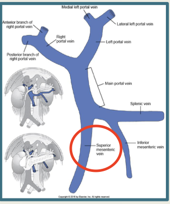
• Splenic vein
• Courses laterally from the spleen and then medially, directly posterior to the pancreas neck
• Joins the superior mesenteric vein to form the main portal vein (at portal confluence)

• Main portal vein
• Formed by union of the splenic vein and superior mesenteric vein posterior to the neck of the pancreas
• Courses superiorly approximately 5-6 cm, then divides into the intrahepatic right and left branches

Smaller veins also empty into the main portal vein including
left and right gastric veins, pancreaticoduodenal veins, and gastroepiploic veins
• Inferior mesenteric vein
• Courses from inferior to superior
• Drains the large intestine via several smaller branches
• Usually joins the splenic vein but can vary widely as to where it actually joins the portal venous system

• Right portal vein
• Courses right from the main portal vein and branches into anterior and posterior divisions

• Left portal vein
• Courses left, horizontally, from the main portal vein and branches into medial and lateral divisions

Distinguish PV from HV
• Hepatic veins flow into the inferior vena cava. Hepatic veins get larger as they course towards IVC
• Portal veins are larger at their origin at the porta hepatis and get smaller as they course through out liver and branch off. Portal veins have more echogenic borders than the hepatic veins because they have a thicker collagenous sheath.
COMMON HEPATIC ARTERY
• Branches from the Celiac Axis off of Aorta
• Runs along the upper border of the pancreas
• Divides into: Hepatic Artery
Proper continues into liver & divides into INTRAHEPATIC RT & LT hepatic arteries

Fissures (4)
1) Main lobar fissure *See
2) Right intersegmental fissure *Don’t see
3) Left intersegmental fissure *Don’t See
4) Fissure for ligamentum venosum fissure *See
Ligaments (7)
1) Coronary
2) Falciform
3) Gastrohepatic
4) Hepatoduodenal
5) Left triangular (lateral)
6) Right triangular (lateral)
7) Round (ligamentum teres)
MAIN LOBAR FISSURE
boundary between left and right lobes of the liver. Identifies the gallbladder fossa. On a sagittal scan it extends from the portal vein to the neck of the GB.
RIGHT INTERSEGMENTAL FISSURE
Right hepatic vein courses within this fissure, and it divides the anterior and posterior segments of the right lobe.
LEFT INTERSEGMENTAL FISSURE
Left hepatic vein courses within this fissure, and it divides the medial and lateral segments of the right lobe.
LIGAMENTUM VENOSUM/Fissure for ligamentum venosum
Remnant of the fetal ductus venosus, which shunted oxygenated blood from the umbilical vein to the IVC. Signifies the anterolateral border of the caudate lobe
LIGAMENT
double fold of peritoneum
FALCIFORM LIGAMENT
(sagittal) from umbilicus to diaphragm, (anterior posterior) from right rectus muscle to the BARE AREA.
Contains the ligamentum teres. Both ligamentum teres and falciform ligament divide the left lobe of the liver into medial and lateral segments.

ROUND LIGAMENT/LIGAMENTUM TERES
oblilateral umbilical vein. round hyperechoic, anterior structure with possible shadow
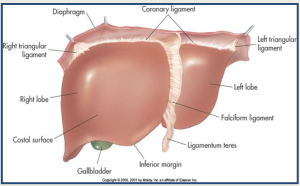
CORONARY LIGAMENT
connects the posteriosuperior surface of the liver to the diaphragm (bare area margins) Is continuous with the rt & lt triangular ligaments and falciform ligament (anteriorly)
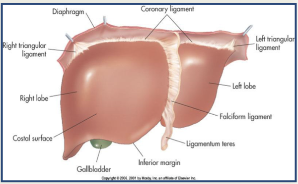
RIGHT TRIANGULAR LIGAMENT
extension of the coronary ligament, starts at the right margin of the bare area. Connects the posterior surface of the right lobe to the right under surface of the diaphragm.

LEFT TRIANGULAR LIGAMENT
left extension of the falciform ligament, connects the posterior surface of the left lobe to the diaphragm

LIGAMENTUM VENOSUM
Remnant of the fetal ductus venosus, which shunted oxygenated blood from the umbilical vein to the IVC. Signifies the anterolateral border of the caudate lobe.
HEPATODUODENAL LIGAMENT
surrounds the portal triad (portal vein, hepatic artery, common duct). Where hepatic artery and portal vein enters and hepatic duct exits.

GASTROHEPATIC LIGAMENT
inserts into the fissure of the ligamentum venosum. Caudate lobe is posterior to where the gastrohepatic ligament inserts into the fissure of the ligamentum venosum
VARIANTS: REIDELS LOBE
• Congenital anomaly & a normal variant of the the right lobe of the liver
• Mostly seen in women
• Is a tongue-like projection which extends inferior to the lower pole of the rt kidney
• Is ISOECHOIC with the rest of the liver
• Should not be confused with pathology
VARIANTS 2 AND 3
2) Situs Inversus
3) Diaphragmatic Indentations
Anomalies of the Liver: AGENESIS
• incompatible with life.
• right, left, or caudate lobes.
• hypertrophy of the other lobes develops
Anomalies of the Liver: ANOMALIES OF POSITION
• The liver may be found in other locations in two conditions:
situs inversus, in which the organs are reversed, with the liver on the left and spleen on the right;
or in a congenital diaphragmatic hernia or omphalocele, where varying amounts of liver tissue may herniate into the thorax or outside the abdominal cavity.
Anomalies of the Liver: ACCESSORY FISSURES
• uncommon and caused by infolding of peritoneum.
• The inferior accessory hepatic fissure is a true accessory fissure that stretches inferiorly from the right portal vein to the inferior surface of the right lobe of the liver.
Anomalies of the Liver: VASCULAR ANOMALIES
• The hepatic artery may have many variations as it arises from the celiac axis.
• At least 45% of patients may have the following variations:
• (1) replaced left hepatic artery originating from the left gastric artery
• (2) replaced right hepatic artery originating from the superior mesenteric artery
• (3) replaced common hepatic artery originating from the superior mesenteric artery
• Variations in the portal venous anatomy are uncommon but include atresias, strictures, and obstructing valves.
• Variations in the branching of the hepatic veins are common, with the most common being when the accessory vein drains the superoanterior segment of the right lobe.
• It may empty into the middle hepatic vein or join the right hepatic vein.
Lab Values 1

Lab Values 2

Lab values 3

Hepatic Enzymes
• protein catalysts used for the metabolic processes.
• can escape into the bloodstream in the presence of damaged or diseased hepatocytes.
• The increase presence of these enzymes in the blood stream are therefore indicators of hepatocellular disease or disorder.
• Liver enzymes:
AST, ALT & Alkaline Phosphate
LIVER FUNTION TESTS (LFT’S): AST / SGOT
• an enzyme present in tissues that have a “high metabolic rate” (not just liver), damage to the heart, musculoskeletal system and mononucleosis. AST can be higher in alcoholic hepatitis.
• released into blood stream in abnormally high levels when there is death or injury (eg HEPATITIS & CIRRHOSIS) to the producing cells.
• Moderate to Severe increase with hepatocellular disease & Mild increase with obstruction
LIVER FUNTION TESTS (LFT’S): ALT / SGPT
• Enzyme that is more specific than AST for evaluating liver function
• Elevated in acute cirrhosis , hepatic metastasis, pancreatitis & obstructive jaundice. Can also be elevated in damage to the heart and musculoskeletal system.
• Moderate to Severe increase with hepatocellular disease & Mild increase with obstruction
LIVER FUNTION TESTS (LFT’S): LDH
• Enzyme found in kidneys, heart, skeletal muscle, brain, liver & lungs
• Cellular injury & death cause this enzyme to increase
• Mildly elevated in hepatitis, cirrhosis & obstructive jaundice. Moderately elevated with mononucelosis.
• Primary use is in detection of mycardial or pulmonary infarction.
LIVER FUNTION TESTS (LFT’S): ALKALINE PHOSPHATASE
• Produced by liver, bones, intestine & placenta.
• Indicator of intrahepatic or extrahepatic obstruction, hepatic carcinoma & abscess
• Moderately elevated in hepatitis & cirrhosis OBSTRUCTION!!!
LIVER FUNTION TESTS (LFT’S): PROTHROMBIN TIME
• Liver enzyme that is part of blood clotting mechanism
• Production of prothrombin depends on adequate intake of VITAMIN K
• Prothrombin time is INCREASED with cellular damage
LIVER FUNTION TESTS (LFT’S): ALBUMIN & GLOBULINS
• Is the assessment of depressed synthesis of proteins, sensitive test for metabolic derangement of the liver
**Serum albumin & plasma coagulation factors
• A LOW serum albumin suggests decreased protein synthesis. Chronic Liver Disease = elevation of gamma globulins. Gamma globulins are a class of proteins in the blood.
LIVER FUNTION TESTS (LFT’S): BILIRUBIN & JAUNDICE
• Bilirubin is the breakdown product of hemoglobin.
• The liver detoxifies bilirubin and excretes it into the gut via the biliary tree.
• Hyperbilirubinanemia (JAUNDICE) - yellow coloration of the skin, sclera and body secretion. Due to excess presence of
SERUM BILIRUBIN Often found first in the whites of the eyes.
• Present in massive blood breakdown, biliary obstruction &
hepatocellular disease
• PRURITIS - is itching that is associated with jaundice
LIVER FUNTION TESTS (LFT’S): Hepatic Detoxification. BUN & Ammonium
• The liver detoxifies waste products.
• Ammonium is a toxic product of nitrogen metabolism. It is converted into nontoxic urea in the liver. Urea is then excreted through the kidneys.
• The level of urea in blood is measured by blood urea nitrogen. BUN can be low with decreased urea production in liver dz patients.
• This can result in increased ammonium (NH4) accumulation. Patients can have fetor hepaticus (fruity pungent breath).
• Urea is essentially a waste product but helps transfer some property of a fluid from one flowing current of fluid to another across a semipermeable membrane. (Helps in nephrons). Urea is highly soluble in water and is, therefore, an efficient way for the human body to expel excess nitrogen.
• Excessive amounts or toxic of (NH4) can lead brain dysfunction (confusion, coordination, disturbances, tremor and coma). Hepatic Encephalopathy, “ Ptis encephalopathic
Associated Tests
• Computed tomography (CT)
• Angiography
• Magnetic resonance imaging (MR, MRI)
• Radionuclide scintigraphy
• Liver biopsy
Common Pathologies
• Fatty Infiltration
• Hepatitis (acute andchronic)
• Cirrhosis
• Storage Diseases
• Liver cell adenoma
• Cavernous Hemangioma
• Focal Nodular Hyperplasia (FNH)
• Hepatocellular Carcinoma (HCC)
• Pyogenic Abscess (bacterial)
• Amebic Abscess (parasitic)
• Echinococcal Cysts
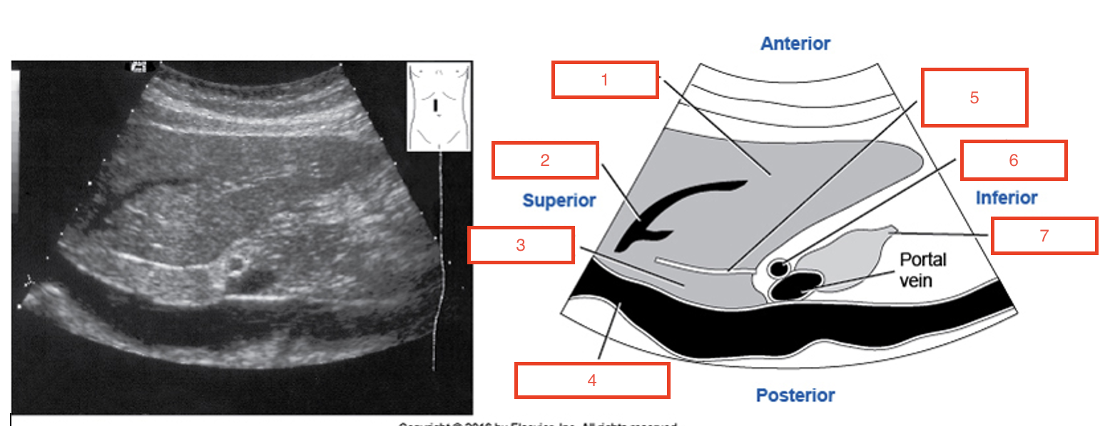
1
left lobe liver

2
left hepatic vein

3
caudate lobe

5
fissure for ligamentum venosum

6
hepatic artery

7
pancreas head
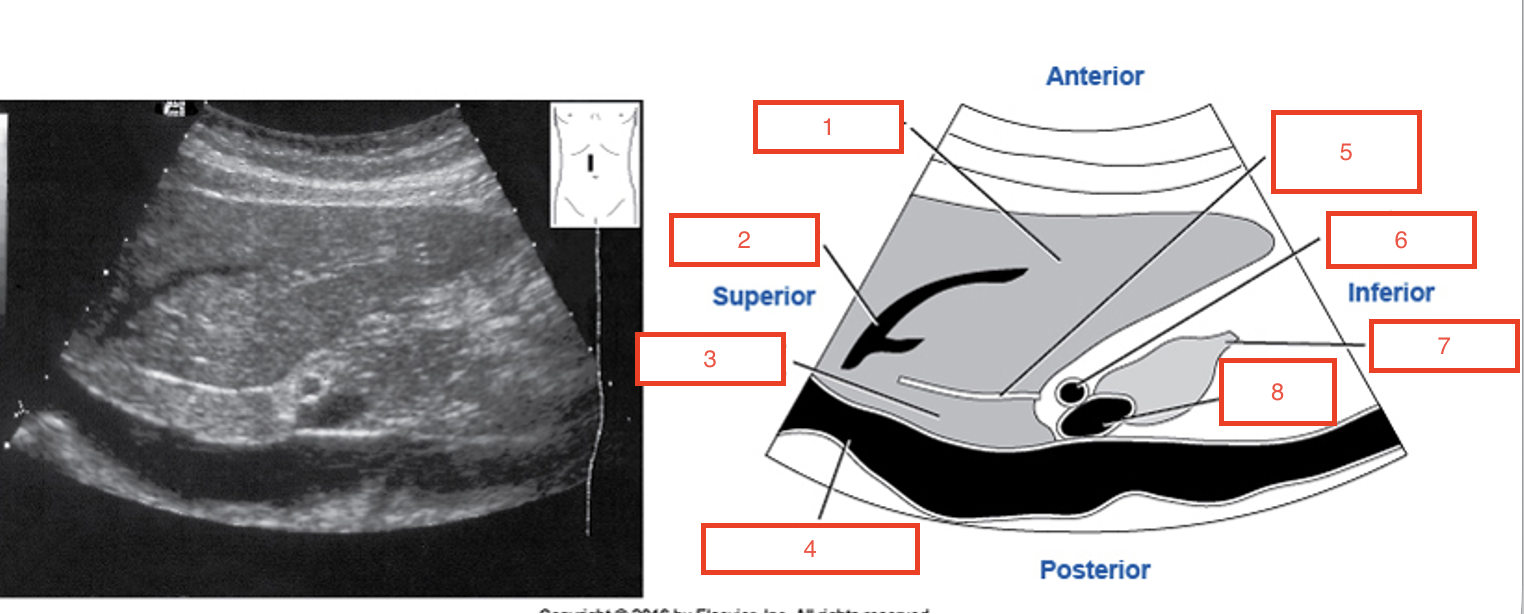
8
portal vein
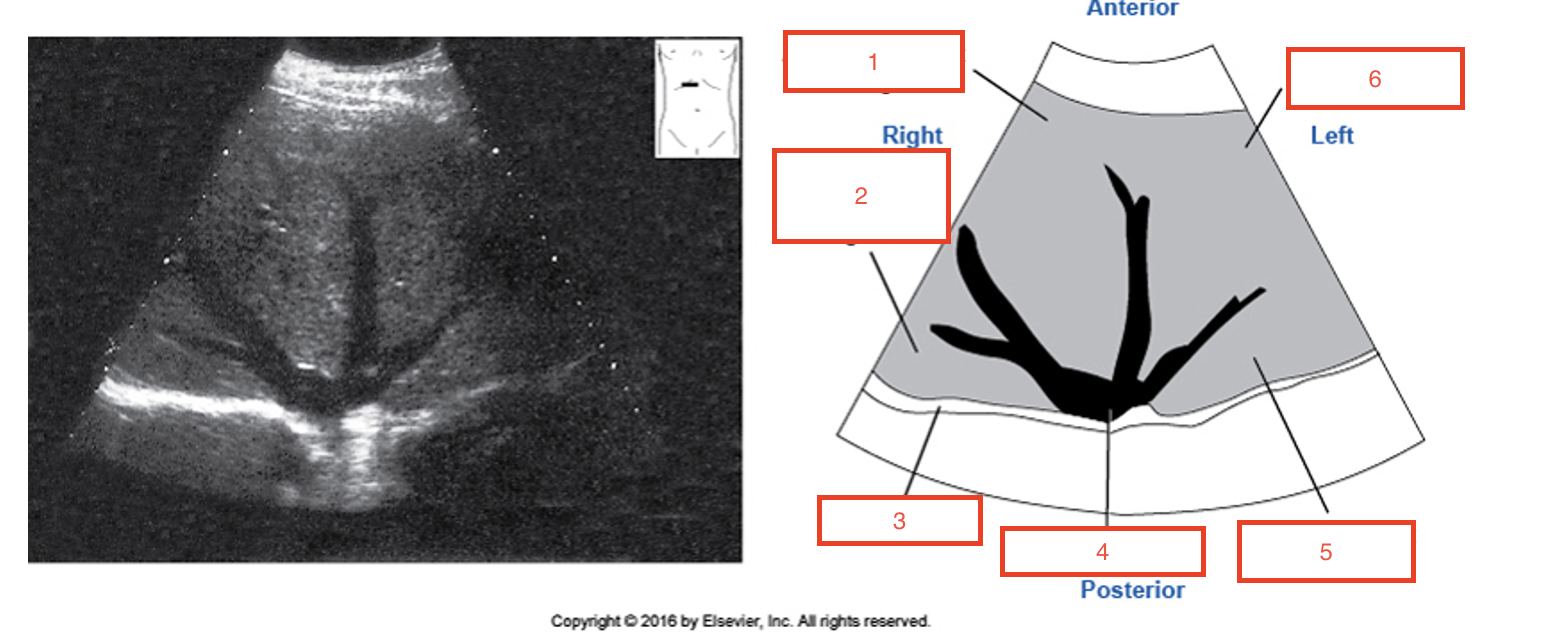
1
anterior segment of right lobe

2
posterior segment of right lobe

3
diaphragm

4
inferior vena cava

5
lateral segment of left lobe

6
medial segment of left lobe