Anatomy: Exam 1 st
 Call Kai
 Learn
Learn Practice Test
 Spaced Repetition
 Match
Match Flashcards
 Knowt Play
1/94

There's no tags or description
Looks like no tags are added yet.
Name | Mastery | Learn | Test | Matching | Spaced | Call with Kai | Chat |
|---|
No analytics yet
Send a link to your students to track their progress
95 Terms
Define Anatomy
Anatomy is what and where something is
Define physiology
Physiology is the function of that thing
Explain the relationship between anatomy and physiology
The anatomy of something contributes to its function and the function of something can tell you its anatomy.
Ex. Long bones structure provides a stable place for muscles to attach —> (function affecting structure)
What is the cell membrane composed off, what are its properties
phospholipid bilayer —> heads are polar, tails are non-polar
The cell membrane is SELCTIVELY Permeable
only allows for small non-polar materials to pass through without any help (or super small polar molecules like water)
Large polar molecules need some assistance (they can’t bc they repel the tails)
charged molecules and ions (K+, Cl-, etc) also CAN’T
Define the membrane proteins and their functions
Peripheral proteins —> found at the surface of the lipid bilayer
Integral proteins —> fully integrated; something can enter and exit
a) Channel protein —> allows particular ions to pass through the cell (PASSIVE like carrier)
b) Recognition Protein —> allows for the binding of a ligand to its receptor to activate a receptor (ACTIVE)
c) Glycoprotein —> protein with carbohydrate molecules found on its surface (ACTIVE)
What sort of cells CAN and CANNOT pass through the lipid bilayer
CAN —> Small, nonpolar molecule + small polar molecules (Water)
CAN’T —> Large polar, ions, charged molecules
Name the different ways to pass through the cell membrane:
Passive transport —> movement of things across the membrane without the use of cellular energy (ATP)
a) Diffusion —> High to low concentration
Osmosis —> Water moves from high to low concentration (know Hyper + Hypotonic)
b) facilitated diffusion
Active transport —> movement of things across the membrane through using ATP
Explain the path of a protein destined for secretion OR insertion into a membrane:
Nucleus → ribosome/Rough ER → transport vesicle → Golgi → vesicle → destination
Nucleus —> where transcription occurs
a gene within the DNA that codes for a specific protein is turned into mRNA.
The protein
Rough ER —> Protein is synthesized on the ribosomes on the Rough ER and modified
ER might: add carbohydrate (glycosylation), disulfide bonds check whether the protein folded correctly assemble multiple protein subunits
After thats done the ER buds off and ships the protein (inside the vesicle, for a secreted protein, or inside the vesicle membrane, for a membrane protein)
Smooth ER (not rlly involved in protein shipping)—> synthesizes lipids (phospholipids, steroid hormones)
controls and regulates the concentration of cellular Ca++ —> triggers neurotransmitter release
breaks down certain toxins
Golgi Apparatus —>
responsible for sorting, modifying (e.g. adding carbohydrates, etc), and shipping products delivered from the RER
The cis side —> receives, medial —> modifies, trans —> sorts and ships
The golgi also decides where the package will go a) Secretion —> leaves, Membrane —> combines with the membrane
Name all the organelles associated with a cell and their functions:
Nucleus —>
Store DNA
Ribosomes are synthesized here
Rough ER —>
makes and modifies the proteins and ships them to the
Smooth ER —>
responsible for lipid synthesis
regulates ca++ —> for neurotransmitter release
breaks down toxins
Golgi —>
responsible for modifying, packaging, and shipping protein products
Peroxisomes —>
carry out oxidation reactions to neutralize free radicals (produce H202 and convert them into water and oxygen, which are released into the body)
loss of it causes Tay-Sachs
Lysosomes —>
contains enzymes that break down unneeded cellular components
Mitochondria —>
responsible for converting sugar to ATP for the cell to use as energy
Cytoskeleton —>
group of fibrous proteins that provide structural support for cells
Types —> Microfilaments, intermediate filaments, microtubules
Describe the structural organization of the body from molecules to organ systems, giving examples of each level of organization.
Atoms —> Molecules —> Cells —> Tissues —> organs —> Organ system —> Organism
Ex. Hydrogen, H20, Keratinocytes, Eplitheal, Skin, Integumentary system, Human
Types of Tissues & GENERAL Function
Epithelial —>
Densley apcket togther
includes skin, lines cavities, passageways and certain glands.
avascular (usually gets nutrients through lower layers like dermis)
Connective —>
Sparsely packed together
Binds the cells and organs of the body together and functions in the protection and support
Includes bone, cartilage, blood, dermis, and fat
Muscular —>
Skeletal, cardiovascular, smooth
Striated (except smooth)
responsive to stimuli, excitable, contracts
3 MAIN types: Skeletal, smooth, cardiac
Smooth —> found in digestive tracts, blood vessels, intestine, urinary bladder
Nervous —>
Involves neurons and neuroglia (glial cells)
excitable, allowing for the spread of electrochemical signals in the form of nerve impulses in different bodily regions

List the body systems of the human body
Integumentary
Skeletal
Muscular
Nervous
Endocrine
Cardiovascular
Lymphatic
Respiratory
Digestive
Urinary
Female + Male reproductive
Integumentary system (examples + function)
Ex. Hair, Nails, Skin
Function →
Protects internal organs
Prevents water loss
Has sensory receptors
Skeletal system (examples + function)
Ex. Bones, Joints, Cartilage
Function →
Protects organs
Ex. Ribcage →Protects the heart
Supports the body
works with the muscular system to create movement through muscle contractions
Produces Blood cells (Ex. WBC)
Stores minerals
Muscular system (examples + function)
Ex. Tendons, skeletal muscles
Function —>
provides movement through contraction
produces heat
supports posture
Endocrine system (examples + function)
Ex. Ovaries, testes, Adrenal gland, pancreas, thyroid gland, pituitary gland
Function —>
creates and releases hormones to regulate the body
cardiovascular system (examples + function)
Ex. Heart, blood vessels, and blood
Function —>
pumps blood
Transports oxygen, hormones, and nutrients,
carries waste away from tissues
Lymphatic System (examples + function)
Ex. Lymph nodes, spleen (blood filter), lymphatic vessels, thymus (upper chest)
Function —>
fight pathogen —> bodies immune sustem
returns excess tissue fluid to the blood stream
Respiratory System (examples + function)
Ex. Lungs, airways, trachea, nasal passage
Function —>
bring oxygen to the body
Get rid of CO2
Digestive System (examples + function)
Ex. Small intestine, large intestine, gallbladder (breaks down fat), liver, mouth, esophagus
Functions —>
Poop
Breaks down food
Helps absorb nutrients (small intestine has microvilli)
Urinary System (examples + function)
Ex. Urinary bladder, kidney, ureter, urethra
Functions —>
release waste from the blood
Control of water and ion balance
produces and eliminates urine
Male reproductive system (examples + function)
Ex. Testicles, penis, epididymis, reproductive ducts
Function →
Sperm production → allows for reproduction
Male sex hormones
Female reproductive system (examples + function)
Ex. Ovaries, vagina, uterus, fallopian tubes, mammary gland
Function →
Egg production
Female sex hormones
Mammary gland supports development of offspring
Define Homeostasis:
a process in the body which regulates it to maintain at a stable state/ internal environment. Is able to do this through negative feedback loops
Negative feedback loop —> the body counteracting a change by doing the opposite to bring it back to normalcy
Define negative feedback loop and give an example
Negative feedback loop —> the body counteracting a change by doing the opposite to bring it back to normalcy
Ex. When it’s hot, your blood vessels widen to release heat, and when it’s cold your blood vessels get smaller to conserve heat.
Describe hyponatremia, its causes, symptoms, and its relationship to homeostasis.
Hyponatremia —> sodium in the blood becomes TOO low compared to the amount of water
happens due to excess consumption of water
Context —> ADH is released to tell the kidneys to conserve water, typically during dehydration
Katie was in a marathon and she was sweating a lot and therefore losing electrolytes (like sodium). In this case the body would typically try to release the water by DECREASING ADH so the concentration can balance.
Katie was also drinking so much water, faster than the rate her body could release the water, so instead her ADH increased and the kidneys kept on reabsorbing the water. This causes her blood to become diluted and water to go into her cells
OVERALL → Hyponatremia is a condition in which blood sodium concentration becomes abnormally low because there is too much body water relative to sodium. Exercise-associated hyponatremia can occur when a person loses electrolytes through sweating but drinks excessive amounts of water faster than the kidneys can eliminate it. This dilutes the blood and causes water to enter cells, making them swell. Symptoms can include headache, vomiting, confusion, diarrhea, abnormal movements, and in severe cases coma or death. Hyponatremia represents a disruption of homeostasis because the body fails to maintain normal water and ion balance.
Symptoms:
Weight gain
Diarrhea
Headache/confusion
vomiting
difficulty staying upright
abnormal, repetitive leg movements

Types of Epithelial Tissues —> draw each
Simple squamous
Simple cuboidal
Stratified Squamous
Simple coumnar
stratified cuboidal
stratified columnar
Function of each layer of epithelial tissue:
Simple squamous —> very thin, good for diffusion & filtration (Ex. air sacs in lungs, lines capillaries)
Simple cuboidal —> Slightly thicker, good for absorption and secretion (found in ducts and ovaries)
Stratified squamous —> can handle abrasions (skin, Esophagus (friction))
Simple Columnar —> Often with microvilli or cilia; good for absorption, secretion, and protection (small intestines)
Stratified Cuboidal —> Protection and Secretion (glands and ducts)
Stratified Columnar —> Protection and secretion (glands and ducts)
Tay-sachs disease - what it is/symptoms, how its caused
Tay Sachs is a disease that’s caused by a dysfunction in the lysosome (specifically it misses an enzyme that breaks up fat) and that causes deterioration/damage of nerve cells
Symptoms include:
Brain damage
catarcat
Jaundice
Enlarged liver
Kidney damage
epidermolysis bullosa; what is it, symptoms, what tissues does it affect
Epidermolysis bullosa is caused by a mutation in the COL7A1 gene.
This gene codes for collagen
Collagen gives structural tissues their functions
Collagen holds together the epidermis and dermis (at basement membrane) —> without that it causes blistering and sores
The skin condition also affects his nutrition as epithelial tissue also lines the mouth, esophagus, and stomach —> needs a feeding tube
Mostly effects —>
eplithealeal tuissue
Main glandular tissue
lines inside and outside
Connective tissue
Dermis (connective) is connected to epidermis
Tissues in the integumentary system
All 4
In the dermis there are muscles (arrector pilli)
dermis is connective
have nerve fibers in dermis
Epidermis is epithelial
what 2 are the layers of the skin (+ layer below)
Epidermis —> (epithelial)
keratinocytes → make keratin (structural protein)
stratified squamous
avascular —> gets nutrients by diffusion from the dermis
dermis —> (connective tissue)
hypodermis (adipose/fat —> connective tissue)
What are the layers of the epidermis and their functions
Bottom (New)
Stratum Basal
Newest Layer
attached to basement membranes
Nursery: RAPID division
Contains melanocytes —> responsible for creating melanin in the melanosome which gets delivered to neighboring cells
Contains Merkel cells —> responsible for sensory
Single layer of cells primarily made up off basal cell
Basal cell is a cuboidal shaped stem cells that is a precursor of the keratinocytes of the epidermis
Dermal Papilla → a fold/projection in the dermis that increases the strength of connection between dermis and epidermis
Stratum Spinosum
have tiny connections with other cells
production of keratin is ramped up
Under a microscope they dry up and look star-shaped.
Stratum Granulosum
contain granules of keratin
Keratin is a fibrous protein that makes the cell tougher and conserves water
CELL DEATH
Stratum Lucidum
Dead keratinocytes densely packed
looks clear (eledin)—> appears on the palms of the hands and feet (need an extra layer because they are more exposed)
cells are less permeable + water proof
Stratum Corneum
Dead flat keratinocytes
covers the whole body
stays on the surface for 2 weeks before coming off
Top (Old)
What are the layers of the dermis and their functions (+ hypodermus)
Dermis is a connective tissue
Within the dermis we have fibers like collagen and elastin + fibrocytes
Core of integumentary system
Contains blood vessels, nerves, hair follicles, sweat glands, etc
Made of 2 layers of connective tissue
These two layers compose an interconnected mesh of elastin and collagenous fibers produced by fibroblasts
Layers:
Papillary dermis
loose connective tissue
named for dermal papillae in the region
Fibroblasts, fat cells, and lots of blood vessels
Contains phagocytes
Contains lymphatic capillaries, nerve fibers, touch receptors
Reticular layer
Dense irregular connective tissue
Well vascularized and sensory and synthetic nerves
Appears net like due to elastin fibers → for elasticity and movement
collagen extending into both the papillary layer and the hypodermis → collagen provide strength
Hypodermis
composed of adipose tissue (fat) —> insulates us
protection against mechanical stress
Function of Epithelial Tissue
Physical & Chemical Protection:
serves as the body's first line of defense against physical wear and tear, chemical damage, and biological pathogens.
Selective Permeability & Barrier Control:
Cells form a selectively permeable barrier that regulates the absorption and transport of materials into and out of underlying tissues.
All substances entering or exiting the body must cross an epithelial boundary.
Secretion:
Specialized cells synthesize and release mucus, enzymes, hormones, and sweat.
Ex. Ciliated cells lining the respiratory tract secrete mucus to trap incoming pathogens and foreign particles.
Sensation & Environmental Sensing:
Specialized epithelial cells (neuroepithelia) contain sensory nerve endings to detect touch, pressure, temperature, and chemical stimuli (e.g., taste buds, olfactory epithelium).
Excretion & Absorption:
Facilitates specialized chemical transport, such as nutrient absorption in the small intestine microvilli and metabolic waste filtration/excretion in renal tubules.
Functions of Connective Tissue
Support and Connection:
Connects tissues and organs together (e.g., muscle sheaths, tendons attaching muscle to bone, and the skeletal frame).
Protection:
Forms fibrous capsules around delicate organs and mineralized bone structures for skeletal defense
Immune Defense:
Contains specialized phagocytic cells that engulf microorganisms, pathogens, and cellular debris.
Ex. Blood WBC !!! → immunology
Transport:
Fluid connective tissues (blood and lymph) transport nutrients, fluid, waste, and chemical messengers throughout the body.
RBC transport oxygen from the lungs to body tissues and carry carbon dioxide back to the lungs for exhalation
Energy Storage & Thermal Insulation:
Adipose (fat) cells store surplus energy and contribute to thermal insulation.
Where did skin color come from?
Melanin (pigment in our skin) absorbs/reflects different wavelengths of light
More melanin (darker) —> absorbs more light (UV) and reflects less light
Less melanin (lighter) —> absorbs less light (UV) and reflects more light
(genetic inheritance determines amount of melanin)
Eumelanin vs Pheomelanin
Eumelanin is a brown to black pigment and gives stronger UV protection.
Pheomelanin is yellow to reddish pigment and gives less UV protection.
When did darker shades of Human skins evolve?
we probably lost hair so our bodies could cool
MC1R gene
gene that codes for a receptor involved in signaling within melanocytes, this influences the TYPE of melainin produces (Eumelainin vs Pheomelanin)
part of the regulation of pigmentation
the melanin controls UV absorption though
Reduced MC1R variation in African population indicates there is a strong selection against any alleles that would alter dark skin
More Melanin Vs Less Melainin (pros vs cons)
More melanated
Melanin Absorbs More UV
Greater UV protection —> less UV reached epidermal cells
Folic acid does not break down —> we need it
Because they can’t absorb UV, they get less UVB radiation for vitamin D production
Need to take vitamin D supplements
Less Melanin
Less UV protection —> More UV reached epidermal cells
More folic acid breakdown —> BAD
more UVB absorption —> producing sufficient vitamin D
*Folate (vitamin B9) helps your body make and repair DNA, divide cells, and produce healthy red blood cells.
How does Melanin protect the DNA from damage from UV
Melanin absorbs UV light before it can reach and damage living epidermal cells.
Explain why mutations in MC1R gene are more common in light-pigmented populations than dark-pigmented populations
due to evolutionary pressure. There is more selection for the mutated MC1R as an active MC1R gene pushed towards eumelanin. In places with lighter people, evolutionary pressure to maintain high eumelanin levels is weaker. MC1R variants associated with lighter pigmentation are therefore more likely to persist and spread through the population.
High UV environment → strong selection for functional MC1R → more eumelanin → darker pigmentation
Low UV environment → weaker selection against MC1R variants → less eumelanin and relatively more pheomelanin → lighter pigmentation
Explain how the integumentary system interacts with other body systems to contribute to organism-level functions and homeostasis.
Nervous system
Skin contains nerves, and sensory receptors that detect any touch
Cardiovascular system
Blood vessels in the skin regulate the temperature (dilations)
Muscular system
Small muscles called arrector pili attach to hair follicles.
When you are cold, these muscles contract and cause goosebumps.
Immune system
Skin is the first physical barrier against pathogens
Endocrine system
Skin helps produce vitamin D when exposed to UV
Vit D is important for calcium
Skeletal system
Calcium is needed for bone mineralization
Excretory glands
Skin has sweat glands that remove water, salt, and waste products through sweat
Respiratory and cardiovascular system
Respiratory system brings in oxygen and cardiovascular system delivers it to the skin
Ultraviolet (UV) light exposure triggers - so we become darker?
triggers keratinocytes to release signaling molecule
this signals for more melanin, which goes back to the keratinocytes
Why Tans Fade:
Melanosomes are eventually broken down by lysosomes
Folic acid and Vitamin D in relation to Melanin
Folic Acid Protection (Shielding):
UV radiation destroys circulating folic acid (folate), a crucial B vitamin
Accumulation of melanin shields epidermal DNA from UV damage and prevents this nutrient breakdown.
Higher UV → Higher Melanin → Shields Folic Acid from Breakdown → PREVENTS Vitamin B Deficiency.
Vitamin D Synthesis (Absorption):
Vitamin D production requires UV light interaction in the skin.
While higher melanin levels protect against radiation, excessive pigmentation in low-sunlight environments can hinder Vitamin D synthesis
Vitamin D synthesis is vital for calcium absorption.
Higher UV → Higher Melanin → blocks vitamin D synthesis
Vit D relation to calcium
Vit D helps absorb calcium
need calcium for nervous system and muscle contractions
Rickets
The body takes calcium from the bones causing bone deformity
Functions of the skeletal system (Bones, Joints, Cartilage)
Protecting internal organs
Skull surrounds brain; lungs protected by ribcage
Stores and releases fat
Yellow marrow (bone marrow) stores fat
Fat is released a s source of energy when body needs it
Produces blood cells
Red marrow (bone marrow) is a site of production for blood cells → red, white, platelets
Stores and releases minerals
Reservoir for minerals and ions → calcium, phosphorus → needed for functioning of body
Any time you contract a muscle, that relies on a supply of calcium ions
Facilitates movement + Supports Body
Framework for soft body tissues
Site of attachment for muscle
That muscle working together with skeletal system allows for movement
Axial Skeleton
skull
thoracic
Ribcage

Appendicular skeleton
appendages (upper and lower limbs)
Pectoral girdle
Pelvic girdle

Types of Bones
Flat Bones
Long Bones
Short Bones
Irregular bones
Sesamoid Bones
Flat Bone Function
Flat surface (thin and curved)
typically for muscle attachments
typically protect soft tissue/internal organs
Ex. Skull, Sternum, Ribs
Function —> protection
Long Bones Function
Longer than it is wide
can vary in size (tiny like a finger bone)
Ex. Upper/lower limb: femur, tibia, fibula, humerus, radius, ulna
Function —> Supports weight, movement, function as levers (move when muscle contracts)
Short Bones
have lengths equal to their widths
external is compact, the internal is spongy
Ex. Bone in wrist (carpal), foot (tarsal)
Function —> Stability, movement
Irregular bone
Elaborate, complex shape
Doesn't fit in any categories
compact bone covered in internal spongy bone
Ex. vertebrae, hip bones (pelvis), and certain skull bones, spinal cord
Function → Protects organs
Sesamoid Bones
Small nones, sesame seed like; reinforce tendons (form in tendons)
help tendons overcome compressive forces
Ex. patella (knee cap), and have some in the foot
Function → Reinforce Tendons
Tissues found within the bone:
Epithelial tissue → blood vessels (like capillaries inside the Haversian canals). The inner lining of these blood vessels consists of simple squamous epithelium
Connective tissues → marrow, blood, adipose (fat)
Nervous tissue → have nerves that run through them that respond to pain
Muscle → blood vessels in the bone are lined with smooth muscle
Fibrodysplasia ossification progressiva (FOP), Symptoms, Tissues if effe ts
Whats going on with jasmine?
Observations:
Jasmine's bones were bulging out
Immobility in her shoulders
Her spine and torso were stiffer bt her limbs had decent movement
Issues with her Axial skeleton (skull, spine, ribs) → Even though her lungs were fine themselves and her respiratory system had no problem, THEY relies on the skeletal system so because of the ossification that shes having, she’s having trouble breathing
Jasmin's Condition → FOP (skeletal disorder)
Insert image
Fibrodysplasia ossification progressiva (FOP)
Ossification is happening where it shouldn't be → causes soft tissue like muscles, tendons, and ligament to turn into bones
If patient has trauma in certain places bone will form
This would eventually result in death → her organs will get semented in place (ex. Can prohibit lungs from expanding and breathing)
Cells responsible for jasmin's condition:
The mutation lies in the Mesenchymal Stem cell → mutation in this cell
Progenitor cells found in connective tissue
Differentiate into variety of cell type inside bone, cartilage, muscle, and dat
In FOP their is a mutation in the ACVR1 gene causes these stem cells to transform into osteoblasts leading to inappropriate bone formation
Which tissue does Jasmine have a problem with?
primarily Connective Tissue → bones
There is bone where there shouldn't be
Muscle tissue →
turns into connective tissue and into bone
Basic Unit of a bone
Esteon
Cells within the Osteon
Osteoprogenitor, Osteoblasts, Osteocytes, Osteoclasts (not from progenitor)
ACVR1 gene Mutation
causes mesenchymal stem cells to transform into osteoblasts, leading to inappropriate bone formation (muscle tissue turns into bone)
Osteoprogenitor (Osteogenic) Cells → Stem cells
Bone STEM cells
can undergo mitosis
Living in the membrane that lines the bone (periosteum and endosteum)
Differentiate and become osteoblasts
Ostoblasts
Build bone
Laying down a collagen matrix (unmineralized matrix)
This gets impregnated with minerals
Essentially get buried/embredded in the hardened tissue
After entrapment, becomes osteocyte
Osteocytes
Bone cells
Inside hardened matrix
Survives through the Canaliculi
Canaliculi → allows cell to connect to neighboring cells AND blood supply
Osteoclast
Large, multinuclear, phagocytic cells
Consuming cells
Function in the bone matrix too remove the bone matrix
Consume bone cells
Osteoblasts and osteoclasts come together to remodel bones
Cleidocranial Dysplasia
Some mesenchyme cells are prevented from turning into osteoblasts —> less bone

Anatomy of the long bone:

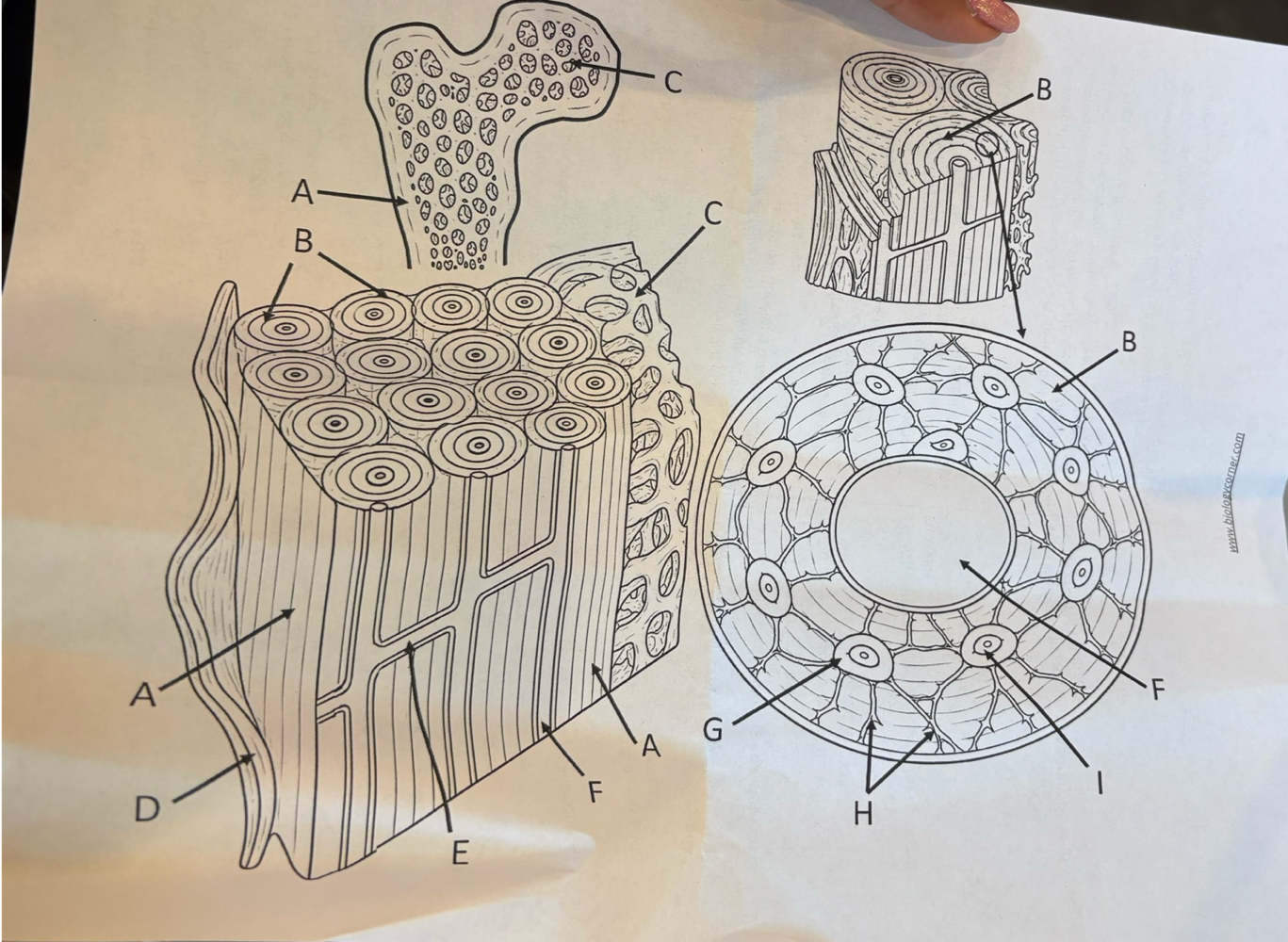
Anatomy of bone cross section including osteon:

Epiphysis Function
knobby region at the end of the bone
strengthens the join
serves to connect tendon and ligament
padded with articular cartilage
has spongy bone —> contains red marrow (makes RBC)
Diaphysis Function
shaft/long portion
contains a medullary cavity with yellow marrow (fat/adipose)
Articular cartilage Function
cartilage at the ends of long bones
To reduce friction and absorb shocl
Periosteum Function
tough sheath that covers the outer layer of bone
covers every area except articular cartilage
contains blood vessels and nerves —> plays a role in bone growth and repair
Medulla Function
hollow, cylindrical space within the diaphysis
stores adipose —> yellow marrow
Proximal vs Distal Epiphysis
Relative to body center
Proximal means closest to the center of the body
Endosteum Function
lines medullary cavity
thin membrane of cells that covers ALL internal surfaces of bone including medullary cavity
In the endosteum, bone growth, repair and remodling occur
Red Marrow Relevant Info?
Found in spongy bone
contains stem cells that form blood cells and platelets
for children its located in spongy bone and medullary cavity
In adults, most red marrow becomes yellow marrow
only in some parts of the AXIAL skeleton
Yellow Marrow
Fat storage
Types of bone tissues in bones and theur properties:
Compact Bone
very solid/dense
present in the external walls of bones
Spongy Bone
more porous
located internally, mostly in epiphysis
Haversian (Central) Canal characteristics
the haversion canal is in the center of the osteon.
Within it are blood vessels, nerves, and connective tissue that supplies the bone with nutrient and oxygen
Concentric lamellae
rings of calcified bone that surround the central canal
Contains collagen fibers and mineral deposits
gives bone strength and resistance
Osteocytes
Bone cells found in the lacunae within the osteon
They are connected together via canaliculi, which allow the ostocytes to exgnge waste, nutrient, and chemical signals.
maintaining bone matrix, bone health, and communicating with neighboring cells
Canaliculi Function
Tiny microscopic channels that Connect lacunae together and to the haversian channel
allow the ostocytes to exgnge waste, nutrient, and chemical signals throughout the bone.
Lacunae Function
tiny spaces where the osteocytes sit
These spaces protect the bone cells and allow them to maintain the tissue
Whats Ossfication, what are types, Name them:
Ossification —> process of bone formation
2 Types:
Endochondral ossification —>
Bone is produced from a cartilage model
typically forms the long bones
Intramembranous ossification —>
Bone forms from via embryonic tissue
produces flat bones (skull, facial bones, jaw, etc)
metaphysis
Metaphysis is the narrow area that contains the epiphyseal (growth) plate, a layer of cartilage growing in the bone
When the bone stops growing (reaches adulthood) that cartilage is replaced by osseous tissue AND the epiphyseal plate become epiphyseal line
What does collagen fiber do for the bone
make it flexible so its not brittle
Spongy bone functional unit
trabeculae!!
the lacunae and osteocytes are found in a lattice-like network of matrix spikes called trabeculae
trabeculae is NOT a random network → each trabecula forms along lines of stress to provide strength to the bone.
spaces of the trabeculated network provide balance to the dense and heavy compact bone by making bones lighter

Nutrient foramen
Spongy bone and medullary cavity receive nourishment from arteries that pass through compact bone through the Nutrient foramen.

Cartilage —> What is it, it’s relation to bone:
Bone is a replacement tissue —> it replaces cartilage (template) as we become older
cartilage provides a template of where bones would be
Cartilage is avascular —> has no blood vessels
Cartilage gets its nutrients by diffusion through the matrix
therefore cannot repair itself as readily
Intramembranous ossification
Overview —> Made directly from connective tissues. Mesenchymal cells turn directly into bone-building cells called osteoblasts, making woven bone that later remodels into compact and spongy bone.
Flat bones (ranial, skull, faaial)
Mesenchymal cells (STEM cells) turn into other cells —> osteoblasts
gather to form an ossification center
secret osteoid —> uncalcified extracellular matrix
after it calcifies the cells turn into osteocytes.
In Addition
Mesenchymal connective tissue on the outer surface condenses to form the periosteum. Entrapped blood vessels within the trabecular network condense to form red bone marrow
Endochondral Ossification
Overview —> Made from a cartilage templaye which blood vessels invade and is broken down so bone can take its place
Bone types —> long bones
Mesenchemal cells dufferentiate into chondrocytes (cartilage cells)
These cells lay down in a cartilage model
connective tissue membrane called the perichondrium forms around the outside of the hyaline cartilage model.
Chondrocytes in the central area enlarge (hypertrophy) and matrix begins to calcify —> blocks diffusion —> they die leaving empty cavities
Blood vessels invade these empty spaces, bringing along osteogenic cells
The expanding and merging cavities form the central medullary cavity
Capillaries penetrating the outer cartilage membrane trigger the perichondrium to convert into the bone-forming periosteum
Osteoblasts in the periosteum build a periosteal collar of compact bone around the diaphysis shaft.
As bone replaces cartilage in the diaphysis, cartilage continues growing at the ends (future epiphyses) to lengthen the bone.
After birth, the exact same sequence (calcification chondrocyte death blood vessel invasion osteoblast activity) repeats in the epiphyses.
These regions are known as secondary ossification centers.
How Bones Grow in Length
The epiphyseal plate is the area of growth in a long bone (found in kids that are growing)
layer of hyaline cartilage where ossification occurs in immature bones.
On the epiphyseal side of the epiphyseal plate → cartilage is formed.
On the diaphyseal side → cartilage is ossified, and the diaphysis grows in length. (Process of cartilage replaced by bone→ Endochondral Ossification!!)
Explain how the skeletal system interacts with other body systems to contribute to organism-level functions and homeostasis.
Muscular System
Muscles contract and they pull in bones producing movement
Nervous System
We have nerves within our bones in the periosteum and bone marrow that sense pain signals and control healing
Nervous system controls skeletal muscle contractions
Skull protects the brain, vertebral column protects the spinal cord
Cardiovascular System
RBC are produced inside the red marrow
Cardiovascular system transports blood cells throughout body
Immune System
WBC develop in bone marrow → fights pathogens
Endocrine System
Hormones help regulate bone growth and calcium levels
Digestive System
Digestive system absorbs calcium and phosphate from food which are used to build bone
Vitamin D helps the intestine absorb calcium
Integumentary System
Skin helps produce vitamin D after exposure to sun
Vitamin D helps the digestive system absorb calcium which creates stronger bones
Urinary System
The kidney help regulate calcium and phosphate levels
They also activate vitamin D into its active form
Helps maintain calcium balance
Respiratory System
Rib cafe protects the lungs; protects respiratory organ