CB.20 Genetic mutations
 Call Kai
 Learn
 Practice Test
 Spaced Repetition
 Match
 Flashcards
 Knowt Play
1/39

Earn XP
Description and Tags
• Types of DNA sequences in the human genome • Types of DNA mutations • Causes and effects of DNA mutations • Effects on gene/protein expression/function • Role of DNA mutations in human evolution and disease
40 Terms
What is the difference between a genome, gene and allele?
Genome: The complete genetic material of an organism or cell.
Gene: A functional region of DNA associated with producing a functional product, such as a protein or non-coding RNA, and its relevant regulatory sequences.
Allele: One of the alternative versions of a gene at a particular locus.
What is a genetic mutation?
A genetic mutation is a change in the DNA sequence. Mutations can affect a single nucleotide, part of a gene, an entire gene or the structure or number of chromosomes.
A mutation can be defined by affected cell type. Define germline vs somatic mutation.
Germline mutation: Occurs in a cell of the germline that contributes to reproduction. It can be passed to offspring and, if present from conception, may be present throughout the offspring's body. Example: an inherited mutation causing a developmental disorder.
Somatic mutation: Occurs in a non-germline cell during development or later life. It can be passed to daughter cells as that cell divides but is not normally passed to offspring. Example: mutations accumulating in a population of colon cells during cancer development.
Mutations can be classified by their effect on gene function. In simple terms what is the difference between loss of function, gain of function and dominant-negative mutations?
1. Loss-of-function mutations (LOF): mutation reduces or abolishes the activity of a gene product.
2. Gain-of-function mutations (GOF): mutation increases or alters the activity of a gene product.
3. Dominant-negative mutations: mutation produces a mutant protein that interferes with the function of the normal protein.
What are the two types of loss of function mutations?
Hypomorphic allele: the gene product retains some function, but its activity is reduced.
Null allele: the gene product is absent or has no meaningful function.
What is haploinsufficiency?
Humans have two copies of an autosomal gene, one inherited from each parent. In some cases, one functioning allele is not enough to maintain normal development or function. Losing the contribution of one copy can therefore produce a clinical phenotype.
The underlying issue is gene dosage: some genes require full output from both alleles, and reducing that output by half leads to dysfunction.
Gain-of-function mutations (GOF) increase or alter the activity of a gene product – in what ways does this occur to the protein?
GOF mutations the protein becomes:
more active than normal;
gains a new activity;
s produced in excessive amounts;
is expressed at the wrong time or in the wrong tissue.
Dominant-negative mutations produce a mutant protein that interferes with the function of the normal protein – how?
A dominant-negative mutation produces a mutant protein that interferes with the function of the normal protein. E.g. type I collagen, which is built from three polypeptide chains arranged into a triple helix: two α1 chains and one α2 chain. If a mutation changes the structure of one of these chains, the abnormal chain can interfere with assembly or stability of the collagen molecule. This is different from simply losing half the normal protein: the mutant product actively disrupts the normal product.
Which type of mutation is a PAX9 mutation?
PAX9 mutation-associated hypodontia is a loss of - PAX9 activity is insufficient during tooth development, some teeth may fail to form.

What type of mutation is FGR mutation?
Certain activating FGFR mutations alter signalling involved in bone development and can cause premature fusion of cranial sutures. FGFR mutations associated with craniosynostosis.

What type of mutation causes osteogenesis imperfecta?
Osteogenesis imperfecta is a caused by a dominant negative mutation. The mutation disrupts the normal folding and assembly of type 1 collagen/ a disorder characterised by bone fragility.

Mutations can also be classified by the DNA change. What are the four broad categories of types of DNA change?
Chromosomal abnormalities.
Nucleotide substitutions.
Nucleotide deletions.
Nucleotide insertions.
Chromosomal abnormalities can either be numerical or structural. What are each?
Numerical abnormalities when non-disjunction occurs (chromosomes fail to separate properly during meiosis/mitosis). Instead of the somatic cells containing 46 chromosomes arranged in 23 pairs they will have too few/too little. Aneuploidy = Gain or loss of individual chromosomes.
Structural abnormalities occur when chromosomes break and are re-joined in an abnormal arrangement, or when unequal recombination changes the amount or position of DNA.
Numerical chromosomal abnormalities can be classified as aneuploidy vs polyploidy what is the difference?
Aneuploidy = Gain or loss of individual chromosomes.
Monosomy = One chromosome of a pair is missing; e.g. Turner syndrome 45,X.
Trisomy = An extra copy of a chromosome; Trisomy 18 or trisomy 21
Polyploidy = Extra complete sets of chromosomes; e.g. Triploidy: three complete chromosome sets. Polyploidy is usually de novo mutation and often not compatible with life.
What is the difference between trisomy and triploidy?
Trisomy 21 means three copies of chromosome 21 = resulting in a total of 47 chromosomes.
Triploidy means three complete sets of chromosomes, usually 69 chromosomes in total.
Structural chromosomal abnormalities can be caused by translocation, inversion, deletion/insertion, ring chromosomes or isochromosome - what are each?
Structural abnormalities occur when chromosomes break and are re-joined in an abnormal arrangement, or when unequal recombination changes the amount or position of DNA.
Translocation = A segment moves to another chromosome. In a reciprocal translocation, two chromosomes exchange segments. Robertsonian translocations involve particular acrocentric chromosomes joining near their centromeres.
Inversion = A chromosome segment breaks off and reinserts in the opposite orientation. Can be Pericentric: includes the centromere. OR Paracentric: does not include the centromere.
Deletion or insertion = A chromosome segment is lost or an extra segment is inserted. Deletions can remove genes and their regulatory elements.
Ring chromosome = a chromosome end breaks and join into a ring.
An isochromosome has one arm lost and the other duplicated.

What is Robertsonian translocation?
Structural chromosome abnormality where particular acrocentric chromosomes joins near their centromeres.
Acrocentric means short arm is present but cannot be visualised (because it is so small). There are five acrocentric chromosomes: 13,14,15,21,22.
In Robertsonian translocation they break at their centromeres and fuse to form a single large chromosome (and short arms fuse but are lost because they are so small).

What is pericentric vs paracentric inversion?
In inversion a chromosome segment breaks off and reinserts in the opposite orientation.
It can be Pericentric: includes the centromere.
OR Paracentric: does not include the centromere.

What is a balanced bs unbalanced chromosomal mutation?
Mutation caused by chromosomal abnormality.
Balanced: there is no net gain or loss of chromosomal material - may have no obvious clinical effect
Unbalanced: there is a gain or loss of genetic material- may alter the dosage of multiple genes, causing a more substantial phenotype.

Example of balanced chromosomal translocation causing disease?
The Philadelphia chromosome, t(9;22), which creates the BCR::ABL1 fusion gene and causes chronic myeloid leukaemia.

Mutations caused by nucleotide substitutions are further sub classified into transition or transversion nucleotide substitutions – explain the difference.
A nucleotide substitution replaces one DNA nucleotide with another. It is one of the smallest types of DNA change.
Two types according to the chemical class of the bases: A and G are purines, whereas T and C are pyrimidines in DNA.
Transition: a purine is replaced by another purine (A ↔ G), or a pyrimidine by another pyrimidine (C ↔ T).
Transversion: a purine is replaced by a pyrimidine, or vice versa.
What are the “Mutation hot spots” in genome where transition of nucleotides is common?
Mutation hotspots (regions of DNA where mutations occur more frequently) such as CpG sites — places where a cytosine (C) is followed by a guanine (G) on the same DNA strand. Often found in promotor region of transcribed genes.
CpG sites are often methylated in the genome (to silence the gene). Methylation adds a chemical group to cytosine, forming 5-methylcytosine.
If there is chemical damage to 5-methylcytosine causes amino acid group to be replaced with oxygen = DEAMINATION of cytosine. If cytosine loses its amino acid group it becomes uracil.
Uracil does not normally belong in DNA. So DNA repair enzymes usually recognise and remove it.
BUT if it is not repaired, U pairs with A instead of GIf unrepaired, this can produce a permanent C:G → T:A mutation.

If one nucleotide is changed how can that affect a protein?
Genetic code is read in codons (groups of three nucleotides). Each codon specifies an amino acid or a stop signal. A substitution can therefore have different consequences depending on where it occurs.

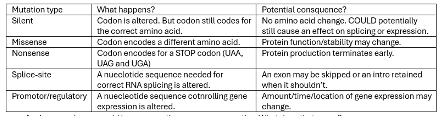
Nucleotide substitutions can cause silent, missense, nonsense, splice-site and promotor/regulatory mutations. What happens in each and what is the potential consequence of this mutation type?
Silent mutations usually caused by third nucleotide being incorrect.
Remember utter NONSENSE so its STOPPED.
A missense change could be conservative or non-conservative. What does that mean?
A conservative substitution replaces an amino acid with one having similar chemical properties; a non-conservative substitution changes it to one with substantially different properties.
Conservative change is not guaranteed to be harmless, and a non-conservative change is not guaranteed to cause disease. The location and role of the amino acid matter.
Mutations caused by nucleotide insertion/deletion have what impact?
Alter the codon reading frame.
In-frame deletion/insertion: nucleotides are added/removed in multiples of three. Downstream reading frame is preserved. However an amino acid is either added/removed.
Frameshift: is when the number of nucleotides added or removed is not a multiple of three.
What are trinucleotide repeat expansions? And what is a repeat expansion?
A trinucleotide repeat = sequence of three bases repeated consecutively; e.g. CAG-CAG-CAG-CAG
A repeat expansion = mutation in which the number of repeated units increases beyond the usual range (e.g. CAG-CAG-CAG-CAG-CAG- CAG-CAG-CAG-CAG). Some repeat sequences are relatively stable, while others are prone to expansion when DNA is copied or passed between generations.
Being prone to expansion when copied/passed between generations is called dynamic mutations because the length of the repeated sequence can change over time or across generations.
How do repeat expansions cause disease?
The effect depends on where the repeat is located, which triplet is repeated, and how large the expansion becomes.
A. Expansion within a protein-coding region : abnormally long chain of amino acids = altered protein may become toxic to cells. E.g. Huntington disease.
B. Expansion in a non-coding region: A large repeat expansion can switch off gene expression, so little or none of the required protein is produced. E.g. Fragile X syndrome
C. Abnormal RNA function: Some expansions produce RNA that interferes with normal cellular processes, including the correct processing of other RNA molecules. E.g. Myotonic dystrophy type 1
Why are trinucleotide repeat expansion mutations called dynamic? And what is anticipation?
Repeat sequences can change length because the DNA-copying and repair machinery sometimes handles repetitive sequences incorrectly. One proposed mechanism is DNA polymerase slippage, in which the newly copied strand misaligns with its template. The repeat can therefore expand or contract when cells divide or when genetic material is passed to offspring. Each disorder has its own repeat-size thresholds, and larger expansions are often less stable.
Anticipation occurs when a genetic disorder tends to appear at an earlier age, or with greater severity, in successive generations of a family. Usually because more repeats can be associated with earlier onset or more severe disease, depending on the condition. E.g. Huntington disease and myotonic dystrophy.
What is an indel nucleotide mutation?
Indels’ = mutations that involve both insertion and deletion of nucleotides.

How do DNA replication errors cause DNA mutations?
DNA polymerase proof reads, removes the the incorrect nucleotide and insert the correct nucleotide.
Proofreading should detect these mutations - however if a mutation escapes it allows it to become a table mutation.
How does chemical damage cause DNA mutations? What is a DNA adduct, depurination and deamination?
Potential chemicals– smoke, tobacco, drugs, cellular metabolism. Depends on the type and dose of the damaging agent, the cell and the effectiveness of DNA repair. If a chemical group is covalently ADDED to a base, it is a “DNA adduct”. The chemical binding causes a DNA mutation…
If a purine base (A/G) is lost it is called depurination.
If a chemical group is REMOVED to any of the nucleotide bases its called deamination. By deaminating cytosine you convert int to uracil. Uracil is often removed.

How does physical damage cause DNA mutations?
Ionising radiation = can break DNA strands (single/double). If repaired incorrectly, they may cause deletions, translocations and other chromosomal abnormalities.
Ultraviolet (UV) radiation = Can cause adjacent pyrimidine bases to bond abnormally, producing lesions such as thymine dimers. These distort DNA and interfere with replication and transcription.

DNA repair mitigates against DNA mutations. If repair fails, the damage may persist and become a mutation. If the damage is too extensive, the cell may stop dividing or die instead. What are the following repair pathways? Mismatch repair, nucleotide excision repair, base excision repair and double-strand break repair.
Mismatch repair =Corrects mismatched bases and small insertion/deletion errors that escape replication proofreading.
Nucleotide excision repair =Removes bulky DNA lesions, including UV-induced pyrimidine dimers.
Base excision repair =Removes damaged or abnormal individual bases.
Double-strand break repair = Repairs double-strand breaks through mechanisms including non-homologous end joining and homologous recombination.
What are the two main mechanisms for repairing double-strand breaks? And how do they allow mutations?
Two repair mechanisms that can remove and repair double stranded breaks.
1: Non homologous end joining: ends of broken chromosomes are processed by nucleuses and joined by DNA ligases. This can lead to deletion of DNA sequences.
2. Homologous recombination: additional enzymess to process the ends of chromosomes – producing staggered ends. Then uses undamaged sister chromatids as a template to repair the double stranded DNA. The break is repaired with no loss of nucleotides at the repair site.

Multigene families arose by gene duplication and evolutionary divergence. Define gene family vs gene superfamily.
Gene families – high sequence similarity. They often have related functions. E.g. HOX genes.
Gene Superfamilies – A broader group of evolutionarily related genes. Their overall DNA or protein sequences may have limited similarity, but they retain evidence of shared ancestry, such as conserved structural domains. Example: the immunoglobulin superfamily, which includes many proteins involved in cell recognition and immune responses.
What are pseudogenes and how were they formed?
Pseudogenes resemble known structural genes but are generally not expressed.
Arose by gene duplication and then became silent through mutation.
OR arose from an insertion into the genome by reverse transcription event but they lack a promotor.
Explain the pathophysiology of sickle cell disease in terms of gene mutation. How is it an example of mutations causing disease but also an evolutionary advantage?
Sickle cell disease is caused by: missense mutation. Autosomal recessive. Alteration in the beta-globin gene which encodes a component of haemoglobin.
The missense mutation changes the codon (A to T transversion: CAG to CTG) means that glutamate (polar) is replaced by valine (non-polar) in the beta-globin protein. This substitution changed the chemical properties of haemoglobin. Hydrophobic addition of valine reduces the solubility of haemoglobin.
Under low-oxygen conditions, the altered haemoglobin can polymerise, deforming red blood cells into characteristic sickle shapes. These cells can break down prematurely and obstruct small blood vessels, causing anaemia and other complications.
Heterozygotes: healthy carriers. Homozygotes: severely anaemic due to malformed RBC shape however survive.
In regions where malaria is endemic, people with one copy of the sickle cell allele can have increased protection against severe malaria because the parasite cannot reproduce in the damaged erythrocytes. This creates a selective advantage that can maintain the allele in the population despite the serious consequences of inheriting two copies.

What are oncogenes and tumour suppressor genes?
Oncogenes: genes that, when abnormally activated, promote cell proliferation or survival.
Tumour suppressor genes: genes that normally restrain cell growth (p53) preserve genomic integrity or help eliminate damaged cells.
What is negative and positive genetic selection in evolution?
Negative selection: a variant that substantially reduces reproductive success may become less common.
Positive selection: a variant that increases reproductive success in a particular environment may become more common.