Anat and Phys Exam 2
 Call Kai
 Learn
Learn Practice Test
 Spaced Repetition
 Match
Match Flashcards
 Knowt Play
1/242

There's no tags or description
Looks like no tags are added yet.
Name | Mastery | Learn | Test | Matching | Spaced | Call with Kai | Chat |
|---|
No analytics yet
Send a link to your students to track their progress
243 Terms
Contraction
Function of the muscle, to move the body and things within the body
3 types of muscles
Smooth, skeletal, and cardiac
Smooth muscle
Involuntary, not striated ; gut, glands, organs, eye chamber, hair; central located nucleus; regulated by ANS
Skeletal
Voluntary, striated; bones; do not branch and lack intercalated disks, multinucleated cells, require nerve impulse for stimulous
Cardiac
Involuntary, striated; found in heart, regulated by ANS; elongated branching cells
Intercalated disks
Highly specialized cell membranes that join cardiac muscle cells, facilitates transmission of electrical impulse (basically makes sure muscle fibers beat in unison thus heart beat)
Fiber
One muscle cell
Sarcolemma
Plasma (cell) membrane of a muscle cell/fiber
Sarcoplasm
Cytoplasm of muscle cell/fiber
Sarcoplasmic reticulum
Smooth ER of muscle cell, storage area for fiber
Sacro and Myo
Prefixes meaning muscle
Origin
Closest to body trunk, least movable attachment, usually proximal and/or medial
Insertion
Most movable attachment, usually distal and/or lateral
Contraction of the muscle brings what together
Origin and Insertion, only way for a muscle to exert force
Extensors
Muscles extend or increase angles between body parts; forearm
Flexors
Flex or decrease angle between body parts; underam
Adductors
Pulls limb towards median plane
Abductors
Pulling limb away from median plane
Constrictors (sphincters)
Surround circular opening, closes when they contract (decreases lumen of something, like iris or sphincters in digestive glands)
Dilators
Surround circular opening, opens when contracts (increases size of lumen, uncommon in mammals)
Agonists
Muscles that promote a given movement, like 4 muscles of the quad
Antagonists
Muscles that inhibit a given movement, prevent hyperextension, etc
Red muscle fibers (dark meat)
Used for endurance; more myoglobin, glycogen, and mitochondria, slow twitch (slower to contract but fatigue slowly)
White muscle fibers (light meat)
Not made for endurance, fast twitch (contracts quickly but fatigues faster)
Myoglobin (mb)
Oxygen storage, in red muscle fibers
Glycogen
Glucose storage, in red muscle fibers
Mitochondria
ATP production, in red muscle fibers
Muscle fiber arrangements in muscle
Parallel, Pennate, Fleshy, and Oblique
Parallel muscle fibers
Tendon that comes off at end of fibers and attach to bones (all muscles of tendons); pectoralis muscle
Pennate
Feather-like fibers with central tendon, wide at the top and converges at the tendon, biceps
Fleshy
Appears to attach directly to bone (no gross tendon), but has short microscopic tendons between muscle fibers and bones, brachialis
Oblique
Circular or convergent, abdomen, at an angle
Connective Tissue (CT) harnessing muscles
Endomysium, Perimysium, Epimysium; All meet at ends (or center for pennate muscles) to attach to bones which transfers movement and energy of contraction to bones
Endomysium
Loose CT around every muscle cell
Perimysium
Loose CT around bundles (fascicles) of muscle cells
Epimysium
Dense irregular to regular CT around whole muscle
Myofibrils
Strands that make up muscle cells, composed themselves of myofilaments, which were arranges in units called sarcomeres along these
Myofilaments of Myofibrils
Myosin and actin
Myosin
Thick filament
Actin
Thin filament
Sarcomeres
Functional and contractile unit of striated muscle tissue (microfibers), made up of actin and myosin, extend from one z line to the next, each tube contracts itself
Striations
Banding patterns of muscle fibers
Parts of the sarcomere
A band, I band, H band, Z line, M line (A band always stays same length, H and I get smaller)
A band
Length of myosin filament, includes overlap with actin, stains dark, yellow in graph

I band
Actin only, does not include area of overlap with myosin, stains light, purple in graph

H band
Myosin only without actin overlap, dark blue

Z line
Ends of sarcomere, connects actin filaments, teal

M line
Middle of sarcomere, connects myosin filaments, green

Myosin filament
Thick protein filaments that grabs onto actin and pull, heads target g-actin molecules

Actin filaments
Protein myofilament (thin), has 3 parts (tropomyosin, troponin, g-actin molecules)
G-actin molecules
Globular molecules that fit like beads on a string, two twist around to make actin filament (red)

Tropomyosin
Groove of actin filament (between actin strands) that have a different strand protein,
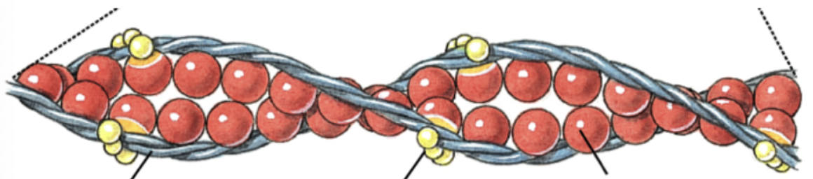
Troponin
Globular protein of tropomyosin that is spaced along it, what calcium binds to that. causes tropomyosin to relax, exposing bonding cite to myosin, causing contractions.

Actin bonding and calcium
Actin has binding sites for myosin heads along filament. in relaxation tropomyosin covers these binding sites so that actin and myosin do not interact. Calcium binds to troponin -> relaxes tropomyosin -> exposes binding sites on actin for myosin to bind
Muscle fiber organelles (on outside)
Nuclei, mitochondria, sarcoplasmic reticulum (SR), terminal cisterna, sacrolemma, T-tubules
Mitochondria (muscle fibers)
Some between myofibers
Sarcoplasmic reticulum
Organelle with terminal cisternae at the end stores fibers and calciums
Terminal Cisternae
At end of SR and A-band, actually store fibers and calcium
T-tubule
One between 2 TC (the ends of SR), electrical impulse propagates through line, free up impulse for calcium for contraction, has many CHAMBERS and bumps
Sarcotubular sytem
Includes Terminal Cisternae, Sarcoplasmic Reticulum, and T-tubules. Help control muscle contractions and electrical signals
Synapse
Junction between nerve/muscle, do not physically touch but filled with fluid, 1 nerve can synapse thousands of muscle fibers
Motor unit
The motor neuron and all skeletal muscle fibers it innervates (causes to move)
Ventral Root
Motor neuron with synapses at the motor end plate
Motor endplate
Folded region of muscle cell membrane (sarcolemma) that makes “contact” with motor neuron
Synaptic Bulb
Nerve impulse (electrical) in motor neuron travels down axon to this, causing the voltage gated gate to open up and let calcium into axon
Voltage gated gate
Opens in synaptic bulb after protein channel reaches threshold, lets calcium enter axon
Acetylcholine (ACH)
Released from synaptic bulb, diffuse across cleft, binds to sacroemma receptor on muscle cell, lets Na+ channels enter and K+ leaves cell, increase voltage across sarcolemma, triggers action potential
Action potential
Electronic impulse released across sacrolemma and down T-tubules once voltage threshold reached, releases calcium, binds to muscle, causes contractions
Action potential initiating contraction
T-tubules next to terminal cisternae of S.R (triad, calcium stored in SR), act as copper wiring like with calcium channels, AP causes them to open in terminal cisternae, release calcium into sacroplasm, lets it contract
Sliding filament theory of muscle contraction (calcium)
Calcium binds to troponin and undergoes conformation change, unlocks tropomyosin by pulling it off binding sites, actin-binding sites exposed to myosin heads, pulls to center and contracts, uses energy ATP → ADP; initiated by Ca++ release into sarcoplasm
Sliding filament theory (ADP and ATP steps)
Heads release ADP and bind to New ATP → heads detach from actin → heads attach to new actin binding site further along; continues as long as calcium and ATP available
ATP
From glycolysis and krebs cycle, more efficient aerobic (reserves used up first) but can be anaerobic but can result in lactic acid build up
Red vs White muscle fibers
Red has more oxygen
Creatine Phosphate (CP)
Can be used to regenerate ATP from ADP by adding phosphate group
Contracture vs Contraction
(1) muscle shortening in absence of impulse; happens because ATP runs out and filaments release actin Ca++ (2) normal muscles shorten
Rigor mortis
CONTRACTURE after death, 12-24 hours
When impulse stops…
Ca++ re-sequenced into SR terminal cisternae → tropomyosin re-shields actin → muscle relax
What do H and I band do during contraction
Get narrower as sarcomere shortens with more overlap of actin and myosin
What do H and I band do during relaxation
Slide back apart
Muscle twitch
Contraction of one motor unit (motor neuron and all muscle fibers it sends singles to)
Muscle contraction phases
Latent, Contraction, Relaxation
Latent phase
No tension or movement, cross-bridges (myosin and actin) are interacting
Contraction phase
Rising part of graph, tension developed, myosin moves actin filaments closer together, muscle fibers get shorter
Relaxation phase
Calcium released back out to SR, K leaves cell, cross bridges become inactive, tension decreases, returns to latent phase
What grades contraction strength
Frequency of stimulus and strength of stimulus
Strength of stimulus
Stimulating one motor unit leads to weak contraction, while stimulating lots makes a large contraction (number of motor units)
Frequency of contractions
Faster nerves fires the more strength you get in contractions (like with lifting heavy things), if muscle stimulated to contract before muscle has relaxed strength of subsequent contraction increases, if goes past threshold muscle twitched becomes fused in SINGLE PROLONGED CONTRACTION
Tetany
Maximum Contraction Strength
Treppe
Muscles “warm up” to max contraction state through the application of stimuli of equal intensity a few seconds apart from the muscle, each twitch slightly stringer that the pervious one, successive stimulations increase calcium concentration in sarcoplasm, can lead to fatigue if too much
Fatigue
Decrease ATP, weakens contraction; Decrease oxygen, lactic acid buildup
Cardiac Contraction
Striated with myosin and actin, has same sarcomere arrangement and sliding muscle contraction mechanism as skeletal (differs in sarcotubular arrangement and number of triads), AP originates in HEART ITSELF, sinoatrial node acts as a pacemaker by contracting at intervals, ANS controls it involuntary, intercalated discs spread action potential cell to cell, Purkkinje fibers in heart spread it very fast
Smooth Muscle Contraction
Not striated (no sacromere or bands), actin/myosin perpendicular in long cells, helps move things, more actin to myosin, has DENSE BODIES where actin attached to
Purkinje fibers
In cardiac muscle contractions (heart), spreads AP very fast
Dense bodies
In smooth muscle tissue, actin attached to the, corresponds to Z-lines of skeletal muscle, connected by proteins and surrounds myosin making contractile unit
Acetylcholinesterase
Enzyme that recycles acetylcholine by breaking it down. Important since it needs to go to away after it binds or else constant contraction
Acetylcholinesterase inhibitors
Prevents ACH from being broken down, muscle keeps contracting until runs out of ATP (muscle sparms and parasympathetic effects, death caused to to respiratory system contracting without relaxation)
Acetylcholinesterase inhibitors examples
Nerve gas, insecticides, venom, black widows, sea kraits, poison (too much Ca+)
Botulism Toxin
Cleaves specific proteins so that synaptic vesicles cannot release acetylcholine at motor endplates, leads to flaccid paralysis
Flaccid paralysis
Limp
Tetanus Toxin
Absorbed by nerves and is transported to the spinal cord, spasmodic tonic contraction of voluntary muscles by interfering with inhibitor release, constant nerve firing, spasms, cause respiratory failure