Muscles Practical
 Call Kai
 Learn
 Practice Test
 Spaced Repetition
 Match
 Flashcards
 Knowt Play
1/70

There's no tags or description
Looks like no tags are added yet.
71 Terms
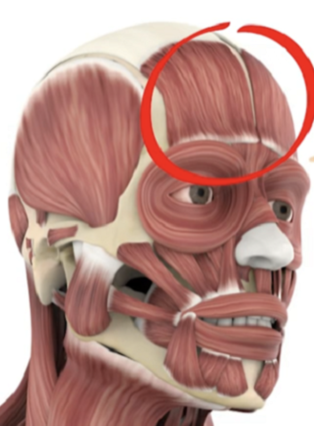
frontalis
Forehead covering the frontal bone; raises eyebrows, wrinkles forehead.
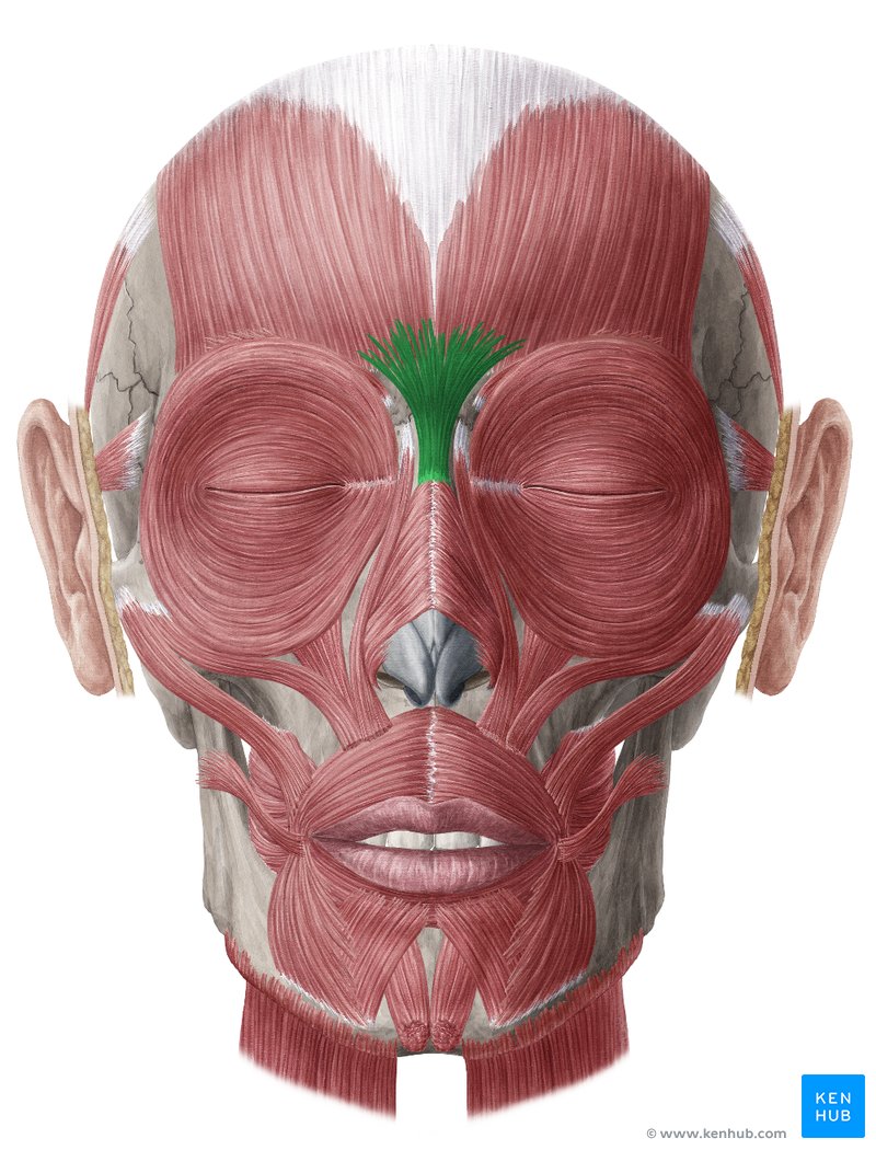
procerus
Bridge of nose, between the eyebrows; Runs vertically down the nasal bridge; wrinkles skin over nose
Orbicularis oculi
Circling the orbit / eye; Circular sphincter muscle; closes eyelids tightly.

Nasalis
Across bridge & flared cartilage of nose; Directly covers the bridge and ala of the nose.

Levator labii superioris
Between lower orbit and upper lip; Lies immediately medial to the zygomaticus muscles; elevates upper lip.

Zygomaticus minor
Upper, thinner diagonal cheek strap; originates on zygomatic bone superior/medial to major; elevates upper lip.

Zygomaticus major
Lower, thicker diagonal cheek strap; Runs down toward corner of mouth; "smiling muscle".

Orbicularis oris
Circling the lips; Circular sphincter muscle; closes/puckers lips ("kissing muscle").

Buccinator
Deep wall of the cheek (under zygomaticus); Horizontal fibers compressing cheek against teeth (whistling/chewing).

Mentalis
Prominence of the chin; Paired vertical fibers on the chin apex; elevates/protrudes lower lip.

Temporalis
Elevates and retracts the mandible (closes jaw).

masseter
Superficial, thick rectangular muscle over the ramus of the jaw; Primary jaw elevator.

Lateral (External) Pterygoid
Visible when the masseter muscle is removed. Runs horizontally; Protracts mandible and opens jaw.

Medial (Internal) Pterygoid
Deep and vertical, running along the medial (interior) surface of the mandibular ramus; Synergist with masseter to elevate mandible.

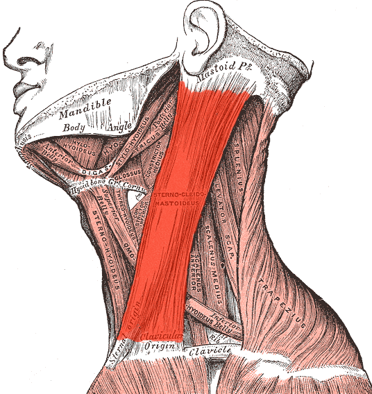
Sternocleidomastoid (SCM)
Prominent strap crossing diagonally from the sternum/clavicle to the mastoid process behind the ear. Divides the neck into anterior and posterior triangles.
Scalene Group (Anterior, Middle, Posterior)
Deep muscle bundle exposed right in the posterior triangle, lateral to SCM and running down to Ribs 1 and 2.

skeletal muscle
Long, cylindrical, unbranched fibers with distinct cross-striations and multiple peripheral nuclei pressed against the edge.

cardiac muscle
Long, cylindrical, unbranched fibers with distinct cross-striations and multiple peripheral nuclei pressed against the edge.

smooth muscle
Non-striated, spindle-shaped (tapered) fibers with a single, elongated central nucleus; packs closely in sheet form.

Axon Terminal & Synaptic Cleft
dark brown part is the axon, wavy part is the synaptic cleft

Schwann Cell

Sarcolemma
clear outer membrane covering the fiber

Sarcoplasmic Reticulum (SR)
The lace-like/tubular blue netting wrapping each myofibril; forms enlarged sac ends called terminal cisternae that store calcium.

Transverse (T) Tubule
The vertical tubule running down between two cisternae; aka the blue part
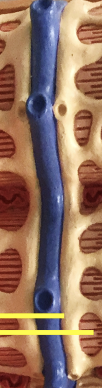
triad
the three lines with the blue in the middle; One central T-tubule flanked by two terminal cisternae of the SR.

sarcomere

piriformis
the first arrow; largest one

Gemellus Superior
the second arrow
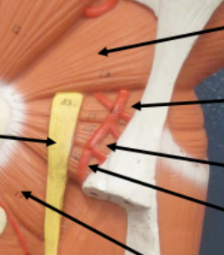
Obturator Internus
the third arrow

gemellus inferior
the fourth arrow

quadratus femorus
the fifth arrow

iliopsoas
combination of the psoa major (first arrow) and the iliacus (second arrow)

sartorius
1st arrow at the top; anterior view of the thigh/quad

rectus femoris
2nd arrow from the top

vastus medius
3rd arrow from the top

vastus lateralis
fourth arrow from top

vastus intermedius

Fibularis (Peroneus) longus
middle arrow

Fibularis (Peroneus) brevis
first and third arrows

superior rectus
directly above eye

inferior rectus

medial rectus

lateral rectus

superior oblique

inferior oblique

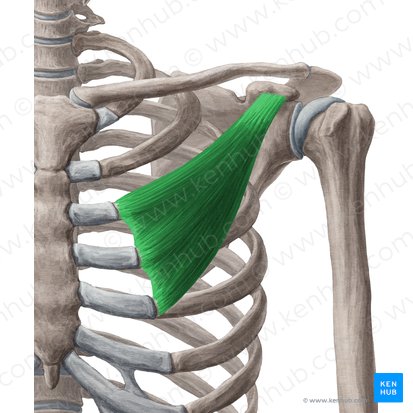
pectoralis minor
combination of three muscles
subclavius
right under the clavicle

Serratus anterior

trapezius

levator scapulae

rhomboid minor
1st line from the top

rhomboid major
2nd line from the top

supraspinatus
1st line at the top

infraspinatus
2nd line from the top

teres minor
3rd line from the top
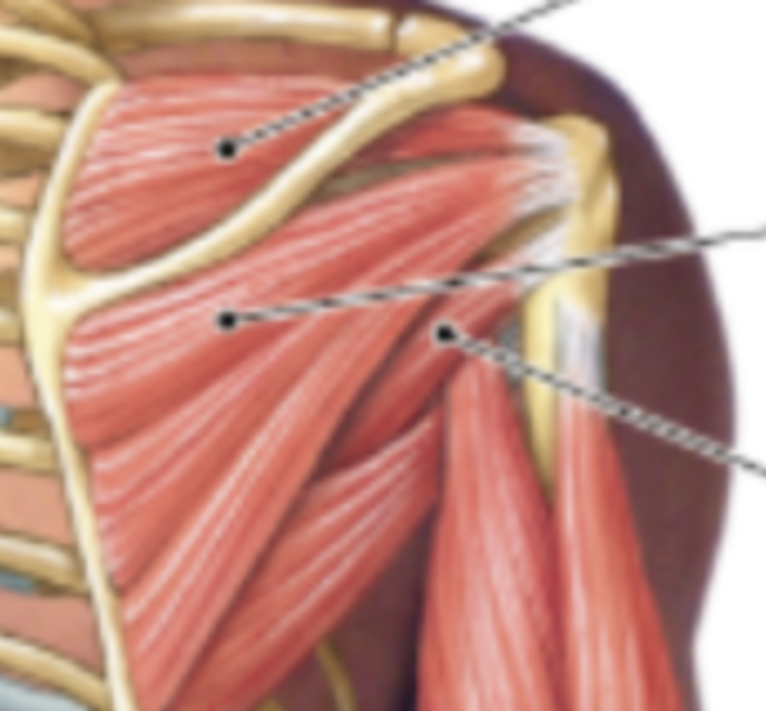
subscapularis

deltoid

pectoralis major
left arrow

Latissimus dorsi

teres major

pronator teres
1st line at the top

flexor carpi radialis
2nd line from the top

palmaris longus
3rd line from the top

flexor carpi ulnaris
4th line from the top
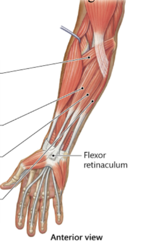
brachioradialis
1st line at the top on the right
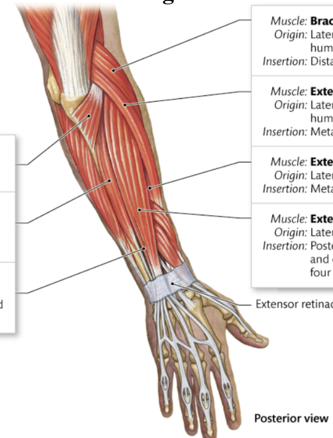
Extensor carpi radialis longus
2nd line from the top on the right

Extensor carpi radialis brevis
3rd line from the top on the right

Extensor digitorum
4th line from the top on the right

Extensor digiti minimi
3rd line from the top on the left
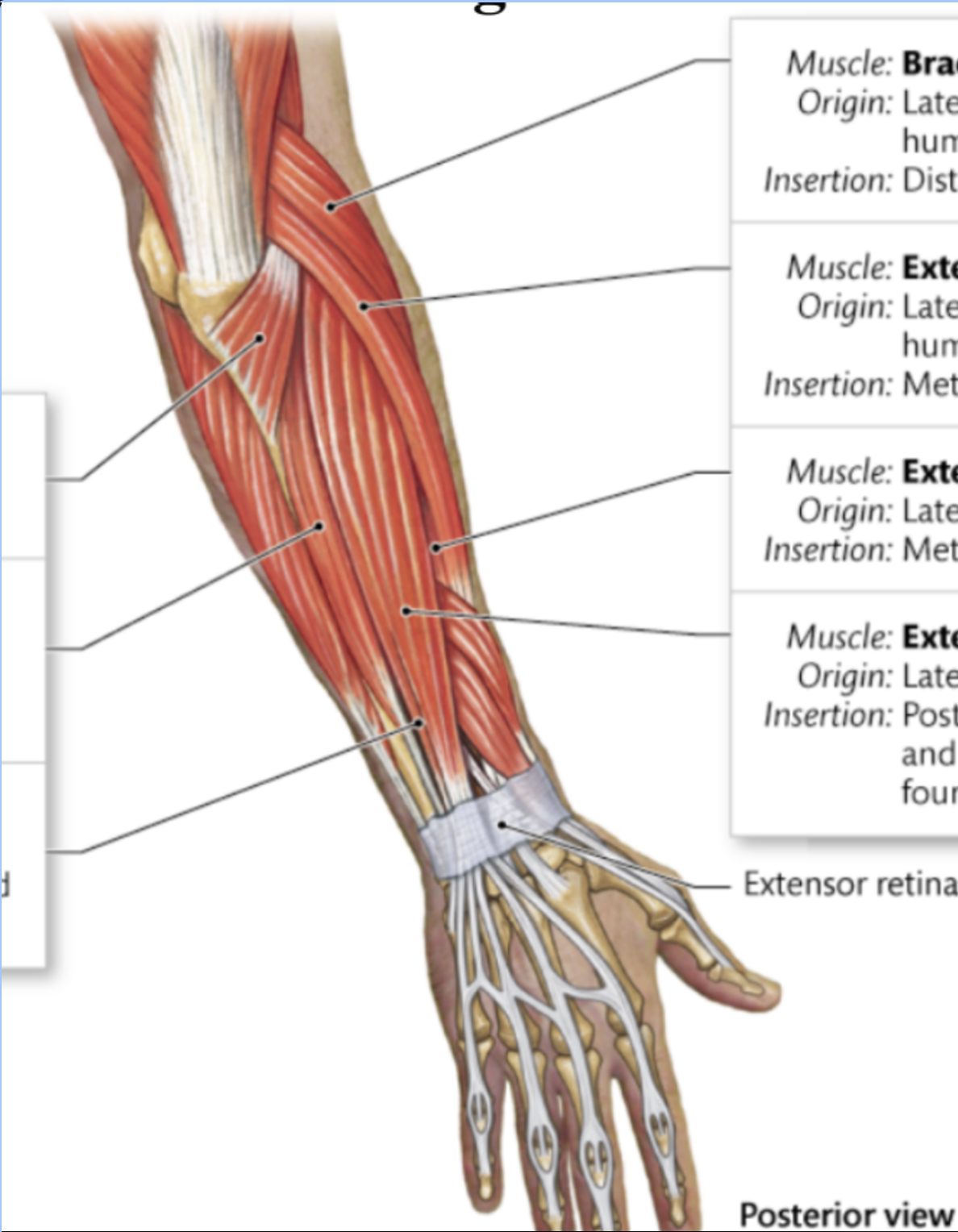
Extensor carpi ulnaris
2nd arrow from the top on the left
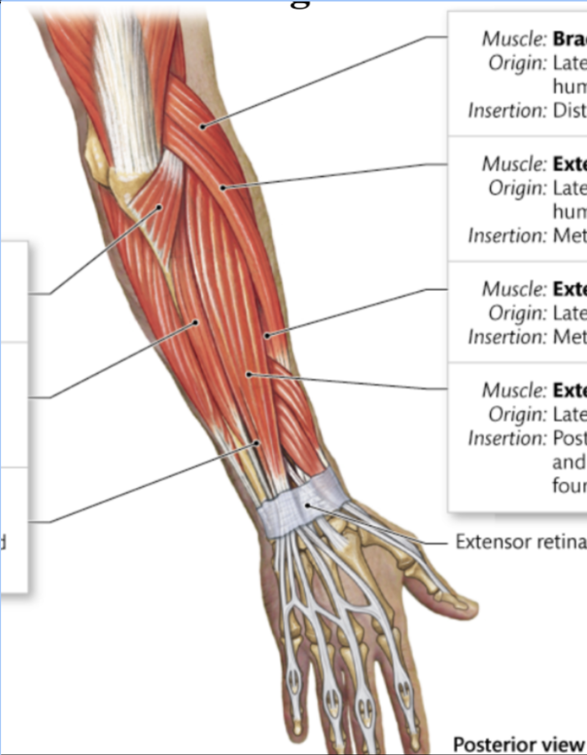
anconeus
1st arrow at the top on the left
