18. Spinal Cord & nerves

0.0(0)
Studied by 0 people
call kaiCall Kai
learnLearn
examPractice Test
spaced repetitionSpaced Repetition
heart puzzleMatch
flashcardsFlashcards
GameKnowt Play
Card Sorting

1/15

encourage image

There's no tags or description

Looks like no tags are added yet.

16 Terms

1
New cards

Spinal cord

  • 31 pairs of Spinal nerves

  • long structure

  • made of nervous tissue

    • neurons

    • glial cells

  • part of CNS

  • connects brain to PNS

  • intergrates info/produces responses too

  • Extends from foramina magnum to L2


<ul><li><p>31 pairs of Spinal nerves</p></li><li><p>long structure</p></li><li><p>made of nervous tissue</p><ul><li><p>neurons </p></li><li><p>glial cells</p></li></ul></li><li><p>part of CNS</p></li><li><p>connects brain to PNS</p></li><li><p>intergrates info/produces responses too </p></li><li><p>Extends from foramina magnum to L2</p></li></ul><p></p>
2
New cards

Foramina magnum

  • Large hole in base fo skull (occipital bone)


3
New cards

Boeny structures around Spinal cord

  1. vertebrae

    • stacked to create vertebral column


  • smooth side โ†’ anterior side

  • spiney side โ†’ posterior side


  • At region of spiney proceses each vertebrae has an opening called veretebral foramina

    • succesive holes make a canal

      • called veretbral canal


4
New cards

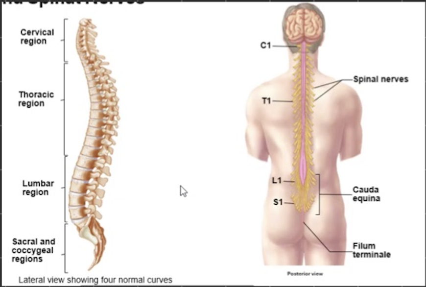
Curves in SC

  • Divided into regions

    • Dictake names of vertebrae + spinal nerves in that region


Superior to inferior:

  1. cervical region

  2. thoracic region

  3. lumbar region

  4. sacral/ coccygeal region


  • due to these curves the SC must also curve to maintin its position in the middle of the VC

    • meninges (connective tissue) used to help surround Sc to help keep it in proper position

<ul><li><p>Divided into regions</p><ul><li><p>Dictake names of vertebrae + spinal nerves in that region</p></li></ul></li></ul><p></p><p>Superior to inferior:</p><ol><li><p>cervical region</p></li><li><p>thoracic region</p></li><li><p>lumbar region</p></li><li><p>sacral/ coccygeal region</p></li></ol><p></p><ul><li><p>due to these curves the SC must also curve to maintin its position in the middle of the VC</p><ul><li><p>meninges (connective tissue) used to help surround Sc to help keep it in proper position</p></li></ul></li></ul><p></p>
5
New cards

Spaces between vertebrae

  • called intervetebral foramina

    • where SC will exit VC

      • extend out to periphery


<ul><li><p>called intervetebral foramina</p><ul><li><p>where SC will exit VC </p><ul><li><p>extend out to periphery</p></li></ul></li></ul></li></ul><p></p><p></p><p></p>
6
New cards

VC longer than SC?

  • Due to period of growth the VC is longer than the SC

    • SC stops hgrowing aronud age 4-5

    • VS elongates until until adulthood


NOTE: SC is not uniform in diameter

  • flat on one side


<ul><li><p>Due to period of growth the VC is longer than the SC</p><ul><li><p>SC stops hgrowing aronud age 4-5</p></li><li><p>VS elongates until until adulthood</p></li></ul></li></ul><p></p><p>NOTE: SC is not uniform in diameter</p><ul><li><p>flat on one side</p></li></ul><p></p>
7
New cards

2 elonments of SC

  1. Cervical enlargement

    • Between C4-T1 vertebrae

  2. Cumbosacral enlargement

    • between T9 and T12


  • BOth these structures assicated iwth extranervous tissue needed to supply aditional structutres of upper/lower limbs

    • Extra

      • boens

      • muslce

      • tissue

        • that need to be suppiled with neruons


  • Thus why these structures become enlarged to hold all these


<ol><li><p>Cervical enlargement</p><ul><li><p>Between C4-T1 vertebrae</p></li></ul></li><li><p>Cumbosacral enlargement</p><ul><li><p>between T9 and T12</p></li></ul></li></ol><p></p><ul><li><p>BOth these structures assicated iwth extranervous tissue needed to supply aditional structutres of upper/lower limbs</p><ul><li><p>Extra</p><ul><li><p>boens </p></li><li><p>muslce</p></li><li><p>tissue </p><ul><li><p>that need to be suppiled with neruons</p></li></ul></li></ul></li></ul></li></ul><p></p><ul><li><p>Thus why these structures become enlarged to hold all these</p></li></ul><p></p>
8
New cards

Conys medullaris

  • Cone like structure

  • Tip/end of SC

  • nerves of LS enlargement can continue down VC

    • Exit of repatative foremina

      • either intervertebral formina

        • or sacral foramina

          • hole in sacral


<ul><li><p>Cone like structure </p></li><li><p>Tip/end of SC</p></li><li><p>nerves of LS enlargement can continue down VC</p><ul><li><p>Exit of repatative foremina</p><ul><li><p>either intervertebral formina </p><ul><li><p>or sacral foramina</p><ul><li><p>hole in sacral</p></li></ul></li></ul></li></ul></li></ul></li></ul><p></p>
9
New cards

Cauda equina

  • Hair-like nature of neurons coming out in sacral region


<ul><li><p>Hair-like nature of neurons coming out in sacral region</p></li></ul><p></p>
10
New cards

Filum terminale

  • Extension of pia matter CT curving around SC

    • anchors SC to inferior part of VC

      • called caxus

    • Acts as tether for Sc to stop any movment in surperior direciton


NOTE:

  • Periphery nerves exiting SC

    • beging as roots of SNโ€™s

    • then merge to from SNโ€™s


<ul><li><p>Extension of pia matter CT curving around SC</p><ul><li><p>anchors SC to inferior part of VC </p><ul><li><p>called caxus</p></li></ul></li><li><p>Acts as tether for Sc to stop any movment in surperior direciton</p></li></ul></li></ul><p></p><p>NOTE: </p><ul><li><p>Periphery nerves exiting SC </p><ul><li><p>beging as roots of SNโ€™s </p></li><li><p>then merge to from SNโ€™s </p></li></ul></li></ul><p></p>
11
New cards

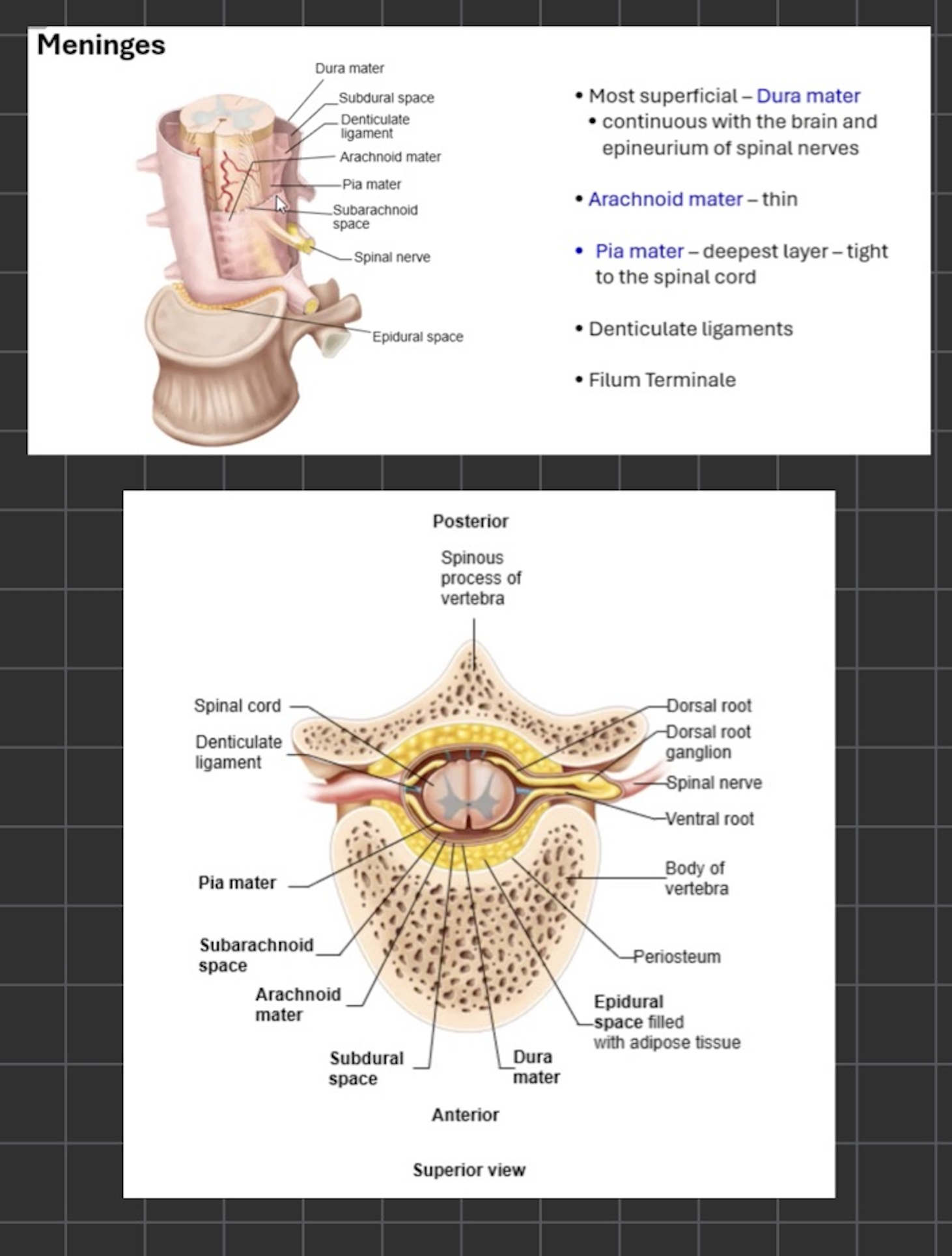
Meninges

  • SC meniges continuous with brain meninges


Mots superior to Inferior

  1. Dura Matwr

    • made of dense irregular CT

    • thickest + strongest of all mengingal layers

      • Difference from brain DURA**

        • A space between dura and bone

          • CT on layer of bone is called periostium

        • space is caled epidermal space

        • In VC space filled with

          • fat

          • Blood vessels

          • Areolar CT

            • (all three help protect SC and hold it in place)


  1. Subdural space

    • contains small amount of serous fluid


  1. Arachnoid mater

    • thin

    • avadcular

    • contain single squamous epithelial cells

    • delicate network of collagen + elastic fibers


  1. Subarachnoid sapce

    • contains CSF which:

      • cushions Sc

      • allows for protection

      • delivers nutrients

      • removes aste


  1. Pia mater

    • Final layer

    • contains

      • blood veseels

        • supplu SC with nutrients and other stuff

    • Tightly adhreres to SC

    • Has small extensions of itself

      • Dectrculiate ligaments

      • move towards dura mater (connet them)

      • occur between each of roots that exit SC

      • help anchor SC laterally


  • Filum terminale

    • another extension of pia mater

    • extends to coccyxl


<ul><li><p>SC meniges continuous with brain meninges</p></li></ul><p></p><p></p><p>Mots superior to Inferior</p><ol><li><p>Dura Matwr</p><ul><li><p>made of dense irregular CT </p></li><li><p>thickest + strongest of all mengingal layers</p><ul><li><p>Difference from brain DURA**</p><ul><li><p>A space between dura and bone </p><ul><li><p>CT on layer of bone is called periostium</p></li></ul></li><li><p>space is caled epidermal space</p></li><li><p>In VC space filled with </p><ul><li><p>fat </p></li><li><p>Blood vessels</p></li><li><p>Areolar CT</p><ul><li><p>(all three help protect SC and hold it in place)</p></li></ul></li></ul></li></ul></li></ul></li></ul></li></ol><p></p><ol start="2"><li><p>Subdural space</p><ul><li><p>contains small amount of serous fluid </p></li></ul></li></ol><p></p><ol start="3"><li><p>Arachnoid mater</p><ul><li><p>thin </p></li><li><p>avadcular </p></li><li><p>contain single squamous epithelial cells</p></li><li><p>delicate network of collagen + elastic fibers</p></li></ul></li></ol><p></p><ol start="4"><li><p>Subarachnoid sapce</p><ul><li><p>contains CSF which:</p><ul><li><p>cushions Sc</p></li><li><p>allows for protection</p></li><li><p>delivers nutrients</p></li><li><p>removes aste</p></li></ul></li></ul></li></ol><p></p><ol start="5"><li><p>Pia mater </p><ul><li><p>Final layer</p></li><li><p>contains</p><ul><li><p>blood veseels</p><ul><li><p>supplu SC with nutrients and other stuff</p></li></ul></li></ul></li><li><p>Tightly adhreres to SC</p></li><li><p>Has small extensions of itself</p><ul><li><p>Dectrculiate ligaments</p></li><li><p>move towards dura mater (connet them) </p></li><li><p>occur between each of roots that exit SC</p></li><li><p>help anchor SC laterally </p></li></ul></li></ul></li></ol><p></p><ul><li><p>Filum terminale</p><ul><li><p>another extension of pia mater</p></li><li><p>extends to coccyxl </p></li></ul></li></ul><p></p>
12
New cards

Internal anatomy of SC

2 portions

  • outer White mater

  • inner gray mater


Posterior median sulcus

  • Narrow groove


Anterior medium fissue

  • large groove


  • weknow about whiteโ€™gray mater

  • this are has as/decnding tracts

  • when NTโ€™s group = columns


3 colums

  1. posterior

  2. lateral

  3. anterior


  • white commisures

    • connect L/R sides of white mater

      • both post and ANT


Horns of gray matter

  1. post (axons of sensory neurons enter SC

  2. Lateral (only found in lumbar/thoracic)

    • associated with neurons of ANS

  3. Ant (motor horn)


Gray commisure

  • Connect R/L sides of gray mater

  • contains central canal

    • continous with ventricles of briain

    • contains CSF


NOTE: All sensory neurons entering into SC through dorsal root


<p>2 portions </p><ul><li><p>outer White mater</p></li><li><p>inner gray mater</p></li></ul><p></p><p>Posterior median sulcus </p><ul><li><p>Narrow groove</p></li></ul><p></p><p>Anterior medium fissue </p><ul><li><p>large groove</p></li></ul><p></p><ul><li><p>weknow about whiteโ€™gray mater</p></li><li><p>this are has as/decnding tracts</p></li><li><p>when NTโ€™s group = columns</p></li></ul><p></p><p>3 colums</p><ol><li><p>posterior</p></li><li><p>lateral</p></li><li><p>anterior</p></li></ol><p></p><ul><li><p>white commisures </p><ul><li><p>connect L/R sides of white mater</p><ul><li><p>both post and ANT</p></li></ul></li></ul></li></ul><p></p><p>Horns of gray matter</p><ol><li><p>post (axons of sensory neurons enter SC</p></li><li><p>Lateral (only found in lumbar/thoracic)</p><ul><li><p>associated with neurons of ANS</p></li></ul></li><li><p>Ant (motor horn)</p></li></ol><p></p><p>Gray commisure</p><ul><li><p>Connect R/L sides of gray mater</p></li></ul><ul><li><p>contains central canal </p><ul><li><p>continous with ventricles of briain</p></li><li><p>contains CSF</p></li></ul></li></ul><p></p><p></p><p>NOTE: All sensory neurons entering into SC through dorsal root</p><p></p>
13
New cards

Spinal nerves

  • 31 pairs

    • names after area of VC where they exit

      • name after vertebrae between their exit until C7

    • next nerve is TA - named afetr tetebrae T1 above its exit

      • this numbering continues for rest of spinal nerves


NERVE BREAKDOWN

8 cervial

12 thoracic

5 lumber

5 sacral

1 coccygeal


<ul><li><p>31 pairs</p><ul><li><p>names after area of VC where they exit </p><ul><li><p>name after vertebrae between their exit until C7</p></li></ul></li><li><p>next nerve is TA - named afetr tetebrae T1 above its exit</p><ul><li><p>this numbering continues for rest of spinal nerves </p></li></ul></li></ul></li></ul><p></p><p>NERVE BREAKDOWN</p><p>8 cervial</p><p>12 thoracic</p><p>5 lumber</p><p>5 sacral</p><p>1 coccygeal</p><p></p>
14
New cards

Structure of peripheral nerves

  • Acons bundled in paralell

    • grouped by function (motor/sensory)

    • schwann cells surround axons + make up myelin sheaths

    • each mylinated axon is surronded by 3 CT layers (deep to superficial)

      1. Endoneurium

        • Separates acons from each other

      2. Perineurium

        • hold together fossicles (axons bundles)

      3. Epenurium

        1. Surrounds SN (bundles of fascicles): dense continuous w/dura mater

  • Surrounding fascicles are arteries + veins (provide blood supply to nervous tissue\


  • Axons form smaler bundles called roolets as axons leave/ before theyr enter the SC

    • Along dorsal + ventral surfaces

      • 6-8 roolets = ventral/dorsal roots pass through subarachnoid space, pierce arachnoid mater + join to form spinal nerve

        • contain motor/sensry neurons

        • each dorsal root contains a gangliion


<ul><li><p>Acons bundled in paralell</p><ul><li><p>grouped by function (motor/sensory)</p></li><li><p>schwann cells surround axons + make up myelin sheaths </p></li><li><p>each mylinated axon is surronded by 3 CT layers (deep to superficial)</p><ol><li><p>Endoneurium</p><ul><li><p>Separates acons from each other</p></li></ul></li><li><p>Perineurium</p><ul><li><p>hold together fossicles (axons bundles)</p></li></ul></li><li><p>Epenurium</p><ol><li><p>Surrounds SN (bundles of fascicles): dense continuous w/dura mater</p></li></ol></li></ol></li></ul></li><li><p>Surrounding fascicles are arteries + veins (provide blood supply to nervous tissue\</p></li></ul><p></p><ul><li><p>Axons form smaler bundles called roolets as axons leave/ before theyr enter the SC</p><ul><li><p>Along dorsal + ventral surfaces</p><ul><li><p>6-8 roolets = ventral/dorsal roots pass through subarachnoid space, pierce arachnoid mater + join to form spinal nerve</p><ul><li><p>contain motor/sensry neurons </p></li><li><p>each dorsal root contains a gangliion</p></li></ul></li></ul></li></ul></li></ul><p></p><p></p><p></p>
15
New cards

Branches of spinal nerve

spinal nerve


  1. Dorsal/ rami: innervating dorsal trunk muscles ( deep back msucles that move VC) + same skin and CT on back


  1. Ventral rami: destination depends on location of VC

    • thoracic region (T2-T12) = form intercostal nerves

    • rest: roots of plexuses (intermingling of nerves @ SC); 5 plexussu total

      1. C1-C4 = cervical plexus

      2. C5 - T1 = Bronchial plexus

      3. L1-L4 = lumbar plexus

      4. L4-S4 = Sacral plexus

      5. S5 - coocygeal nerve = coccygeal plexus


<p>spinal nerve</p><p></p><ol><li><p>Dorsal/ rami: innervating dorsal trunk muscles ( deep back msucles that move VC) + same skin and CT on back</p></li></ol><p></p><ol start="2"><li><p>Ventral rami: destination depends on location of VC</p><ul><li><p>thoracic region (T2-T12) = form intercostal nerves</p></li><li><p>rest: roots of plexuses (intermingling of nerves @ SC); 5 plexussu total</p><ol><li><p>C1-C4 = cervical plexus </p></li><li><p>C5 - T1 = Bronchial plexus</p></li><li><p>L1-L4 = lumbar plexus </p></li><li><p>L4-S4 = Sacral plexus </p></li><li><p>S5 - coocygeal nerve = coccygeal plexus</p></li></ol></li></ul></li></ol><p></p>
16
New cards

More in depth plexuses

Cervial plexus โ†’ innervates superficial neck structures, skin of neck, post portion of head, superior head + chest regions

  • origin of phrenic nerve โ†’ from C3-C5, innervates diaphragm


Brachail plexus โ†’ innervates shoulder + upper limbs

  • 3 trunks โ†’ 6 divisions โ†’ 3 cords โ†’ 5 branches (axillary nerve, radical nerve musculocutaneaus nerve, Ulnar nerve, Median nerve,


Lumbar + social plexuses โ†’ odten associated

  • Lumbar major nerves = pbturator + feumaral (supply parts of lower limb)

  • Social major nerves = tibial + common fibular โ†’ bound together = sciatic nerve

    • supplied upper limb


Coccygeal plexuus โ†’ innervaition of pelvic floor muscles + sensory info from skin over over coccyx


<p>Cervial plexus โ†’ innervates superficial neck structures, skin of neck, post portion of head, superior head + chest regions</p><ul><li><p>origin of phrenic nerve โ†’ from C3-C5, innervates diaphragm</p></li></ul><p></p><p>Brachail plexus โ†’ innervates shoulder + upper limbs</p><ul><li><p>3 trunks โ†’ 6 divisions โ†’ 3 cords โ†’ 5 branches (axillary nerve, radical nerve musculocutaneaus nerve, Ulnar nerve, Median nerve, </p></li></ul><p></p><p>Lumbar + social plexuses โ†’ odten associated </p><ul><li><p>Lumbar major nerves = pbturator + feumaral (supply parts of lower limb)</p></li><li><p>Social major nerves = tibial + common fibular โ†’ bound together = sciatic nerve</p><ul><li><p>supplied upper limb </p></li></ul></li></ul><p></p><p>Coccygeal plexuus โ†’ innervaition of pelvic floor muscles  + sensory info from skin over over coccyx</p><p></p>