skull
 Call Kai
 Learn
Learn Practice Test
 Spaced Repetition
 Match
Match Flashcards
 Knowt Play
1/166

There's no tags or description
Looks like no tags are added yet.
Name | Mastery | Learn | Test | Matching | Spaced | Call with Kai | Chat |
|---|
No analytics yet
Send a link to your students to track their progress
167 Terms

bone/structure
frontal bone

bone/structure
supraorbital margin
(part of the frontal bone)

bone/structure
coronal suture
joins frontal bone to the parietal bones

structure that provides a passage for veins, nerve, and artery through the highlighted feature
supraorbital foramen
(part of the frontal bone)

bone/structure (in pink)
foramen magnum
hole in the cranium where the spinal chord meets the brain
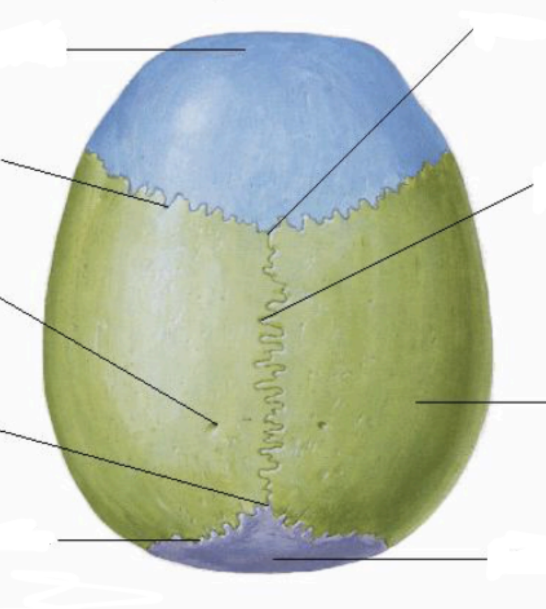
bone/structure
calvaria
the dome of the top of the skull composed of many bone parts
what are the cranial fossa
anterior cranial fossa
middle cranial fossa
posterior cranial fossa
how many cranial bones are there?
8
(1 frontal bone, 2 parietal bones, 2 temporal bones, 1 occipital bone, 1 sphenoid bone, and 1 ethmoid bone)

structure/feature the arrows are pointing to
supraorbital notch (part of the frontal bone)
Only in some people, occurs when the edge of the supraorbital foramen breaks through the margin of the orbit.

structure/feature highlighted
glabella
smooth area of the frontal bone above the root of the nose

structure/feature highlighted in pink
frontal sinus
air filled space in the frontal bone
may not be present in all skulls

structure/feature highlighted in pink
frontal sinus
air filled space in the frontal bone
may not be present in all skulls
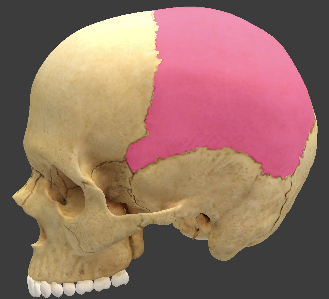
bone/structure highlighted
parietal bones

bone/structure highlighted
parietal bones

bone/structure highlighted
parietal bones

bone/structure highlighted
sagittal suture
between the two parietal bones in the skull.

bone/structure highlighted
lambdoid suture
separates the occipital bone from the parietal bones.

bone/structure highlighted
squamous suture
connects the parietal bone with the temporal bone.

bone/structure highlighted
parietal foramen
sometimes occurs near the corner of the lambdoid and sagittal sutures, allowing blood vessels to pass.

bone/structure labeled 1
superior temporal line
arc of the parietal and frontal bone
mark the attachment of the temporalis muscle.

bone/structure labeled 2
inferior temporal line
arc of the parietal and frontal bone
mark the attachment of the temporalis muscle.
the temporal bone is divided into how may parts?
4 parts
squamous part
tympanic part
mastoid part
petrous part
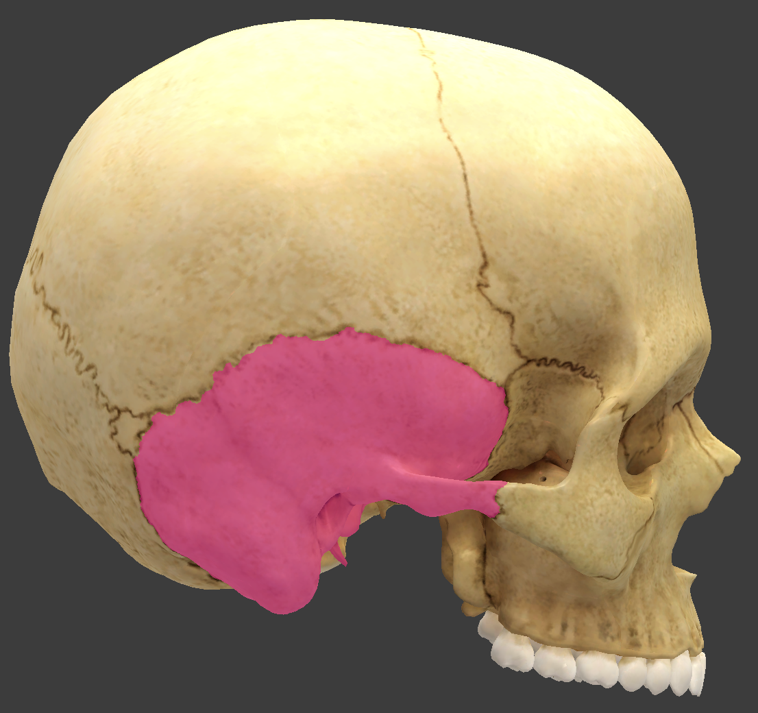
bone/structure highlighted
temporal bones
superior and anterior to the external ear

bone/structure highlighted
temporal bones
superior and anterior to the external ear
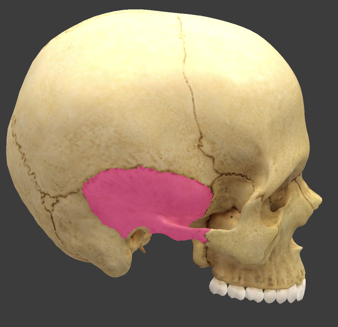
bone/structure highlighted
squamous part of the temporal bone

bone/structure highlighted
zygomatic process of the temporal bone
feature of the squamous part of the temporal bone

bone/structure highlighted
zygomatic arch
part of the temporal bone
includes the zygomatic process

bone/structure highlighted
zygomatic arch
part of the temporal bone
includes the zygomatic process
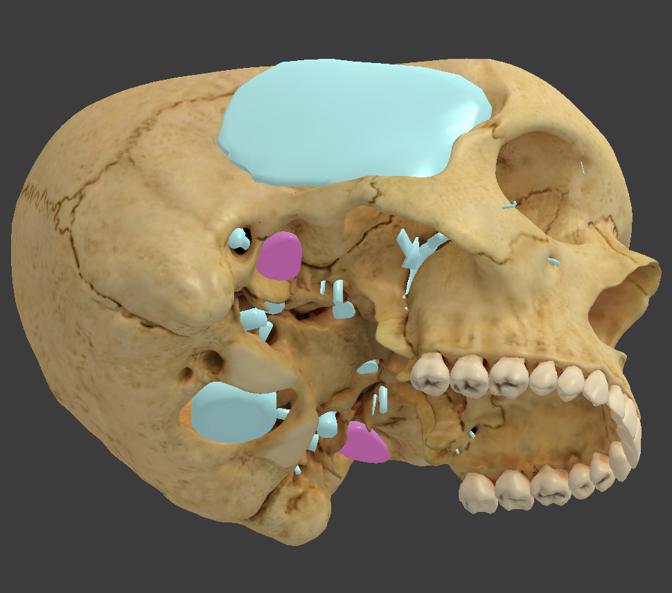
bone/structure highlighted in pink
mandibular fossa
depression where the mandible articulates with the cranium
feature of the squamous part of the temporal bone

bone/structure highlighted in pink
mandibular fossa
depression where the mandible articulates with the cranium
feature of the squamous part of the temporal bone

bone/structure highlighted
tympanic part of the temporal bone
small ring of bone that surrounds the external acoustic meatus (ear canal)

bone/structure highlighted
tympanic part of the temporal bone
small ring of bone that surrounds the external acoustic meatus (ear canal)

bone/structure highlighted in pink
external acoustic meatus
ear canal
feature of the tympanic part of the temporal bone

bone/structure highlighted in pink
styloid process
pointed spine
feature of the tympanic part of the temporal bone
provides attachment point for tongue, pharynx, and hyoid bone
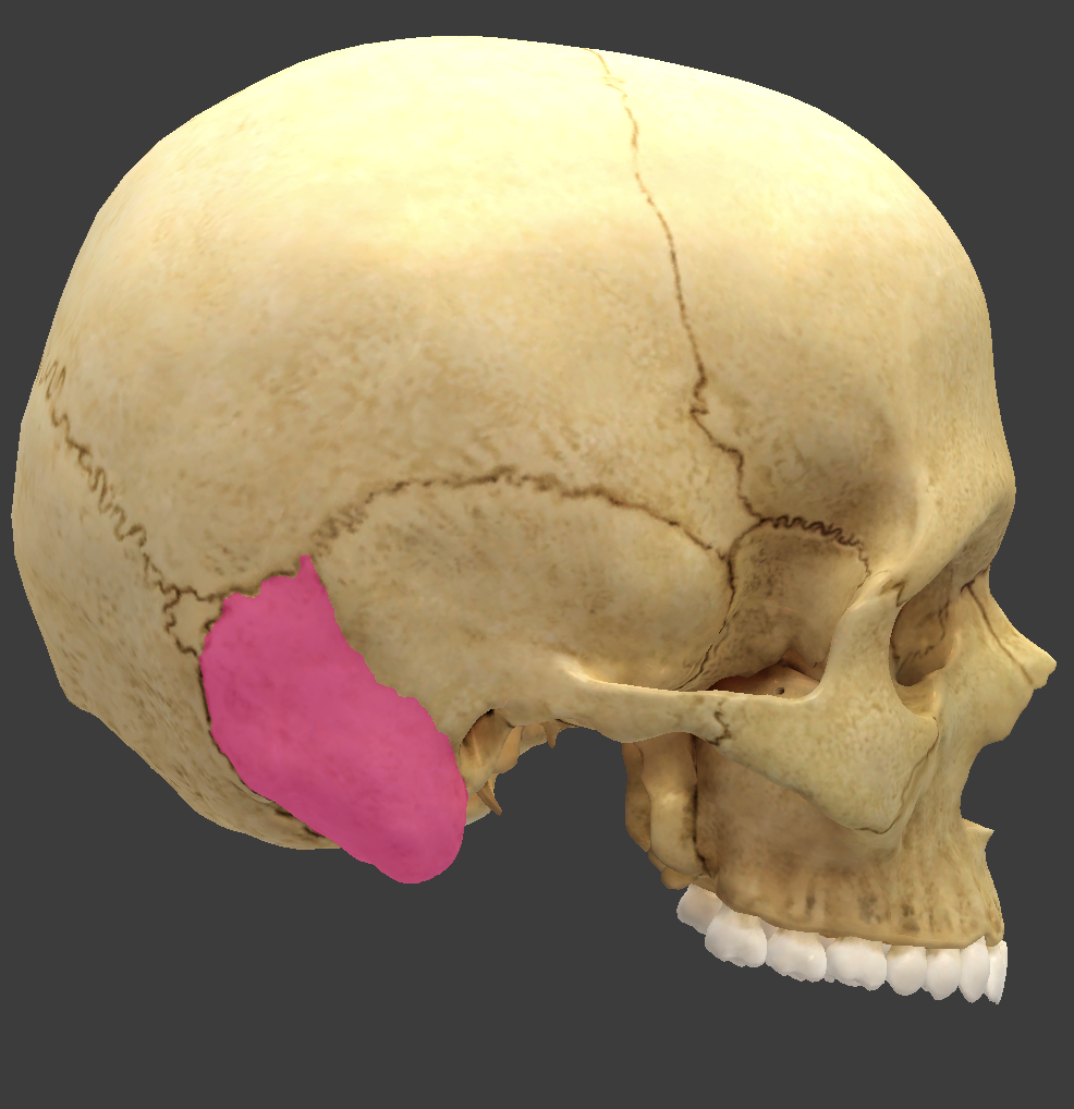
bone/structure highlighted in pink
mastoid part of the temporal bone

bone/structure highlighted in pink
mastoid process
feature of the mastoid part of the temporal bone
filled with small air sinuses

bone/structure highlighted
mastoid notch
indentation located on the underside of the mastoid process
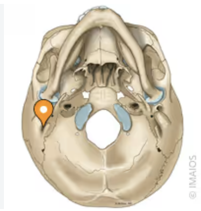
bone/structure highlighted
mastoid notch
indentation located on the underside of the mastoid process

bone/structure highlighted in pink
stylomastoid foramen
feature of the mastoid part of the temporal bone

bone/structure highlighted
mastoid foramen
feature of the mastoid part of the temporal bone
passes small artery and vein from the brain
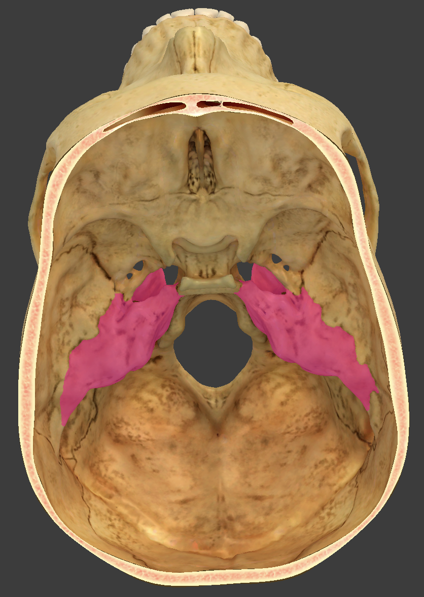
bone/structure highlighted in pink
petrous part of the temporal bone
houses the middle and inner ear cavities

bone/structure highlighted in pink
petrous part of the temporal bone
houses the middle and inner ear cavities

bone/structure highlighted in pink
internal acoustic meatus
located in the petrous part of the temporal bone
passage for nerve that carries signals for hearing and balance

bone/structure highlighted in pink
carotid canal
located in the petrous part of the temporal bone
passage for the internal carotid artery

bone/structure highlighted in pink
carotid canal
located in the petrous part of the temporal bone
passage for the internal carotid artery

bone/structure highlighted in pink
jugular foramen
located in the petrous part of the temporal bone
large irregular opening medial to the styloid process
passage for the jugular vein that drains blood from brain

bone/structure highlighted in pink
occipital bone
forms the rear of the skull

bone/structure highlighted in pink
basilar plate
part of the occipital bone
anterior to the foramen magnum
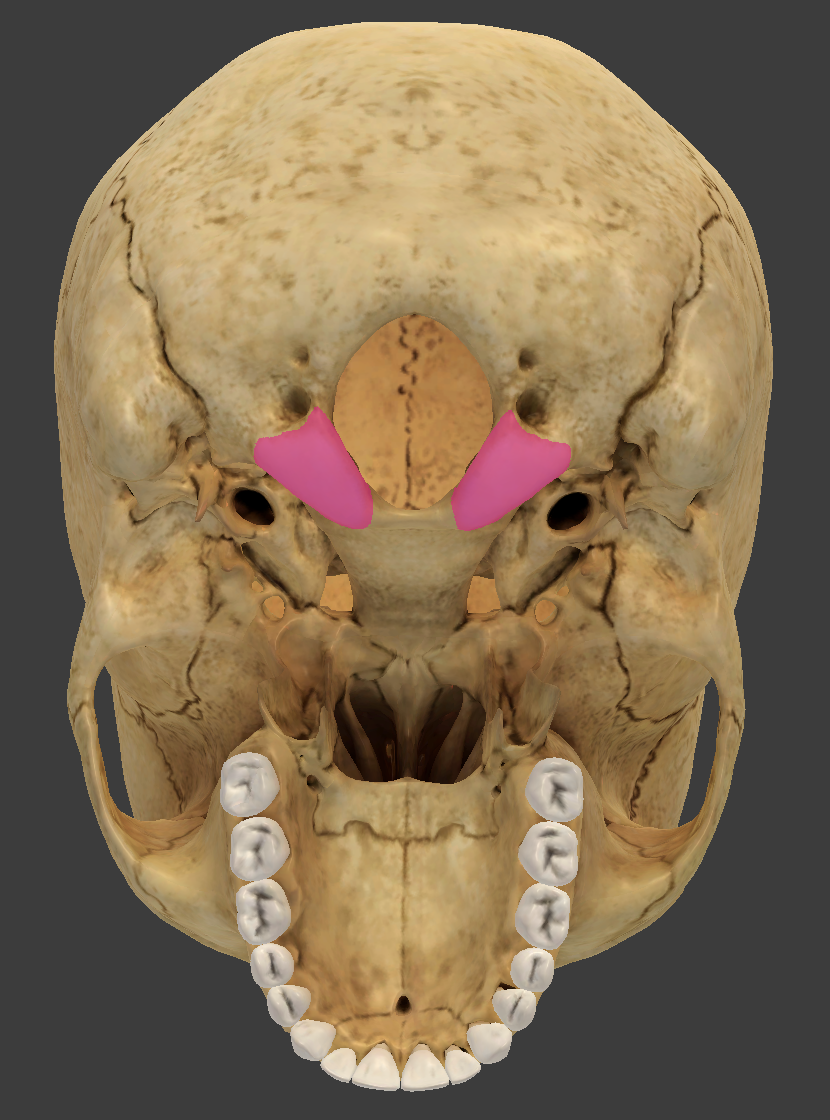
bone/structure highlighted in pink
occipital condyle
smooth knob on either side of the foramen magnum
part of the occipital bone

bone/structure highlighted in pink
hypoglossal canal
passing tunnel beneath each occipital condyle
tunnel for the nerve that innervates the tongue muscle
part of the occipital bone

bone/structure highlighted in pink
hypoglossal canal
passing tunnel beneath each occipital condyle
tunnel for the nerve that innervates the tongue muscle
part of the occipital bone
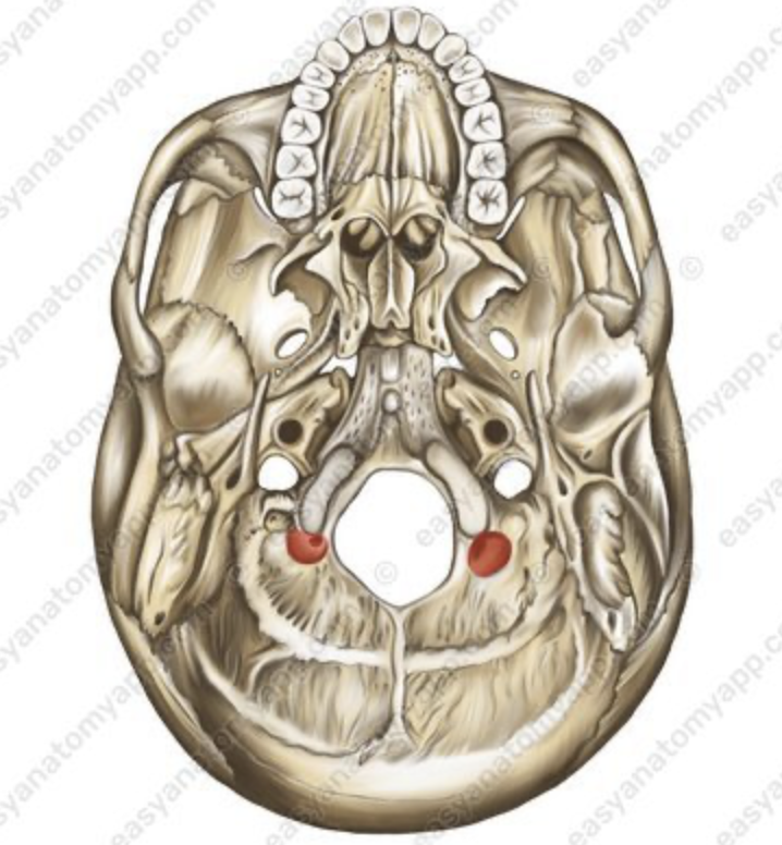
bone/structure highlighted in pink
condylar canal
posterior to each occipital condyle, it serves as a passage for the veins and a nerve.
part of the occipital bone

bone/structure highlighted in pink
external occipital protuberance
prominent medial bump on the occipital bone
serves as the attachment point for the nuchal ligament.
part of the occipital bone

bone/structure highlighted
superior nuchal line
part of the occipital bone
defines the superior limit of the neck
provides an attachment point for neck and back muscles

bone/structure highlighted in pink
inferior nuchal line
attachment point for deep neck muscles
located on the occipital bone
deeper Than the superior nuchal line
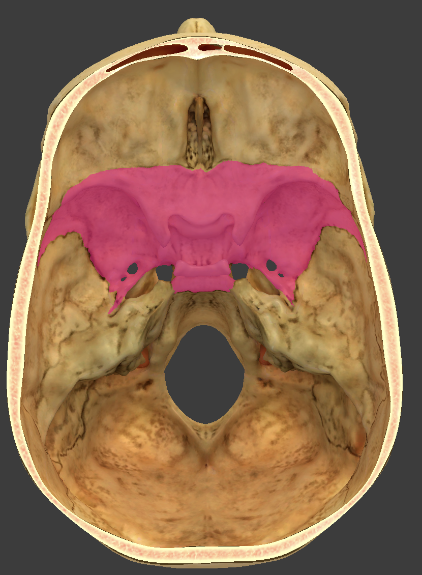
bone/structure highlighted in pink
sphenoid bone
Butterfly shaped

bone/structure highlighted in pink
sphenoid bone
Butterfly shaped

bone/structure highlighted in pink
sphenoid bone
Butterfly shaped

bone/structure highlighted in blue
greater wings of the sphenoid bone
form ½ the middle cranial fossa
form part of the lateral surface of the cranium

bone/structure highlighted in pink
lesser wings of the sphenoid bone
form the posterior wall of the orbit and

bone/structure highlighted in pink
optic canal
housed in the lesser wings of the sphenoid bone
passage of optic nerve
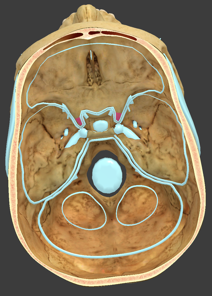
bone/structure highlighted in pink
anterior clinoid processes
boney spins of the lesser wings of the sphenoid bone
protect the optic foramina
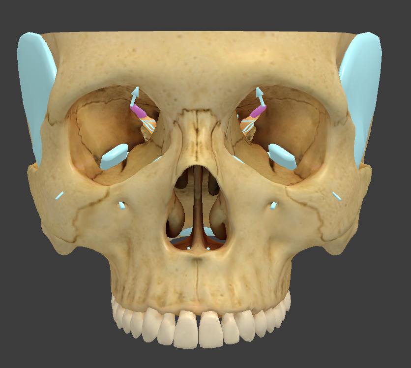
bone/structure highlighted in pink
superior orbital fissure
slit in the posterior wall of the orbit
passage for nerves the move the eyes
part of the sphenoid bone

bone/structure highlighted in pink
sella turcica
a saddle-shaped depression in the sphenoid bone
houses the pituitary gland
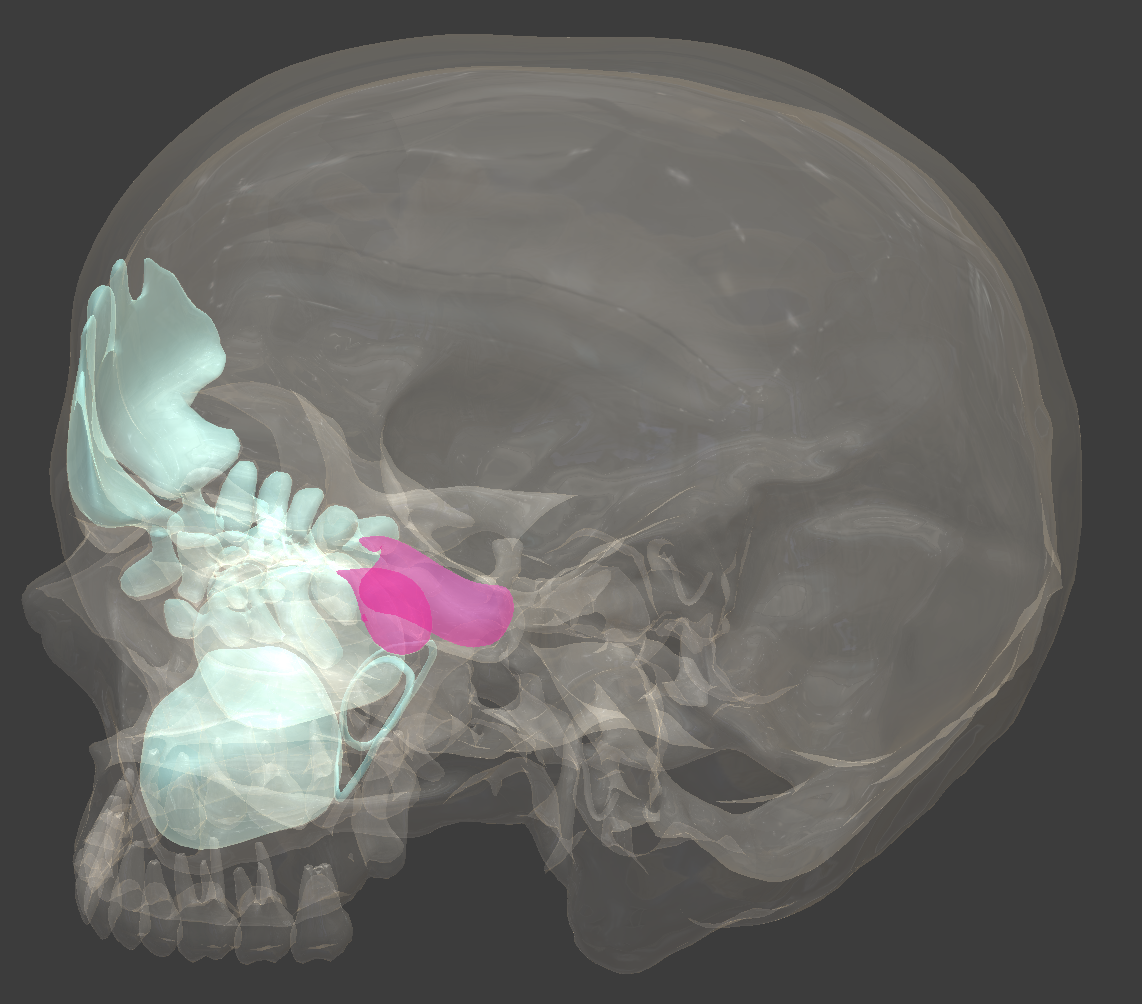
bone/structure highlighted in pink
sphenoidal sinuses

bone/structure highlighted in pink
hypophyseal fossa
deep pit in the sella turcica that houses the pituitary gland

bone/structure highlighted
tuberculum sellae
raised anterior margin of the sella turcica

bone/structure highlighted
tuberculum sellae
raised anterior margin of the sella turcica

bone/structure highlighted
tuberculum sellae
raised anterior margin of the sella turcica

bone/structure highlighted in pink
dorsum sellae
posterior margin of the sella turcica.

bone/structure highlighted in pink
foramen rotundum
passageway in the greater wings of the sphenoid bone for the trigeminal nerve

bone/structure highlighted in pink
foramen ovale
passageway in the greater wings of the sphenoid bone for the trigeminal nerve

bone/structure highlighted in pink
foramen ovale
passageway in the greater wings of the sphenoid bone for the trigeminal nerve

bone/structure highlighted in pink
foramen spinosum
small opening in the greater wing of the sphenoid bone

bone/structure highlighted in pink
foramen spinosum
small opening in the greater wing of the sphenoid bone
next to the foramen oval

bone/structure highlighted in pink
foramen laceram
irregular gash in the junction of the temporal, sphenoid, and occipital bones.

bone/structure highlighted in pink
posterior nasal apertures (also called choanae)
internal openings of the nasal cavity

bone/structure highlighted in pink
medial pterygoid plate
bony plate that project downward from the sphenoid bone, forming part of the pterygoid process.

bone/structure highlighted in pink
lateral pterygoid plate
bony plate that project downward from the sphenoid bone, forming part of the pterygoid process.

bone/structure highlighted
lateral pterygoid plate
bony plate that project downward from the sphenoid bone, forming part of the pterygoid process.

bone/structure highlighted in pink
pterygoid process
narrow inferior extension from the medial and lateral pterygoid plates

bone/structure highlighted in pink
Ethmoid bone
located between the eyes
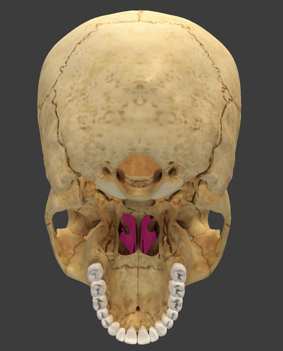
bone/structure highlighted in pink
Ethmoid bone
located between the eyes
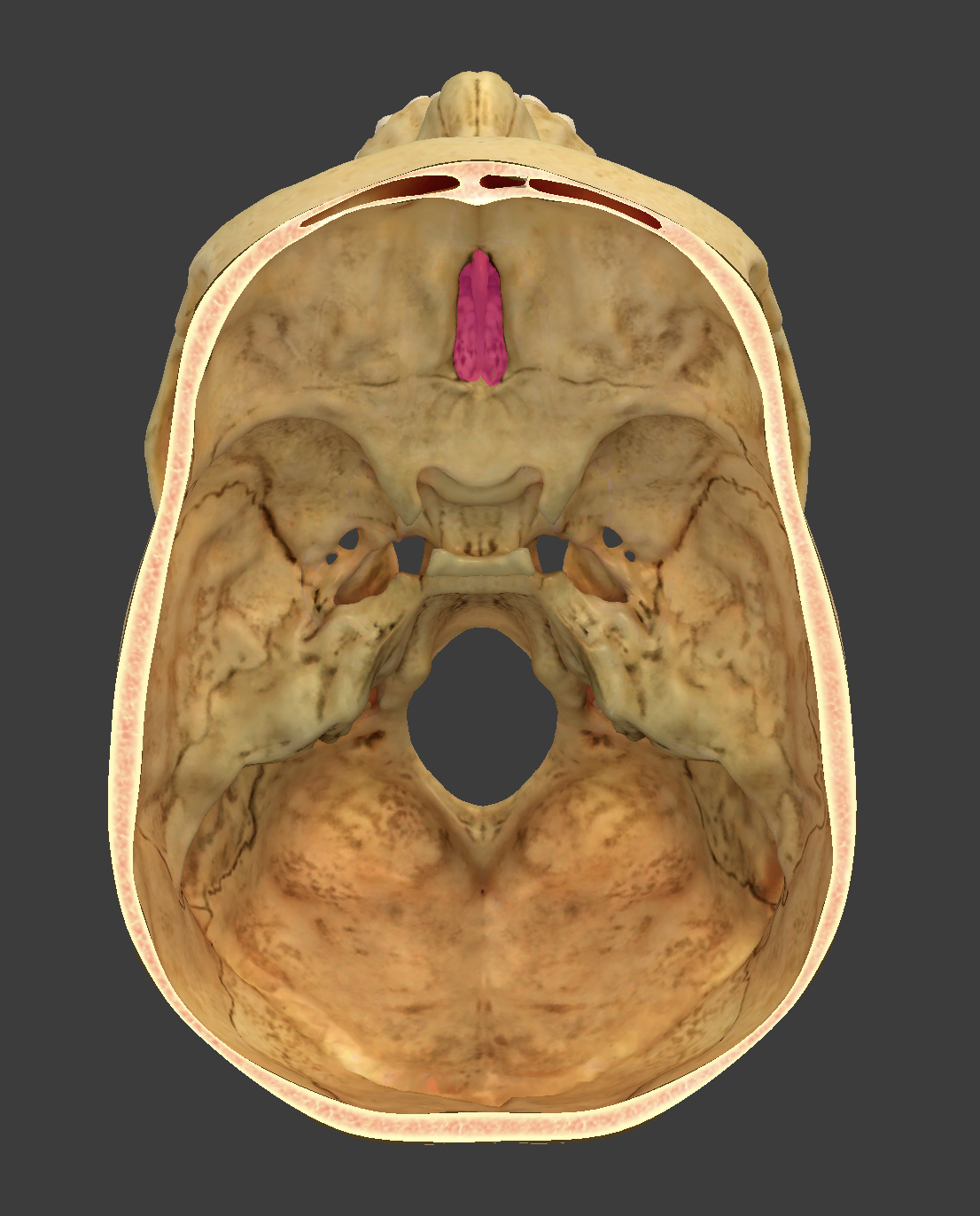
bone/structure highlighted in pink
Ethmoid bone
located between the eyes
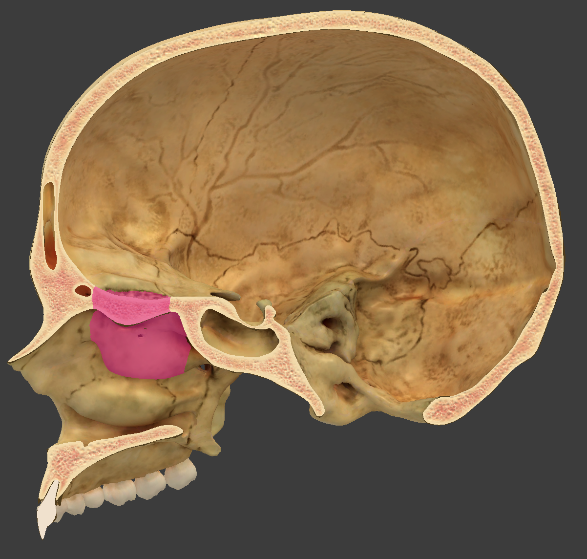
bone/structure highlighted in pink
Ethmoid bone
located between the eyes
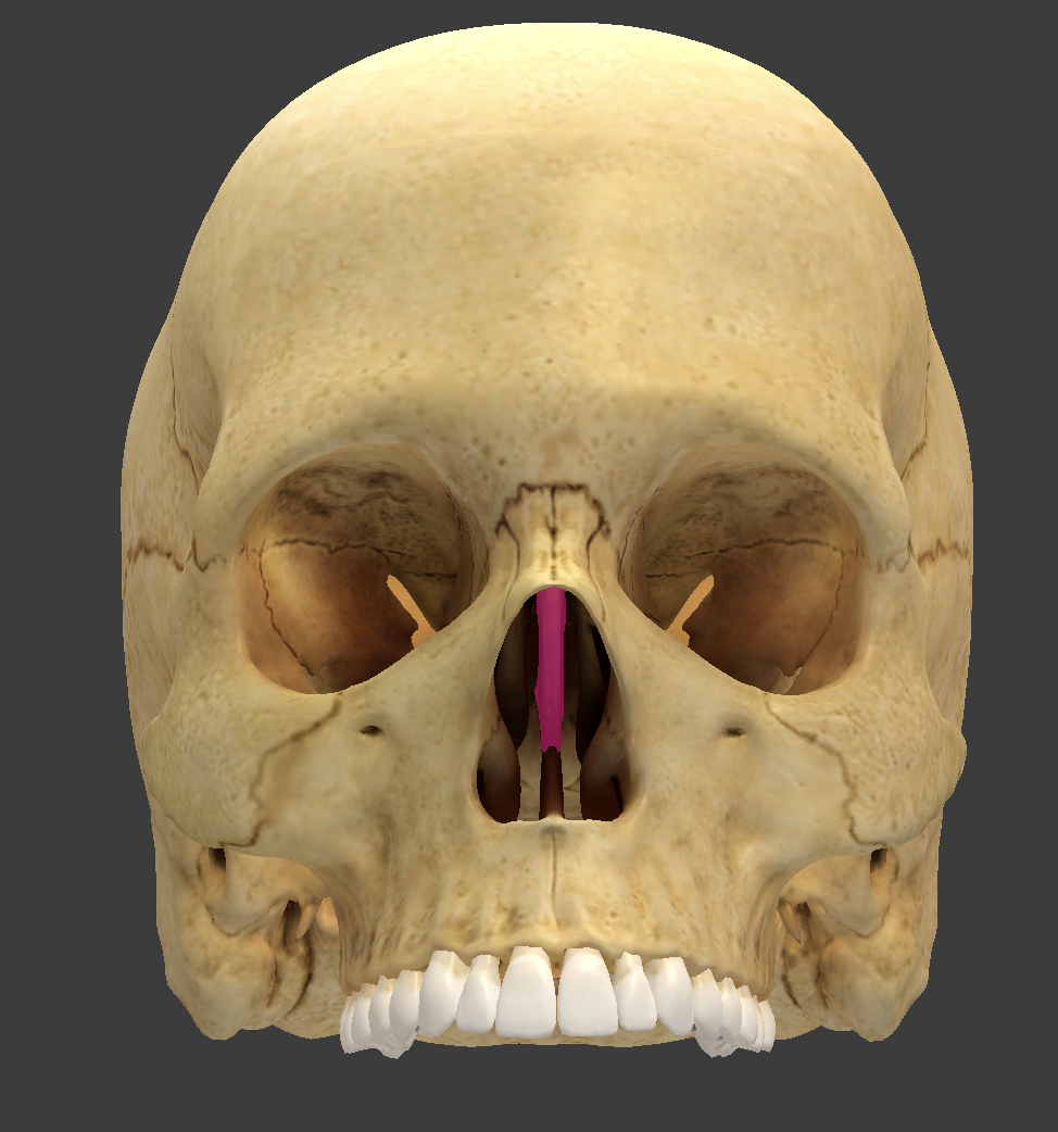
bone/structure highlighted in pink
perpendicular plate of the ethmoid bone
thin median plate that forms the superior part of the nasal septum.
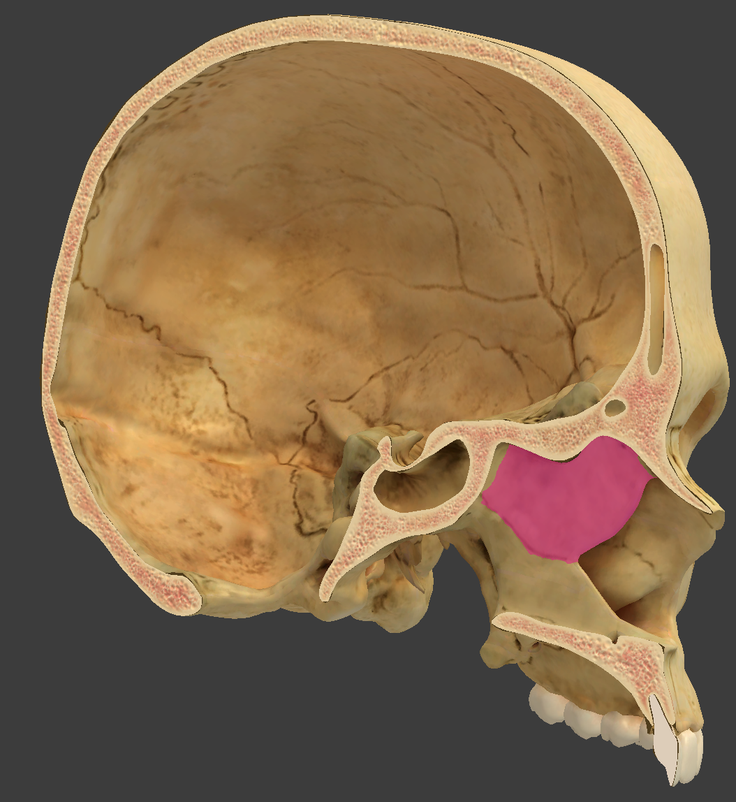
bone/structure highlighted in pink
perpendicular plate of the ethmoid bone
thin median plate that forms the superior part of the nasal septum.
bone/structure highlighted in pink
nasal fossae
right and left airspaces in the nasal cavity separated by the septum
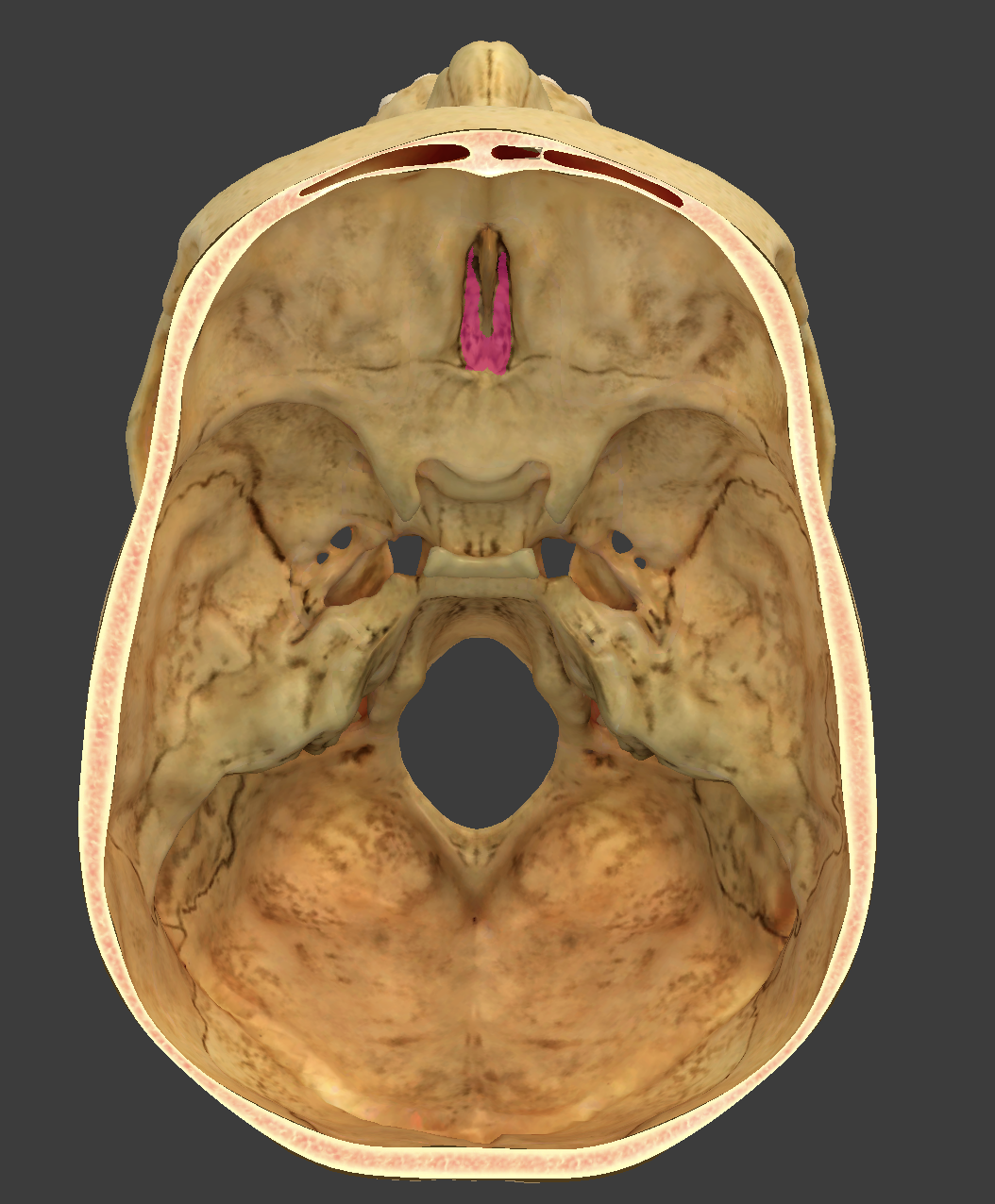
bone/structure highlighted in pink
cribriform plate of the ethmoid bone
thin median plate that forms the superior 2/3 of the nasal septum

bone/structure highlighted in pink
crista galli
the median blade of the cribriform plate of the ethmoid bone

bone/structure highlighted in pink
cribiform (olfactory) foramina
small openings in the cribriform plate that allow passage of olfactory nerves from the nasal cavity to the brain.

bone/structure highlighted in blue
ethmoid labyrinths
large masses on either side of the perpendicular plate that contain the ethmoid sinuses.

bone/structure highlighted in pink
ethmoid cells
air spaces inside of the ethmoid bone that form part of the sinus system and communicate with the nasal cavity.

bone/structure highlighted
ethmoid cells
air spaces inside of the ethmoid bone that form part of the sinus system and communicate with the nasal cavity.

bone/structure highlighted in pink
orbital plate
smooth concave plate
medial wall of the orbit


bone/structure highlighted in pink
medial nasal conchae
scroll like plates on the medial surface of each labyrinththat contribute to the structure of the ethmoid bone.


bone/structure highlighted in pink
Superior nasal conchae
scroll like plates on the medial surface of each labyrinththat contribute to the structure of the ethmoid bone.

bone/structure highlighted in pink
inferior conchae

how many facial bones are there?
14
what bones line anterior to the cranial cavity and do not enclose the brain
facial bones