Micro Lab Midterm (exercises 1-9)
 Call Kai
 Learn
Learn Practice Test
 Spaced Repetition
 Match
Match Flashcards
 Knowt Play
1/344

There's no tags or description
Looks like no tags are added yet.
Name | Mastery | Learn | Test | Matching | Spaced | Call with Kai | Chat |
|---|
No analytics yet
Send a link to your students to track their progress
345 Terms
What do you clean the bench with before lab?
70% EtOH
what do u do if u spill something?
do not try to move it or clean it on ur own!! let a TA and the ppl around u know immediately
what is proper lab attire?
wearing provided lab coat entire time until u leave. lab coats can never leave lab
gloves and goggles worn at all times
long pants down to ankles and closed-toe shoes
long hair tied back
What do you clean the bench with after lab?
Bleach wipes
Where do you label Petri dishes?
Bottom of the plate where medium is. label around the edge for easy observation of growth on plate
label ur seat #, date, and organism u inoculated
How do you store Petri dishes?
Inverted (bottom on top) → this is so condensation that might form on the lid doesnt fall into the plate and ruin ur inoculation
What temperature are Petri dishes incubated at?
37 degrees Celsius
What is used to label test tubes?
China markers
What is the thing called that sterilizes the inoculating loop and openings of test tubes?
Microincinerator. only touch the base
How much of the loop needs to be glowing red hot for sterilization?
¾ make sure to not rest it against the inside of the incinerator
what types of media preparations do we handle?
general purpose, selective, differential
broth
agar slants
agar deep tube
agar plate

Asepsis means
without infection
how do u handle and label test tubes?
never carry by the cap. transport them in the tube rack and hold them in ur non-dominant hand
never put caps down on benchtop → remove cap w ur dominant hand and hold it throughout transfer
label tubes on clear portion, never on the cap. use china marker
What are the 7 (8) steps of a two-tube transfer?
Resuspend if broth culture
1) sterilize loop
2) uncap tubes (avoid touching cap opening w glove)
3) sterilize tubes
4) transfer from culture to fresh medium
5) sterilize tubes
6) recap tubes
7) sterilize loop
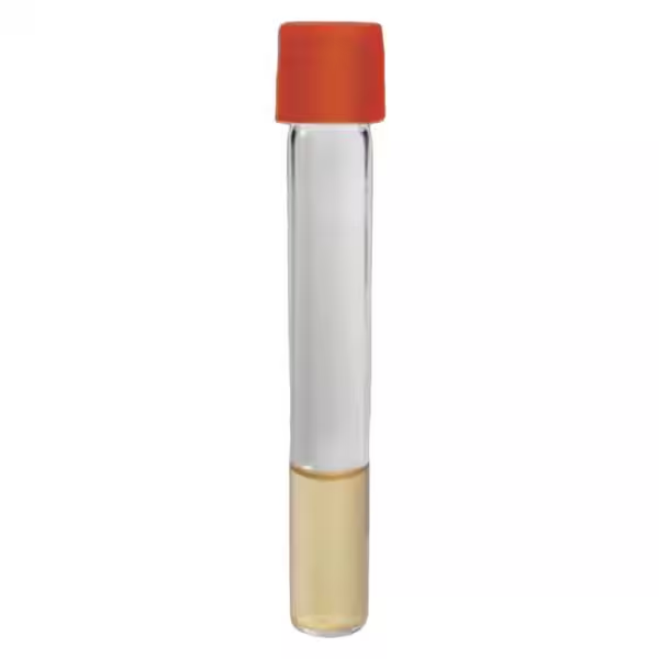
How do you obtain culture from a broth tube?
Stab
liquid
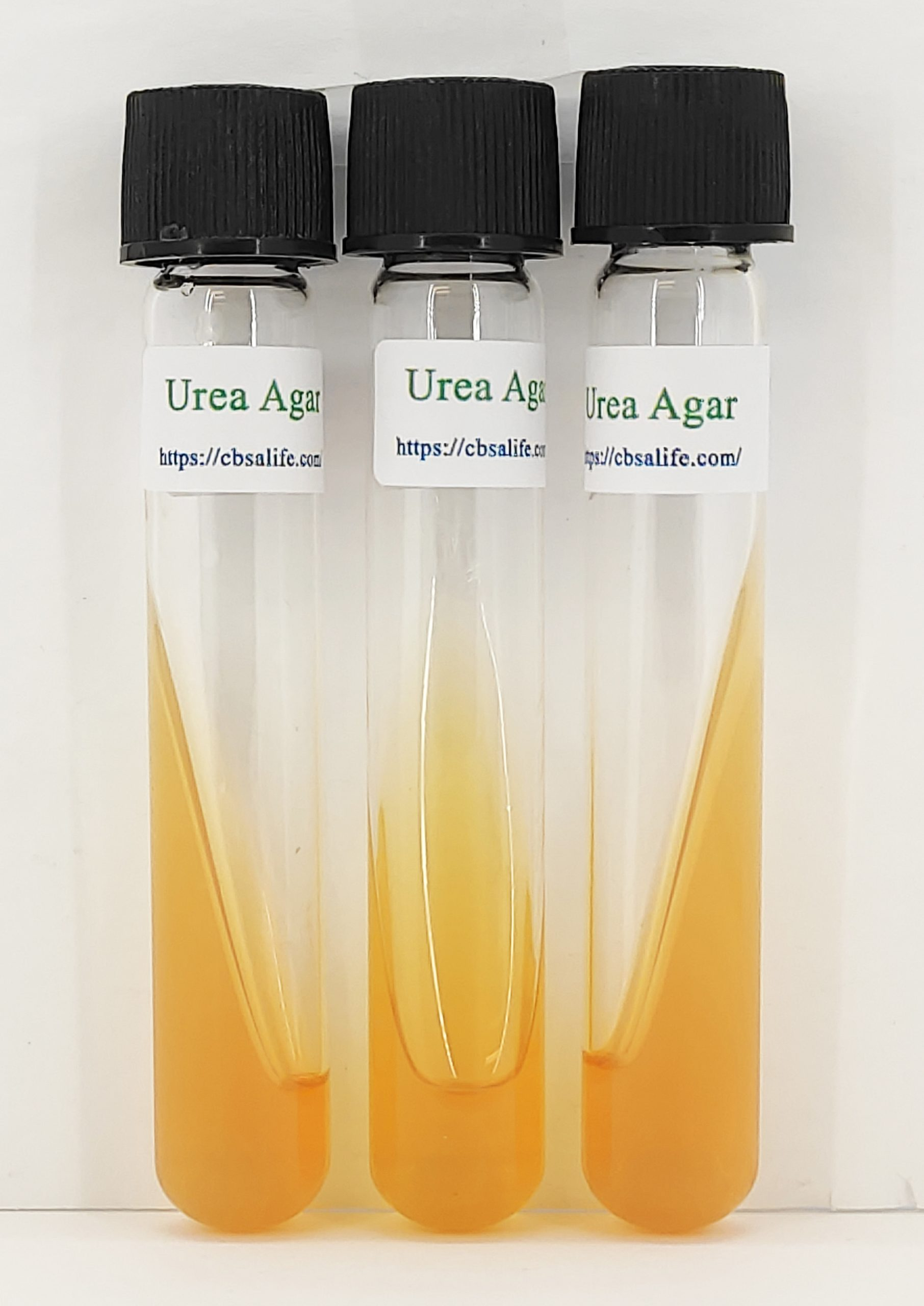
How do you obtain culture from a slant tube?
Fishtail
Staining provides
contrast to identify morphology, size, and arrangement of bacterial sample
Staining is useful for identifying
morphology, shape, and arrangement
What are the different types of staining?
Simple (cationic)
differential
structural
negative
What are the types of dyes used in staining?
Cationic (basic dyes)
positively charged chromophores
ex: Crystal Violet or Methylene Blue
Anionic (acidic dyes)
negative ion exhibits color
ex: Acid Fuchsin, Congo Red, Nigrosin
Cationic dyes are
Basic, positively charged chromophores
positive ion exhibits color
ex: Methylene Blue or Crystal Violet
actually stains the cell → attracted to net negative charge of cell
Examples of cationic dyes
Methylene blue and crystal violet
Anionic dyes are
Acidic, negatively charged
negative ion exhibits color
ex: Acid fuchsin, congo red, Nigrosin
doesn’t penetrate cell, cell is not stained → background takes color
Examples of anionic dyes
Acid fuchsin, Congo red, nigrosin
A negative stain doesn't…
penetrate cell, stains background
negatively charged stain repels negative cell

Are negative stains cationic or anionic?
Anionic
Are negative stains heat fixed?
No
cuz of this its very good for examining morphology and size
no heat = distortion of bacterial cells minimized
negative stain
a anionic dye that doesnt penetrate the cell
repelled by negative charge at cell surface → we stain the background instead so u see transparent cells against colored background
v good to examine size and morphology of cells cuz we do not heat fix! so distortion of bacterial cells is minimized
good for organisms that dont stain easily
ex: Nigrosin (black anionic dye)
Negative stains are useful for organisms that
don't stain easily
it doesnt actually stain cell so it doesnt matter if it doesnt stain easily
Example of a negative stain dye
Nigrosin (black anionic dye)
How does a negative dye work?
Repelled by negatively charged cell surface

simple stains
use cationic dye to stain all cells present
good to distinguish diff morphologies, arrangements, and relative sizes. will have some distortion cuz we need to heat fix
colors the cells and provides contrast when using bright field microscopy
positive charge of the dye interacts w negative charge on bacterial cell wall → colors cell surface
ex: Crystal Violet
Are simple stains cationic or anionic?
Cationic ( + charge will interact w - cell charge to actually stain the surface of cell)
how many dyes are used in a simple stain?
one dye used to stain all cells present
What dye do we use for simple stains?
Crystal violet
How does a simple stain work?
Positive charge interacts with negative charge of bacterial cell wall to color cell surface

Are simple stained slides heat fixed? What does this cause if so?
Yes, some distortion of shape + morphology
how do u smear prep from broth?
1) transfer 1 loopful of culture to clean slide and spread for max thinnness
2) air dry
3) heat fix by holding over microincinerator for 10 seconds - use clothespin!
how do u smear prep from slant or plate?
these r solid media so:
1) add 1 loopfull of water to slide
2) grab bacterial sample by softly tapping loop in media and then mixing it with water on slide to spread
3) allow to air dry
4) heat fix by holding over microincinerator for 10 seconds - use clothespin!
How do you carry the microscope?
With two hands - one holds the handle and other supports base
how do u properly store microscope?
clean oil off 100x using lens cleaner and kimtech wipe
lower stage as low as possible
move 4x objective into position
make sure cord is wrapped up
What is the part of the microscope called that you look through?
Ocular

What are the parts of the microscope called that allow you to see the microbe?
Objectives (4x, 40x, 100x oil)

What is the part of the microscope called that holds your slide?
Stage

What is the focus called that is bigger?
Coarse
moves stage up/down
used for 4x objective

What is the focus called that is smaller?
Fine focus → moves stage up and down

What adjusts the diameter of the cone of light so that is just fills the objective lens?
Iris diaphragm

What is the smallest distance between two objects which can be seen as separate?
resolution
what r the 3 principles of microscopy?
1) illumination
light source and condensor to direct light towards obj. lens.
iris diaphragm to adjust diameter of cone of light
2) magnification
ocular = 10x
Objectives = 4x, 10x, 40x, 100x
total magnification = ocular x objective
3) resolution
the smallest distance btwn 2 objects which can be seen as separate
How do you calculate magnification?
Ocular (10) x objective = total magnification
10x (ocular) x 100x (objective) = 1000x magnification
Which objective is the oil immersion?
100X
for both cationic and anionic stains
cuz oil has same refractive index as glass and oil will stop the refraction of light btwn specimen and lens
oil must be touching lens.
doesnt help w magnification but helps w light!!

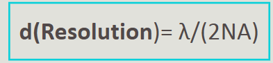
How do you calculate resolution?
d = lambda (wavelength of light nm)/(2NA)
d = resolution
lambda = wavelength of light (nm)
NA = numerical aperture (light gathering capacity of lens)
smaller d = better resolution
What does d stand for?
Resolution

What does NA stand for?
Numerical aperture (light gathering capacity of the lens)

What does lambda stand for?
Wavelength of light (nm)

How do you improve d (resolution)?
Smaller d = better resolution (increase NA or decrease gamma)

Streptococci
Chains of cocci
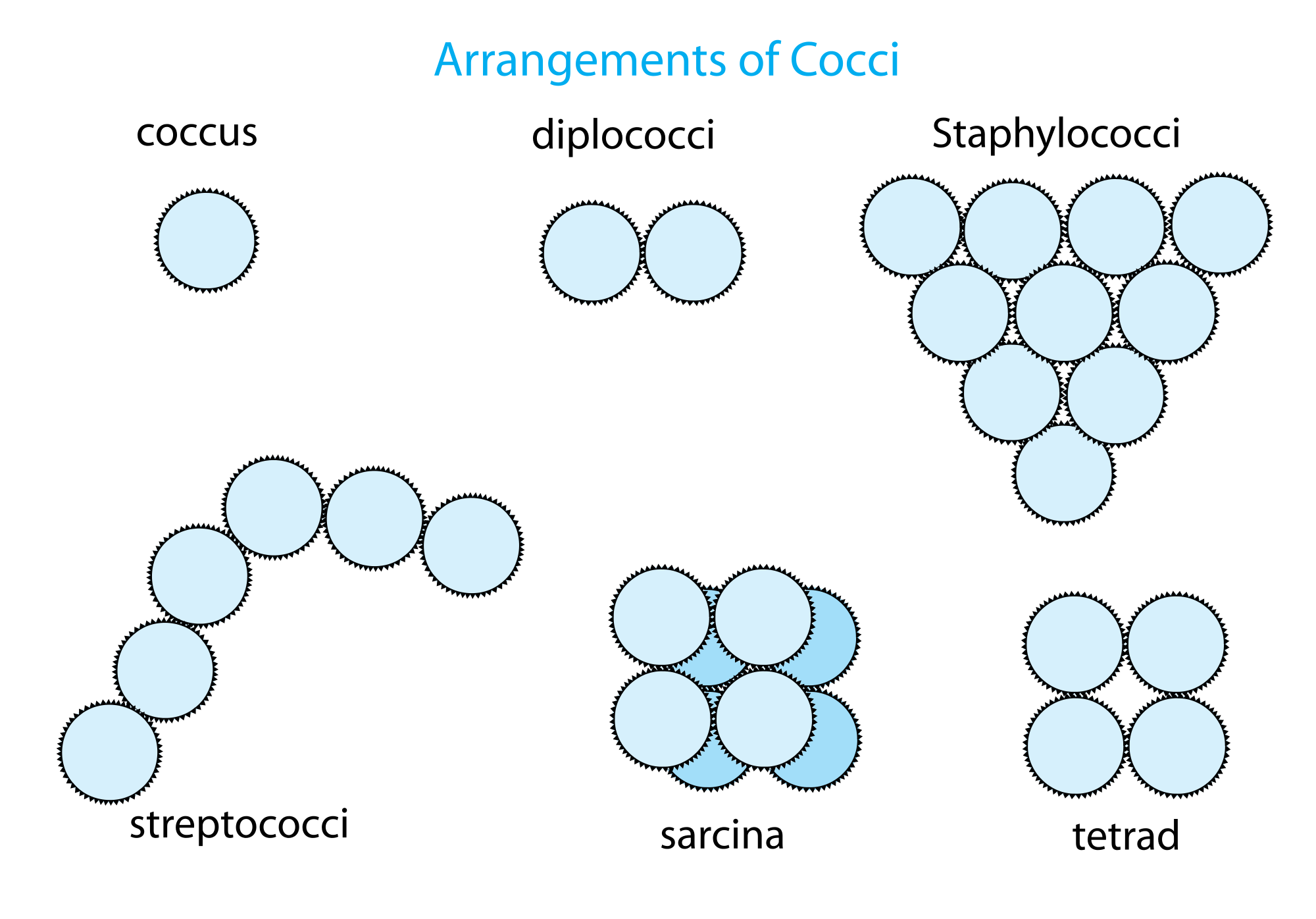
Sarcina
tetrads (square/4 cocci)
4 in front and 4 in back

Staphylococci
grape-like clusters of cocci

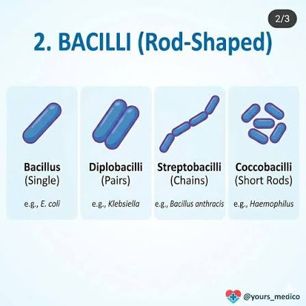
what shape are bacilli?
rod shaped
strepto = chain
diplo = pairs
Vibrio cholerae are what shaped?
Comma shaped

Spirillum volutans are what shaped?
Rigid and spiral

Pleomorphic
Morphology that is not easily categorized
ex: Corynebacterium diptheriae is club-shaped
Which stain uses one dye to stain all cells present?
Simple
Which stain detects differences between organisms through the use of many dyes and reagents?
Differential stain/gram stain
Who developed the gram stain?
Hans Christian Gram
He was trying to differentiate bacteria causing pneumonia from the lung tissue of deceased patients
Gram +
Thick petidoglycan layer
Gram -
Thin peptidoglycan layer, 2 membranes
What is the primary dye in gram staining?
Crystal violet
What is the mordant in gram staining?
Gram's iodine
increases how well crystal violet binds to cell wall
What is the decolorizer/differential step in gram staining?
95% ethanol
What is the secondary dye/counterstain in gram staining?
Safranin
what would happen if we skip the 95% ethanol in gram staining?
all cells would look purple!
after 95% ethanol, gram (-) cells become clear and gram (+) cells remain purple. then Safranin secondary dye stains gram (-) cells pink
gram stain process
1) crystal violet - 1 min
2) grams iodine - 1 min
3) 95% ethanol - 10 secs
4) safranin - 1 min
What color does gram + stain?
Purple
What color does gram - stain?
Red
What types of staining are differential?
Gram stain and acid-fast stain (Ziehl-Neelsen method)
What types of staining are structural?
Endospore stain (Schaeffer-Fulton Method)
Acid fast staining is also called
Ziehl-Neelsen
Acid-fast microbes have what in their cell walls that makes them hard to stain? Why?
Mycolic acid, waxy and resists
What do we do to force the primary dye to penetrate the waxy cell wall in acid-fast staining?
Use steam
fastness
whether or not a organism is going to retain the primary dye when challenged by a decolorizer
What is the primary dye in acid-fast staining?
Carbol fuchsin
bright pinky/red
What is the decolorizer in acid-fast staining?
Acid alcohol
What is the secondary dye/counterstain in acid-fast staining?
Methylene blue
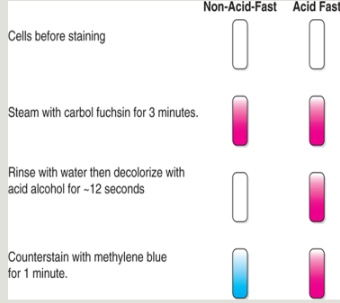
Procedure for acid-fast staining
Heat fixed slide
Steam with carbon fuchsin for 3 minutes and rinse with water
need to steam to penetrate waxy cell wall in microbes w/ high mycolic acid content
Decolorize with acid alcohol for about 12 seconds
Counterstain with methylene blue for 1 minute
Pink/violet = acid-fast
Blue = acid-fast negative
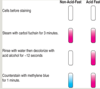
Acid fast cells stain
Cerise/pink/violet
Non-acid fast cells stain
Blue
What is an example of a microbe that is acid fast?
Mycobacterium
ex: M. tuberculosis and M. leprae
have high mycolic acid content
What is the causative agent of Tuberculosis?
Mycobacterium tuberculosis
acid-fast
What is the causative agent of leprosy/Hansen's disease?
Mycobacterium leprae
acid-fast
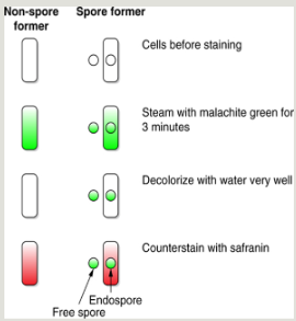
Spore staining is also called
Schaeffer-Fulton method
Endospores release structures called
free spores
How do you force the primary dye into resistant endospores?
Steaming
cuz spores r highly resistant
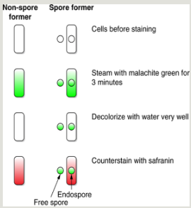
What is the primary dye in spore staining?
Malachite green
What is the decolorizer in spore staining?
Water
What is the secondary dye/counterstain in spore staining?
Safranin (reddish)
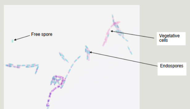
What color do spores and cells stain?
Spores stain green
cells stain pink
spores
produced by some organisms in response to poor environmental conditions. They are a dormant form of bacteria “doomsday bacteria”.
if they release spore its a free spore
if spore is still inside bacteria then its an endospore
if bacteria is yet to produce spores then its a vegetative cell
What color do spores stain?
Green