Phay 0078
 Call Kai
 Learn
 Practice Test
 Spaced Repetition
 Match
 Flashcards
 Knowt Play
1/608

There's no tags or description
Looks like no tags are added yet.
609 Terms
anatomy and function of GI system

oral cavity
contains the teeth and tongue and is closed by the lips and surrounded by the cheeks

the oesophagus
a muscular tube connecting the throat (pharynx) with the stomach
approximately 8 inches long
lined by moist pink tissue called mucosa
with age it ability to contract decreases causing acid reflux
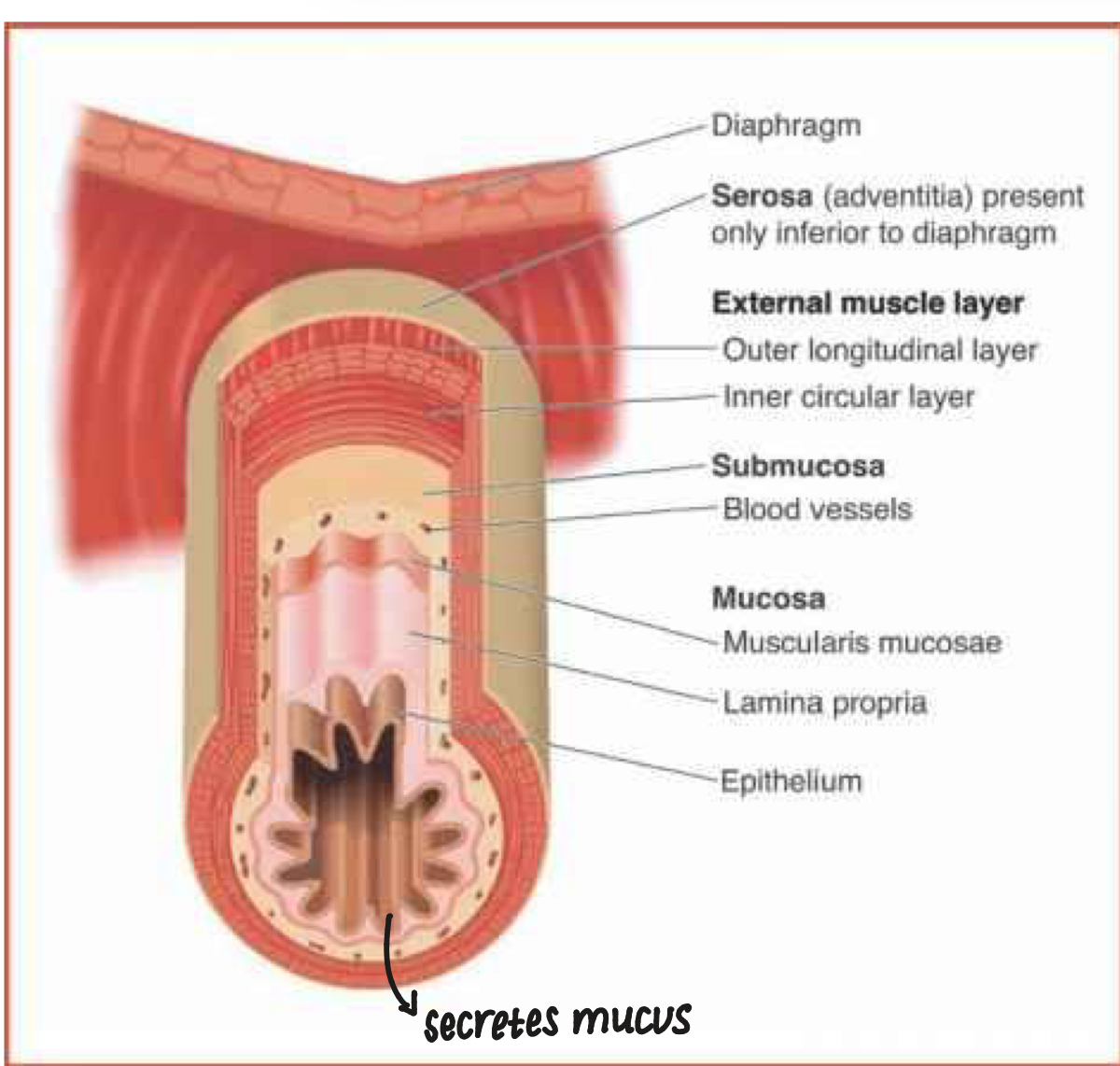
what are the 4 layers of the oesophagus (inner to outer)
mucosa- absorptive and secretary
submucosa
muscular propria (longitudinal/ circular muscle)
serosa (adventitia)- protective layer and secretary
stomach
stores food and controls its release into the duodenum
it churns the food so it mixes with the gastric secretions to make chyme
anatomical features of the stomach
the diagonal line shows the approximate division of the stomach into the 2 secretory regions: the oxyntic secretory area and the pyloric secretory area

the oxyntic secretor area
consists of the fundus and the body
the pyloric secretory area
consists of the pyloric antrum
stomach cells important for acid secretion
HCl is secreted from the parietal (oxyntic) cells together with intrinsic factor (important for the absorption of vitamin B12)
parietal cells
regulate the release of HCl
gastric mucosa secrete
mucus-layer serves to protect the underlying cells from the enzymes and acid present in the body of the stomach
ibuprofen or aspirin in its iconic form can get trapped in the mucus or released to the stomach, which releases extra acid forming ulcers
HCO3- ions trapped in the mucus thus creating a neutral pH environment in the later apposing the gastric mucosa
gastric secretions
comprise HCl
pepsinogen, the precursor of pepsin (a digestive enzyme)
pepsinogen is released from chief cells in response to acetylcholine released from the vagus nerve as well as gastrin and other hormones
gastric mediators
prostaglandins E2 and I2 are important local mediators in regulating mucus secretions
stomach also secretes inartistic factor which is important in vitamin B12 absorption
small intestine
about 6m long and is the site of the most digestion and absorption
the first 25cm comprises the duodenum and the next 2.5m is the jejunum and the remainder is the ileum

duodenum
first 25cm of the small intestine
jejunum
the next 2.5m of the small intestine after the duodenum
ileum
the rest of the small intestine after the duodenum and jejunum
structural features of the villus
the surface of the small intestine is covered by villi, which serve to increase the surface area for nutrient absorption
each villus has a capillary network and is covered by column epithelial cells many which have microvilli
cell types in the intestinal epithelium
A- absorptive columnar cell
B- structure of microvilli
C- planets cell
D- endocrine cell
E- goblet cell
F- undifferentiated columnar cell
G- localisation of the different types of cells in the epithelium of the crypts and the villi

the epithelial cells consist of 4 types
absorptive cells
goblet cells
granular cells
endocrine cells
absorptive cells
produce digestive enzymes and absorb nutrients from the chyme
goblet cells
produce mucus that lubricates the surface and protects it from mechanical damage, migrate up the villi towards the tip
granular cells (cells of paneth)
produce lysozyme and protect the intestinal surface from bacteria
endocrine cells
release peptide hormones that regulate intestinal secretions and motility remain at the base
crypts of lieberkuhn
undifferentiated cells at the base of the villi
large intestine
the last 1.5m of the gastrointestinal tract
function: to store faecal material and regulate its release to the external environment
involved in water and electrolyte reabsorption and provides an environment for bacteria that are important in synthesising certain vitamins
the bacteria is also involved in digesting bile acids
bacteria in the large intestine
lives symbiotically with the host
firmicutes and bacteroidetes being the most common types of bacteria
good bacteria in the large intestine
vitamins: gut bacteria synthesis vitamin B and K and absorbs calcium and iron
immunity: provides a barrier to invasive microbes
bad bacteria in the large intestine
obesity: gut bacteria of obese patients are significantly different from health individuals
inflammation: plays a major role in development of IBD and colitis
liver
provides bile acid and alkaline fluid for the absorption of fats and neutralisation of gastric acids, the conjugation, degradation and excretion of waste products of metabolism and detoxifies of poisonous substances
has hepatocytes- has many tubules

gall bladder
stores bile
below the liver and is connected to the duodenum by the bile duct
the entry of food into the duodenum causes the gall bladder to contract and bile to be released unto the duodenum via the sphincter of oddi
the stimulus for bile secretion is cholecystokinin (CKK) and gastrin
what 3 types of cell is the pancreas made up of
alpha cells
beta cells
delta cells
alpha cells
20%- secretes the hormone glucagon
beta cells
70%- secretes the hormone insulin
delta cells
10%- secrete growth hormone inhibiting hormone or somatostatin which acts as a paracrine to inhibit the secretion of insulin and glucagons
3 main gastric phases
cephalic phase
gastric phase
intestinal phase
cephalic phase
sight, taste and smell (30% of total acid produced)
gastric phase
stimulated by contact of food with gastric mucosa (60% of total acid produced)
intestinal phase
stimulated as food enter duodenum (10% of total acid produced)
enteric nervous system
the GI tract is innervated by both the sympathetic and parasympathetic branches of the autonomic nervous system
autonomic nerves form synapses directly on smooth muscle and secretory cells
however the GI system possesses a nervous system of its own- the enteric nervous system which also receives autonomic innervation
the enteric nervous system comprises various types of neurons organised into the Auerbach’s and Meissner’s

Auerbach’s neurones
situated between longitudinal and circular muscle layers (in the outer layer of the gut)
Meissner’s
situated in the mucosal layer (in the inner layer of the gut)
the arrangement of the ENS
synaptic connections are made with: the enteric neurones with one another and with autonomic nerves
motor neurones- excitatory
interneurones-inhibitory
they act to regulate muscle contraction/ relaxation and secretions
sensory neurones
activated by:
mechanoreceptors (which detect stretching of the muscle)O
chemoreceptors (which detect irritant substances in the gut lumen)
mediators
ATP
5-hydroxytryptamine
substance P
vasoactive intestinal polypeptide
nitric oxide
common symptoms of GI disorders
heartburn
bloating
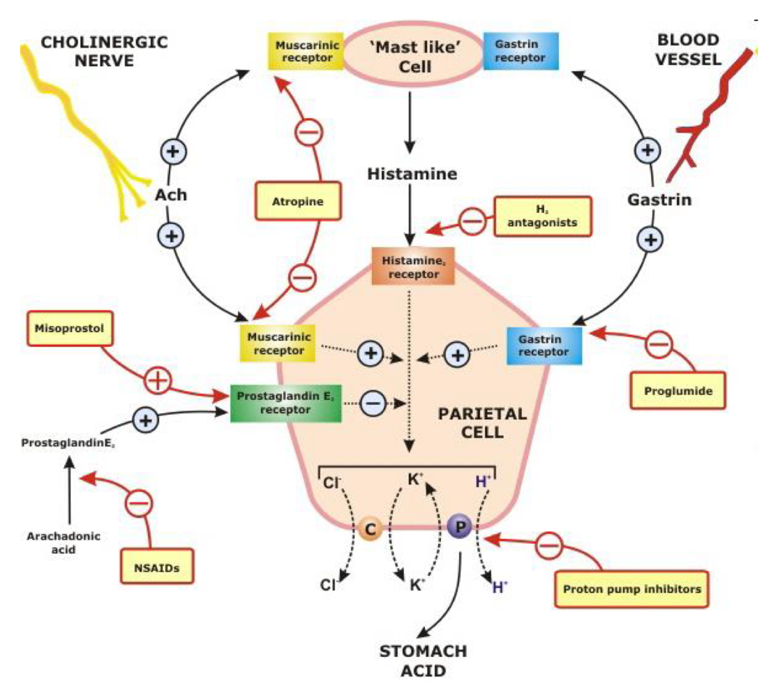
process of acid secretion
carbonic anhydrase in the parietal cell cytoplasm catalyses the conversion of CO2 and H2O to H2CO3
the H+ ions are actively pumped into the lumen of the stomach via H+/K+ ATPases which move K+ ions in the opposite direction
The HCO3- ions are transported into the bloodstream by antiporters in exchange for Cl- ions which are then carried by symporters into the lumen together with K+ ions
picture of acid secretion process

stimulation of acid secretion
when we eat our stomach stretched triggering the production of gastrin from G cells
gastrin goes into the bloodstream so it can be widespread and once it comes back into the stomach it activated various sites of the muscarinic acetylcholine receptors
gastrin will activate CCK cells and the entrochromaffin like cells that releases histamine and H2 receptor
vagus nerve activity and gastrin are also involved in the activation of chief cells to produce pepsinogen

acid secretion control
acid secretion is inhibited by somatostatin released from the D cells in response to high acidity
mechanism of feedback inhibition
somatostatin inhibits both the release of gastrin from G cells and release of histamine from the entrochromaffin like cells

drugs that inhibit or neutralise acid secretion and their targets
histamine H2 receptor
CCK cholecystokinin-B receptor
PG, prostaglandin receptor

summary of control of gastric acid secretion
dyspepsia
disorder associated with the upper GI
symptoms: heartburn, abdominal pain, nausea and belching
often have unknown origin but can be indicative of an underlying disease or ulcer
ulcer
a wound, a discontinuity or break in a bodily membrane that impedes the organ of which that membrane is a part from continuing its normal functions
peptic ulcers
break in the membrane in the stomach, duodenum and occasionally lower oesophagus

difference between gastric or duodenum ulcers
if pain comes straight after eating food, more likely to be a gastric ulcer
risk factors of peptic ulcers
family history
heavy smoking
alcohol consumption
poor diet
symptoms of peptide ulcers
abdominal epigastric pain- strongly correlated with food intake (certain food can trigger pain like acidic foods like peppers or lemon)
bloating and abdominal fullness
nausea and vomiting
weight loss and anorexia- common in those with gastric ulcers
pain associated duodenal ulcerations often experienced at night and resolves overnight or by consumption of food
complications of peptic ulcers
gastrointestinal bleeding is common- triggered by when an ulcer erodes a blood vessel
perforation- a hole in the membrane of the gastrointestinal tract
penetration- perforation extends to other organs
gastric outlet obstruction- ulcers causes narrowing of pyloric canal or swelling of gastric antrum and duodenum
these can become life-threatening if not treated
causes of gastric ulcers
long term use of NSAIDs- these interfere with the mucus layer which protects the gastric lining from the acidic environment in the stomach. NSAIDs block the synthesis of prostaglandins by inhibiting the enzyme cyclooxygenase
an imbalance between the pepsin and stomach acids- reflux of acid due to GORD
increasing evidence that the presence of helicobactor pylori in the guy may also be a predisposing factor
long term use of NSAIDs
about 30-50% of long-term NSAID users have endoscopic lesions
prostaglandins stimulate the release of mucus from the gastric membranes which acts as a protect barrier preventing acid attack
traditional or non-selective NSAIDs inhibit both isoforms of cyclooxygenase enzyme, which is essential for prostaglandin production
COX-2 selective inhibitors that produce less GI damage

helicobacter pylori
spiral shaped micro-organisms in stomach mucosa
found in 2/3 of the world’s population
prevalence is higher in developing countries
western countries H.Pylori infection is correlated with age: 20% at age 20, 30% at age 30
hypothesised to be spread by unclean food and water
major cause duodenal and stomach ulcers and stomach cancers
60% of gastric and 50-75% of duodenal ulcers
life cycle of helicobacter pylori
it survives the acidic conditions of the stomach by growing in the mucus layer
the ammonia serves to buffer the acid in the bacteria’s environment
H.pylori penetrate the mucus layer of host stomach and adhere the surface of gastric mucosal epithelial cells
produces ammonia from urea by the urease and the ammonia neutralise the gastric acid to escape from elimination
proliferate, migrate and finally form the infectious focus
the gastric ulceration is developed by destruction of mucosa, inflammation and mucosal cell death

GORD
heartburn, difficulty in swallowing and regurgitation of gastric contents into the mouth
reflux is associated with reduced peristaltic activity in the oesophagus and transient relaxation of the oesophageal sphincter allowing gastric acid, pepsin and bile to come into contact with the oesophageal epithelium
with age GORD is more common because the oesophagus muscles that contract and relax might be weaker but in pregnancy the hormonal changes may cause GORD
common diagnostic tests for peptic ulcers
tests for H.pylori
non-invasive breath test
direct visual identification
blood tests
tests for H.pylori
basis is the ability of H.pylori to secrete the urease enzyme which catalyses the conversion of urea to ammonia and carbon dioxide
non-invasive breath tests
using radioactive carbon to detect H.pylori and the level of radioactive CO2 can be detected to identify ulcer-causing bacteria
rapid urease tests for biopsy
stool antigen test- small sample is tested for bacteria
direct visual identification
esophagogastroduodenoscopy- used to look inside to see inflammation and examine the severity of the ulcer
blood test
may describe information on red blood cell and white blood cell counts, bacteria presence
not reliable because they may give false negative tests (due to recent medications such as antibiotics)
treatment of peptic ulcers
antacids: aluminium hydroxide, magnesium carbonate, magnesium trisillicate
alginates: gaviscon
histamine H2 antagonist: cimetidine, famotidine, ranitidine, nizatidine
proton pump inhibitors: omeprazole, esomeprazole, lansoprazole, pantoprazole, rabeprazole
antacids
used to overcome the symptoms of the conditions:
they neutralise gastric acidity and are more effective if taken after food but doesn't treat the underlying disease
some antacid preparations contain simeticone, an anti-foaming agent that prevents flatulence and others contain alginates which form a ‘raft’ on the surface of the stomach contents thus reducing reflux
H2 antagonists
classed as ulcer healing drugs are used in the treatment of gastric ad duodenal ulceration
side effects of H2 antagonist
diarrhoea
headache
dizziness
rash
tiredness
Proton pump inhibitors mechanism of action
PPIs are given in an inactive form which is neutrally charged (lipophilic)
PPIs readily cross cell membranes into the intracellular compartments within acidic environments (e.g. parietal cells)
these drugs are substituted benzimidazoles- weak bases, and as a result accumulate in the acidic compartment of the parietal cells close to the H+/K+ ATPase
PPIs are protonated and reconfigured into their active forms in acidic conditions:
the acid environment catalyses their conversion to a sulphonamide intermediate which interacts covalently with the sulphydryl groups on the cysteine residues in the extracellular domain of the H+/K+ ATPase
thus blocking its activity
Proton pumping inhibitors
better than H2 receptor antagonists
they target the final step in acid secretion
their effects can’t be overcome by food-induced acid secretion medicated by the activation of muscarinic or CCK receptor
example of PPI
Omeprazole
duration of action is 24 hrs
its dissociation from the proton pump is probably mediated by glutathione which releases the omeprazole sulphide and allows re-activation of the pump
signs of an allergic reaction: hives, difficulty breathing, swelling
common side effects: stomach pain, gas, headache, nausea, vomiting, diarrhoea
PPIs with antibacterials
1 week triple therapy regime is successful in 85% of cases and 2 week therapy can be affected by side effects reducing patient compliance
typical regime: 20mg omeprazole 2x a day with 1g amoxicillin 2x a day and 500mg clarithromycin 2x a day
if patient has already received clarithromycin for another infection, 400mg metronidazole 3x a day can be given with 500mg amoxicillin 3x a day
other types of ulcers
ulcerative colitis
Crohn’s disease
ulcerative colitis
chronic disease that causes inflammation in the rectum, colon and the last part of the small ileum
the inflammation affects the inner lining of the colon causing ulcers
Crohn’s disease
chronic disease that can cause inflammation anywhere along the digestive tract from the mouth to the anus
symptoms of ulcerative colitis and Crohn’s disease
abdominal pain and cramps
bloody stool
diarrhoea
fever
loss of appetite
constipation
the passage of small, hard stools less frequently than is usual
often caused by a diet low in fibre and/or inadequate fluid intake
can also be a symptom for an underlying disease or a side effect of certain drugs
treatment involves use of bulking agents, osmotic or stimulant laxative
bulking agents
example: wheat bran, ispaghulxa husk, sterculia and methylcellulose
increase faecal mass- promoting peristalsis by stimulating mechanoreceptors in the colon wall
increase retention of water in the lumen of the colon which softens the faeces
takes 24 hrs to work and should be accompanied with intake of fluid
osmotic laxatives include lactulose and magnesium salts
lactulose
disaccharide which is broken down by bacteria into fructose and galactose which release osmotically active lactic and acetic acids upon fermentation
magnesium salts
stimulate the release of CCK in the small intestine promoting motility
constipation treatments
bisacodyl, danthron, Senna and sodium pico sulphate are examples of stimulant laxatives
its unknown how these agents work but it is believed that they stimulate myenteric nerve plexuses thus promoting gut motility and reducing fluid reabsorption
senna
the most gentle and acts within 8-12 hrs
metabolised bu colonic bacteria to senosides A and B which irritate the gut thus stimulating faecal movement
danthron
a carconogenic
used in elderly terminally ill patients either in a formulation with the wetting agent poloxamer 188 or with the detergent decussate sodium
faecal softeners
useful in less severe cases of constipation
glycerol, arachis oil and liquid paraffin are examples
diarrhoea
gastrointestinal infections are the usual cause of acute diarrhoea however this can also be a side effect of certain drugs like broad spectrum antimicrobials
diarrhoea- treatments
opioids like codeine, diphenoxylate and loperamide are used to treat this condition
codeine is a morphine congener whilst the other 2 are congeners of pethidine
their therapeutic effects are due to their action on opioid receptions (mui and k) on enteric neurones, resulting in: reduced gut motility allowing more time for fluid reabsorption and increasing the transit time of intestinal contents
both codeine and loperamide have anti-muscarinic properties which decreases peristalsis
adequate hydration and electrolyte balance should be maintained
irritable bowel syndrome (IBS)
constipation and/or diarrhoea together with abdominal pain, bloating and discomfort is associated with IBS
the cause is usually unidentified
antispasmodic agents help overcome the abdominal pain which is caused by smooth muscle spasms
atropine sulphate, dicloverinehydrochlordie, hyoscine butyl bromide and propantheline bromide are anti-muscarinics
alverine citrate, mebeverine hydrochloride and peppermint oil are direct intestinal smooth muscle relaxants
vomiting
ejection of the gastric contents through the mouth
protective role of the stomach as it results in the removal of toxic substances from the body
controlled by the vomiting centre and the chemoreceptor trigger zone (CTZ) situated in the brain stem
CTZ is relatively permeable and can be affected by various circulating mediators and by drugs that do not cross the blood brain barrier
the main neurotransmitter involved in this reflex are ACh, 5-HT, histamine, dopamine and substance P and it is the receptors they act on that provide the targets for the most commonly used anti-emetic drugs
drugs affecting vomiting
muscarinic antagonist: hyoscine
histamine antagonists: promethazine, cinnarizine, cyclizine
dopamine antagonists: metaclopramide, domperidone
5-HT antagonists: ondansetron, dolasteron, granisteron, tropisetron
neurokinin 1 antagonist: arepitant
cannabinoids: nabilone- used treatment of chemotherapy-induced nausea and vomiting
what drugs are used in the treatment of motion sickness and have no effect on the CTZ
muscarinic antagonists
Histamine antagonists
liver function
involved in processing nutrients from food; providing bile acids and alkaline fluid for the absorption of fats and neutralisation of gastric acids
the conjugation, degradation and excretion of waste products of metabolism
and detoxifies poisonous substances
functions vital to life
the liver is resilient and capable of regenerating new cells
liver
hepatic secretions comprise
various elements that make up bile
bile contains a number of electrolytes particularly HCO3-, which helps to neutralise gastric acid
bile acids which are important in the digestion of dietary fats
pigments mainly bilirubin (breakdown product of haemoglobin)
products of metabolism that are extracted from the blood
signs and symptoms of liver disease
impaired live function may lead to:
digestive problems
impaired blood sugar levels- hypogycaemia
immune disorders
abnormal absorption of fats:
leads to indigestion
reflux
fat soluble vitamin deficits
haemorrhoids
gall stones
abdominal bloating and constipation
nausea and vomiting
nervous system disorders
change in mood resulting in
depression
anxiety
anger/ irritability
poor concentration
cognitive ability
headaches
types of liver disease
hepatitis A,B,C
liver fibrosis/ cirrhosis
fatty liver disease
primary liver cancer
hereditary disease
alcoholic liver disease
hereditary disease
2 most common:
haemochromatosis- excess iron is absorbed from the diet and deposited in the liver
alpha 1 antitrypsin deficiency- this is a liver protein important for normal lung function