The Respiratory System
 Call Kai
 Learn
Learn Practice Test
 Spaced Repetition
 Match
Match Flashcards
 Knowt Play
1/33

There's no tags or description
Looks like no tags are added yet.
Name | Mastery | Learn | Test | Matching | Spaced | Call with Kai | Chat |
|---|
No analytics yet
Send a link to your students to track their progress
34 Terms
What are the primary and secondary functions of the respiratory system?
Functions of the respiratory system:
Primary functions:
Oxygen (O₂) supply: Delivers oxygen to the blood for cellular respiration.
Carbon dioxide (CO₂) removal: Eliminates CO₂ produced by cellular metabolism.
Secondary functions:
Sound production: Air passing through the larynx enables speech.
Blood pH regulation: Controls CO₂ levels to help maintain acid–base balance (homeostasis).
Key point: The respiratory system enables gas exchange and helps maintain blood pH.
What are the main structures of the respiratory system, and what are their functions?
Main structures of the respiratory system:
Nose and nasal cavity: Filter, warm and humidify inhaled air.
Pharynx: Passageway carrying air towards the larynx.
Larynx: Produces sound and helps protect the airway.
Trachea: Conducts air towards the lungs.
Bronchi: Branch into each lung to distribute air.
Lungs: Main organs responsible for gas exchange.
Alveoli: Tiny air sacs where O₂ enters the blood and CO₂ leaves it.
Airflow pathway:
Nose/mouth → Pharynx → Larynx → Trachea → Bronchi → Bronchioles → Alveoli
Key point: The conducting airways transport air, while the alveoli are the primary site of gas exchange.


How should the main respiratory-system diagram be labelled, and how does air travel through the conducting zone?
Conducting zone:
Includes the nose and nasal cavity, pharynx, larynx, trachea, bronchi, bronchioles and terminal bronchioles.
Conducts, filters, warms and humidifies air.
No gas exchange occurs in the conducting zone.
Airflow through the conducting zone:
Nose or mouth → pharynx → larynx → trachea → primary bronchi → secondary bronchi → tertiary bronchi → bronchioles → terminal bronchioles
The terminal bronchioles are the final part of the conducting zone. Air then enters the respiratory zone through the respiratory bronchioles.

What are the four processes involved in respiration?
The four processes involved in respiration:
Pulmonary ventilation (breathing): Movement of air into and out of the lungs (inhalation and exhalation).
External respiration: Exchange of O₂ and CO₂ between the alveoli and pulmonary blood.
Transport of respiratory gases: Movement of O₂ and CO₂ through the bloodstream.
Internal respiration: Exchange of O₂ and CO₂ between systemic blood and body tissues.
Key point: External respiration occurs in the lungs, while internal respiration occurs in the body tissues.
What are the conducting and respiratory zones of the respiratory system, and what are their functions?
Conducting and respiratory sones of the respiratory system:
1. Conducting Zone
Structures: Nose, nasal cavity, pharynx, larynx, trachea, bronchi and terminal bronchioles.
Function: Transports, filters, warms and humidifies inhaled air.
No gas exchange occurs.
2. Respiratory Zone
Structures: Respiratory bronchioles, alveolar ducts and alveoli.
Function: Site of gas exchange, where O₂ enters the blood and CO₂ leaves it.
Key point: Conducting zone = air transport; Respiratory zone = gas exchange.
What are the functions of the nasal cavity, and how does its mucosa protect the respiratory system?
Main functions of the nasal cavity:
Filters, warms and humidifies inhaled air.
Contains olfactory receptors for smell.
Acts as a resonance chamber for speech.
Divided into left and right cavities by the nasal septum.
Two types of mucosa:
Olfactory mucosa: Contains sensory receptors responsible for smell.
Respiratory mucosa: Lined mainly by pseudostratified ciliated columnar epithelium.
Protective mechanisms:
Mucus: Traps dust, bacteria and other particles.
Cilia: Move trapped particles towards the pharynx for removal.
Lysozyme: Antimicrobial enzyme in secretions that helps destroy bacteria.
Key point: The nasal cavity conditions inhaled air and protects the lower respiratory tract from harmful particles.
What are the three regions of the pharynx, and what are their functions?
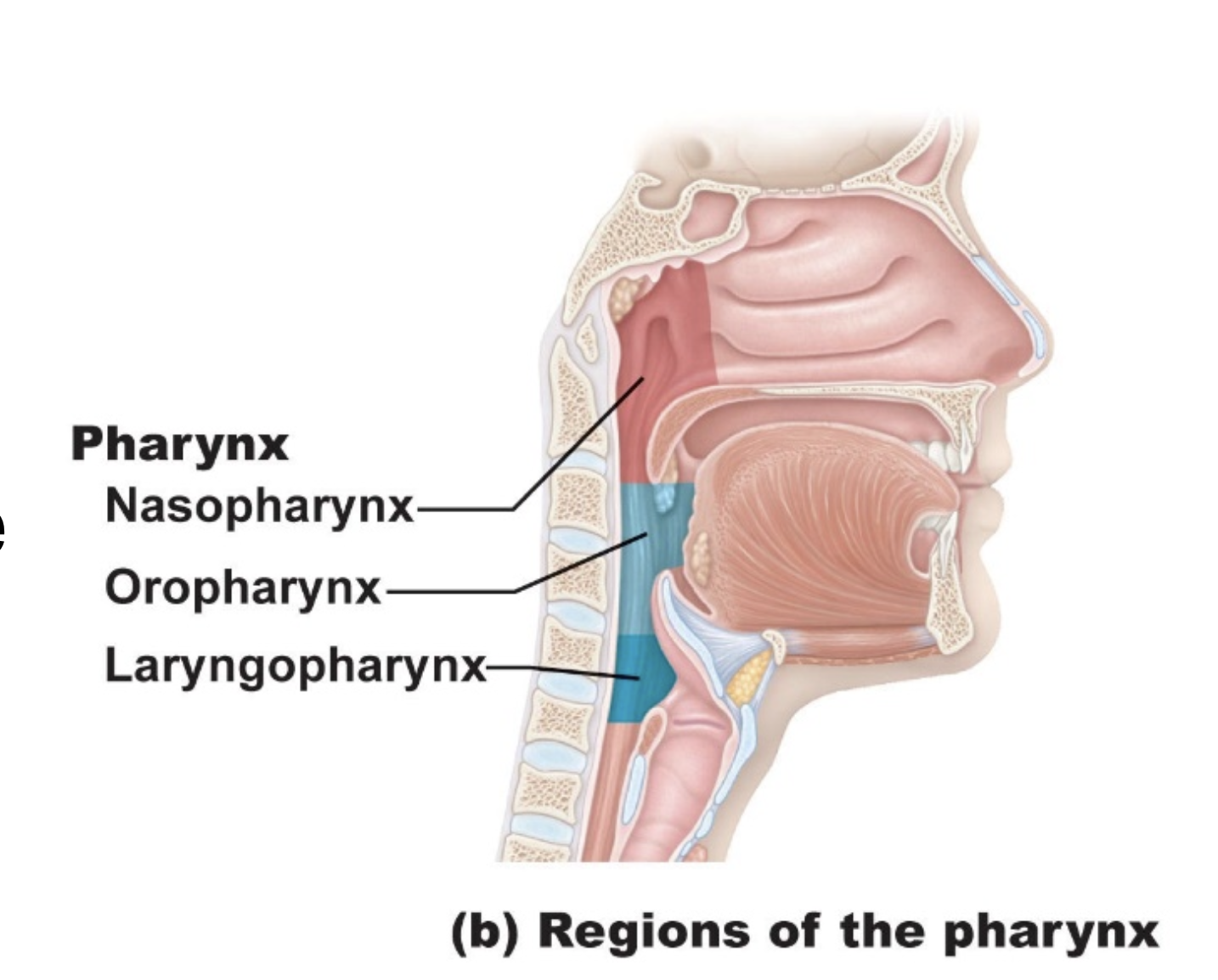
The pharynx (throat) is a muscular passageway connecting the nasal and oral cavities to the larynx and oesophagus.
Three regions:
1. Nasopharynx
Located behind the nasal cavity.
Passageway for air only.
Lined with pseudostratified ciliated columnar epithelium, which helps remove mucus and debris.
Contains the pharyngeal tonsil (adenoids), which helps protect against pathogens.
2. Oropharynx
Located behind the oral cavity.
Passageway for air and food.
Contains palatine and lingual tonsils.
Lined with stratified squamous epithelium for protection against abrasion.
3. Laryngopharynx
Located behind the larynx.
Passageway for air and food, directing them towards the larynx and oesophagus.
Lined with stratified squamous epithelium for protection.
Key point: The nasopharynx carries only air, while the oropharynx and laryngopharynx carry both air and food.
What is the larynx, and what are its main functions?
The larynx (voice box) connects the pharynx to the trachea.
Main functions of the larynx:
Air passage: Allows air to enter the trachea.
Voice production: Contains vocal cords that produce sound.
Airway protection: The epiglottis helps prevent food and liquids entering the airway during swallowing.
Thyroid cartilage: Protects the larynx and forms the Adam's apple.

What is the trachea, and how is it structurally adapted to its function?
The trachea (windpipe) is a flexible tube connecting the larynx to the primary bronchi.
Trachea key structures and functions:
C-shaped cartilage rings: Prevent airway collapse while allowing the oesophagus to expand during swallowing.
Pseudostratified ciliated columnar epithelium: Lines the trachea and helps remove inhaled particles.
Mucus: Traps dust, bacteria and other debris.
Cilia: Move mucus and trapped particles towards the pharynx for removal.
Key point: The trachea maintains an open airway and protects the lungs through mucociliary clearance.

How do the bronchi branch within the lungs, and what are their main functions?
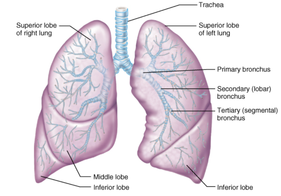
The bronchi and bronchioles conduct and distribute air throughout the lungs.
Branching pathway:
Trachea → Primary bronchi → Secondary bronchi → Tertiary bronchi → Bronchioles → Terminal bronchioles → Respiratory bronchioles → Alveoli
Bronchi key features:
Primary bronchi: One enters each lung; the right is shorter, wider and more vertical.
Secondary bronchi: Supply individual lung lobes (3 right, 2 left).
Tertiary bronchi: Supply bronchopulmonary segments.
Bronchioles: Small airways that regulate airflow through smooth muscle contraction and relaxation.
Terminal bronchioles: Smallest conducting airways; lead into the respiratory zone.
Key point: Bronchi distribute air throughout the lungs, while bronchioles regulate airflow.
What structures make up the respiratory zone, and what is their function?
The respiratory zone: is the region of the lungs where gas exchange occurs.
Airflow pathway:
Respiratory bronchioles → Alveolar ducts → Alveolar sacs → Alveoli
Respiratory zone key structures:
Respiratory bronchioles: Small airways where gas exchange begins.
Alveolar ducts: Passages leading to alveolar sacs, with alveoli opening along their walls.
Alveolar sacs: Clusters of alveoli sharing a common opening.
Alveoli: Tiny, thin-walled air sacs surrounded by capillaries where O₂ enters the blood and CO₂ leaves it.
Key point: The respiratory zone enables gas exchange between air in the lungs and blood in pulmonary capillaries.


How should the respiratory-zone diagram in Workshop 8 be labelled, and how does air travel through this region?
Important distinction:
The terminal bronchiole is the final structure of the conducting zone.
Respiratory bronchioles mark the beginning of the respiratory zone.
Airflow:
Terminal bronchiole → respiratory bronchioles → alveolar ducts → alveolar sacs → alveoli
Functions:
Respiratory bronchioles: Small airways where gas exchange begins.
Alveolar ducts: Passages with alveoli opening along their walls.
Alveolar sacs: Clusters of alveoli sharing a common opening.
Alveoli: Individual thin-walled air sacs where O₂ enters pulmonary blood and CO₂ leaves it.

What are the two types of alveolar cells, and what are their functions?
The alveolar walls contain two main types of epithelial cells:
1. Type I Alveolar Cells (Type I Pneumocytes)
Thin, simple squamous epithelial cells.
Form most of the alveolar surface.
Allow rapid diffusion of O₂ and CO₂ between alveoli and blood.
2. Type II Alveolar Cells (Type II Pneumocytes)
Cuboidal epithelial cells scattered among Type I cells.
Produce pulmonary surfactant, a mixture of lipids and proteins.
Surfactant reduces surface tension, helping prevent alveolar collapse during exhalation.
Key point:
Type I = Gas exchange.
Type II = Surfactant production (prevents alveolar collapse).
What are the main anatomical features of the right and left lungs?
The lungs are paired respiratory organs located in the thoracic cavity, separated by the mediastinum (which contains the heart).
Right Lung:
Larger and wider than the left lung.
Contains 3 lobes: Superior, middle and inferior.
Left Lung:
Smaller due to the space occupied by the heart.
Contains 2 lobes: Superior and inferior.
Has a cardiac notch to accommodate the heart.
Key point: Right lung = 3 lobes; Left lung = 2 lobes
What are the pleural membranes, and what is the function of the pleural cavity?
Pleural membranes, and function of the pleural cavity:
Two layers:
Visceral pleura: Covers the external surface of the lungs.
Parietal pleura: Lines the internal thoracic cavity, diaphragm and mediastinum.
Pleural cavity:
Thin, fluid-filled space between the two pleural layers.
Pleural fluid: Reduces friction during breathing and helps keep the lungs expanded against the chest wall.
Clinical significance – Pleurisy:
Inflammation of the pleura, causing sharp pain during breathing.
May be associated with excess pleural fluid (pleural effusion), which can restrict lung expansion.
Key point: The pleural membranes allow smooth lung movement during breathing and help maintain lung expansion.

What is the difference between pulmonary and bronchial circulation, and how does the autonomic nervous system affect the airways?
The difference between pulmonary and bronchial circulation:
The lungs receive blood from two circulatory systems.
1. Pulmonary Circulation (Gas Exchange)
Pulmonary arteries: Carry deoxygenated blood from the right ventricle to the lungs.
Pulmonary capillaries: Allow O₂ and CO₂ exchange at the alveoli.
Pulmonary veins: Return oxygenated blood to the left atrium.
Low-pressure, high-flow system.
2. Bronchial Circulation (Lung Tissue Supply)
Bronchial arteries: Arise from the systemic circulation (usually the aorta) and deliver oxygenated blood to lung tissues.
Supplies the bronchi and supporting lung structures.
Higher-pressure, lower-flow system than pulmonary circulation.
3. Autonomic Regulation of Airways
Parasympathetic stimulation: Causes bronchoconstriction (airway narrowing).
Sympathetic effects (β₂ receptors): Promote bronchodilation (airway widening), mainly through circulating adrenaline.
Key point: Pulmonary circulation enables gas exchange, while bronchial circulation supplies oxygen and nutrients to lung tissues.
What are atmospheric, intrapulmonary and intrapleural pressures, and how do they help keep the lungs inflated?
Atmospheric, intrapulmonary and intrapleural pressures:
1. Atmospheric Pressure (Patm)
Pressure exerted by the air surrounding the body.
Approximately 760 mmHg at sea level (0 mmHg relative pressure).
2. Intrapulmonary Pressure (Palv)
Pressure inside the alveoli.
Changes during breathing to move air into and out of the lungs.
Equals atmospheric pressure between breaths when airflow stops.
3. Intrapleural Pressure (Pip)
Pressure within the pleural cavity.
Normally negative relative to atmospheric pressure (approximately −4 mmHg at rest).
Helps prevent lung collapse.
Transpulmonary Pressure:
Ptp = Palv - Pip
The difference between intrapulmonary and intrapleural pressures helps keep the lungs expanded.
Key point: Intrapleural pressure is normally lower than intrapulmonary pressure, creating a pressure difference that keeps the lungs inflated.

What are pneumothorax and atelectasis, and how can they cause lung collapse?
Pneumothorax and atelectasis, and how can they cause lung collapse:
Pneumothorax
Air enters the pleural cavity due to injury or rupture of the pleura.
Intrapleural pressure rises towards atmospheric pressure.
Loss of normal transpulmonary pressure can cause the affected lung to collapse.
2. Atelectasis
Partial or complete collapse of alveoli or lung tissue.
May result from airway obstruction, insufficient surfactant or pneumothorax.
Reduces the surface area available for gas exchange.
Key distinction:
Pneumothorax = Air in the pleural cavity.
Atelectasis = Collapse of alveoli or lung tissue.

How do inspiration and expiration occur, and how do pressure and volume changes control airflow?
Pulmonary ventilation is the movement of air into and out of the lungs, driven by changes in thoracic volume and pressure.
1. Inspiration (Inhalation)
Diaphragm contracts and flattens; external intercostal muscles lift the ribs.
↑ Thoracic cavity volume → ↑ Lung volume.
↓ Intrapulmonary pressure below atmospheric pressure.
Air flows into the lungs.
2. Expiration (Exhalation)
Diaphragm and external intercostal muscles relax.
Elastic recoil decreases lung and thoracic volume.
↑ Intrapulmonary pressure above atmospheric pressure.
Air flows out of the lungs.
Key point: Air moves from higher pressure to lower pressure.
Inspiration: ↑ Volume → ↓ Pressure → Air enters.
Expiration: ↓ Volume → ↑ Pressure → Air exits.

What are Boyle's and Charles's laws, and how do they relate to respiration?
1. Boyle's Law
At constant temperature, gas pressure is inversely proportional to volume.
↑ Volume → ↓ Pressure.
↓ Volume → ↑ Pressure.
Explains how changes in lung volume create pressure differences that move air during breathing.
2. Charles's Law
At constant pressure, gas volume increases as absolute temperature increases.
↑ Temperature → ↑ Gas volume.
↓ Temperature → ↓ Gas volume.
Helps explain the physical behaviour of gases as they are warmed in the respiratory tract.
Key point: Boyle's law is particularly important for understanding pulmonary ventilation, because breathing depends on changes in lung volume and pressure.
What are Dalton's and Henry's laws, and how do they relate to respiratory gas exchange?
1. Dalton's Law (Partial Pressures)
The total pressure of a gas mixture equals the sum of the partial pressures of its individual gases.
Each gas contributes to total pressure according to its proportion in the mixture.
Example:
Oxygen makes up approximately 21% of atmospheric air.
Po₂ = 0.21 × 760 ≈ 159 mmHg
2. Henry's Law (Gas Solubility)
The amount of gas dissolved in a liquid depends on its partial pressure and solubility.
↑ Partial pressure → ↑ Amount of gas dissolved.
CO₂ is approximately 20 times more soluble in blood than O₂.
Key point:
Dalton's Law: Explains the partial pressures of gases.
Henry's Law: Explains how much gas dissolves in blood
How do external and internal respiration differ, and what drives the movement of O₂ and CO₂?
Gas exchange occurs through diffusion, with gases moving down their partial pressure gradients.
External versus internal respiration:
1. External Respiration (Lungs)
Gas exchange between alveoli and pulmonary capillaries.
O₂: Alveoli → Blood.
CO₂: Blood → Alveoli.
2. Internal Respiration (Body Tissues)
Gas exchange between systemic capillaries and body tissues.
O₂: Blood → Tissues.
CO₂: Tissues → Blood.
Key point: O₂ and CO₂ diffuse from areas of higher partial pressure to lower partial pressure.

What factors influence the efficiency of gas exchange between alveoli and pulmonary capillaries?
Four main factors that influence the efficiency of gas exchange between alveoli and pulmonary capillaries:
1. Partial Pressure Gradients
Gases diffuse from higher to lower partial pressure.
Larger pressure differences increase the rate of diffusion.
2. Gas Solubility
More soluble gases diffuse more readily into blood.
CO₂ is approximately 20 times more soluble than O₂.
3. Respiratory Membrane
Thin membrane: Allows rapid gas diffusion.
Large surface area: Increases the amount of gas exchanged.
Thickening of the membrane or loss of surface area reduces gas exchange.
4. Ventilation–Perfusion Matching (V/Q)
Efficient gas exchange requires adequate alveolar ventilation (airflow) and pulmonary perfusion (blood flow).
Poor matching reduces oxygen uptake.
Key point: Efficient gas exchange depends on pressure gradients, gas solubility, a thin respiratory membrane, sufficient surface area and appropriate ventilation–perfusion matching.
What are the four respiratory volumes, and what does each represent?
Respiratory volumes measure the amount of air inhaled, exhaled or remaining in the lungs.
Tidal Volume (TV): Air inhaled or exhaled during normal, quiet breathing (~500 mL).
Inspiratory Reserve Volume (IRV): Additional air that can be forcibly inhaled after a normal inspiration.
Expiratory Reserve Volume (ERV): Additional air that can be forcibly exhaled after a normal expiration.
Residual Volume (RV): Air remaining in the lungs after maximum forced expiration; prevents complete lung collapse.
Key point: TV = normal breathing; IRV = extra inhalation; ERV = extra exhalation; RV = air remaining.

What are the four respiratory capacities, and how are they calculated?
Respiratory capacities are combinations of two or more respiratory volumes.
Inspiratory Capacity (IC): Maximum air inhaled after normal expiration.
IC = TV + IRV
Functional Residual Capacity (FRC): Air remaining after normal expiration.
FRC = ERV + RV
Vital Capacity (VC): Maximum air exhaled after maximum inspiration.
VC = TV + IRV + ERV
Total Lung Capacity (TLC): Total air lungs can contain after maximum inspiration.
TLC = TV + IRV + ERV + RV
Key point: Lung capacities describe combinations of volumes, while lung volumes describe individual quantities of air.

What is spirometry, and how does it help distinguish obstructive and restrictive lung diseases?
A spirometry measures the amount and rate of air inhaled and exhaled to assess lung function.
Key measurements:
Forced Vital Capacity (FVC): Maximum volume of air forcibly exhaled after a deep breath.
Forced Expiratory Volume in 1 second (FEV₁): Volume of air exhaled during the first second.
FEV₁/FVC ratio: Proportion of total forced expiration completed in the first second (normally approximately 80% in healthy young adults).
Disease differences:
Obstructive disease: Difficulty exhaling due to narrowed airways (e.g., COPD).
↓ FEV₁ and ↓ FEV₁/FVC ratio.
Air trapping may increase residual volume.
Restrictive disease: Difficulty expanding lungs (e.g., pulmonary fibrosis).
↓ Total lung capacity (TLC) and FVC.
FEV₁/FVC ratio is usually normal or increased.
Key point: Obstructive = difficulty getting air out; restrictive = difficulty expanding the lungs to get air in.

How is oxygen transported in the blood, and what is the role of haemoglobin?
Oxygen is transported in two ways:
Bound to haemoglobin (98.5%): Most oxygen binds to haemoglobin within red blood cells.
Dissolved in plasma (1.5%): A small amount circulates freely in blood plasma.
Haemoglobin (Hb):
Each Hb molecule contains four haem groups, each capable of binding one O₂ molecule.
Oxyhaemoglobin (HbO₂): Haemoglobin bound to oxygen.
Deoxyhaemoglobin: Haemoglobin that has released oxygen.
Key point: Haemoglobin carries most oxygen in blood, binding O₂ in the lungs and releasing it into body tissues.
What does the oxygen–haemoglobin dissociation curve represent, and what factors influence oxygen loading and unloading?
The oxygen–haemoglobin dissociation curve shows the relationship between oxygen partial pressure (PO₂) and haemoglobin oxygen saturation.
Normal relationship:
High PO₂ (lungs): Haemoglobin binds oxygen → ↑ Saturation.
Low PO₂ (tissues): Haemoglobin releases oxygen → ↓ Saturation.
Factors shifting the curve:
Right shift = Increased O₂ unloading
↑ Temperature
↑ CO₂
↓ pH (increased acidity)
↑ 2,3-BPG
These changes decrease haemoglobin's affinity for oxygen, allowing more oxygen to reach active tissues.
Left shift = Decreased O₂ unloading
↓ Temperature
↓ CO₂
↑ pH
↓ 2,3-BPG
These changes increase haemoglobin's affinity for oxygen.
Key point: Right shift = haemoglobin releases O₂ more easily; left shift = haemoglobin holds O₂ more tightly.

What are the three ways carbon dioxide is transported in blood, and what is the role of carbonic anhydrase?
CO₂ is transported in blood in three forms:
Bicarbonate ions (HCO₃⁻): ~70%
Most CO₂ is converted into bicarbonate inside red blood cells and transported in plasma.
Carbaminohaemoglobin (HbCO₂): ~20%
CO₂ binds to the globin portion of haemoglobin.
Dissolved in plasma: ~7–10%
A small amount circulates directly in plasma.
Bicarbonate reaction:
CO₂ + H₂O ⇌ H₂CO₃ ⇌ H⁺ + HCO₃⁻
Carbonic anhydrase: Enzyme that rapidly converts CO₂ and water into carbonic acid (H₂CO₃), and catalyses the reverse reaction.
At tissues: CO₂ is converted into bicarbonate for transport.
At lungs: The reaction reverses, releasing CO₂ for exhalation.
Key point: Most CO₂ travels as bicarbonate, and carbonic anhydrase helps convert CO₂ into a form that can be transported efficiently.

How does breathing rate influence blood CO₂ levels and pH?
The respiratory system helps regulate blood pH by controlling how much CO₂ is exhaled.
1. Increased ventilation (faster/deeper breathing):
More CO₂ exhaled → ↓ Blood CO₂.
↓ Carbonic acid and H⁺ → ↑ Blood pH (more alkaline).
2. Decreased ventilation (slower/shallower breathing):
Less CO₂ exhaled → ↑ Blood CO₂.
↑ Carbonic acid and H⁺ → ↓ Blood pH (more acidic).
Bicarbonate buffer system:
CO₂ + H₂O ⇌ H₂CO₃ ⇌ H⁺ + HCO₃⁻
Key point: ↑ CO₂ → ↓ pH; ↓ CO₂ → ↑ pH.
This is an important homeostatic mechanism for maintaining normal acid–base balance.
How do respiratory centres and chemoreceptors regulate breathing rate and depth?
Breathing is primarily regulated by respiratory centres in the medulla oblongata and pons.
1. Neural Regulation
Medulla: Generates and regulates the basic breathing rhythm.
Pons: Modifies and smooths the breathing pattern.
Phrenic nerve (C3–C5): Stimulates diaphragm contraction.
2. Chemical Regulation
Central chemoreceptors:
Located in the brainstem.
Detect changes in CO₂ indirectly through changes in H⁺/pH in brain extracellular fluid.
↑ CO₂ → ↑ Breathing rate and depth.
Peripheral chemoreceptors:
Located in the carotid and aortic bodies.
Detect ↓ O₂, ↑ CO₂ and ↓ pH.
Signal respiratory centres to adjust ventilation.
3. Higher Brain Centres
Cerebral cortex: Allows voluntary breathing control.
Hypothalamus: Modifies breathing during emotions, stress, pain and temperature changes.
Key point: CO₂ is the main chemical regulator of breathing under normal conditions.

What are hyperventilation and hypoventilation, and how do they affect blood gases and pH?
1. Hyperventilation
Ventilation exceeds the body's need to remove CO₂.
Excess CO₂ is exhaled → ↓ PCO₂.
↓ H⁺ concentration → ↑ Blood pH (respiratory alkalosis).
May cause dizziness, tingling and muscle spasms.
2. Hypoventilation
Ventilation is insufficient to remove CO₂ adequately.
CO₂ accumulates → ↑ PCO₂.
↑ H⁺ concentration → ↓ Blood pH (respiratory acidosis).
May also cause ↓ O₂ levels (hypoxaemia).
Compensatory response:
↑ CO₂ stimulates chemoreceptors to increase ventilation.
↓ CO₂ reduces the chemical drive to breathe.
Key point:
Hyperventilation = ↓ CO₂ → ↑ pH.
Hypoventilation = ↑ CO₂ → ↓ pH.

What is COPD, and how do emphysema and chronic bronchitis affect lung function?
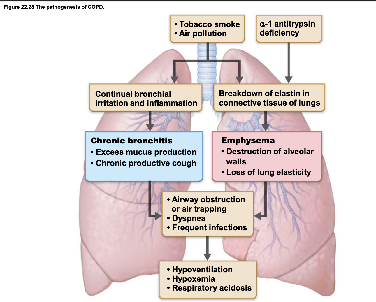
COPD is a chronic lung disease characterised by persistent airflow obstruction, making it difficult to exhale.
1. Emphysema
Destruction of alveolar walls and loss of lung elasticity.
Causes air trapping, hyperinflation and reduced gas exchange.
May result in a flattened diaphragm and increased breathing effort.
2. Chronic Bronchitis
Chronic airway inflammation and excessive mucus production.
Narrows and obstructs airways.
Causes persistent productive cough and frequent respiratory infections.
Common effects of COPD:
Dyspnoea (difficulty breathing).
Reduced expiratory airflow.
Impaired gas exchange.
Possible hypoxaemia and respiratory acidosis.
Key point: Emphysema primarily damages the alveoli, while chronic bronchitis primarily affects the bronchial airways.
What is asthma, and how does it affect airflow through the respiratory system?
Asthma is a chronic inflammatory airway condition characterised by variable, usually reversible airflow obstruction.
Main mechanisms:
Bronchoconstriction: Airway smooth muscle contracts, narrowing the bronchi.
Inflammation: Swelling of airway walls further restricts airflow.
Mucus production: Excess mucus contributes to airway obstruction.
Common symptoms:
Wheezing.
Shortness of breath (dyspnoea).
Coughing.
Chest tightness.
Key point: Asthma causes airway narrowing through bronchoconstriction, inflammation and mucus production, making breathing difficult.
Unlike COPD, asthma-related airflow obstruction is often reversible, either spontaneously or with treatment.