eye anatomy
 Call Kai
 Learn
 Practice Test
 Spaced Repetition
 Match
 Flashcards
 Knowt Play
1/82

There's no tags or description
Looks like no tags are added yet.
83 Terms
what are the 3 eye tunics (layers)
fibrous layer
vascular layer
retina
which layer is the fibrous layer?
outer layer
fibrous layer is made of what?
avascular connective tissue
what does the fibrous layer consist of?
sclera
cornea
cornea function
refracts light
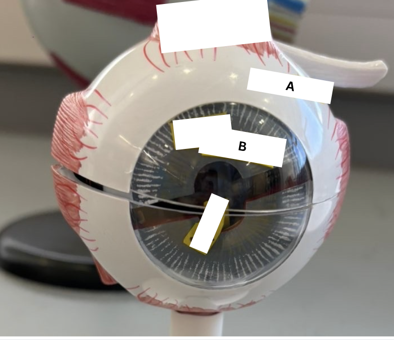
label the fibrous layer in humans (A-B)
sclera
cornea
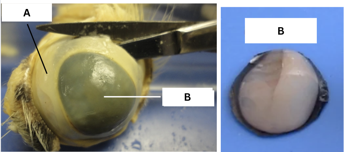
label the fibrous layer in cows (A-B)
sclera
cornea
which layer is the vascular layer?
middle layer
what does the vascular layer consist of?
choroid
tapetum lucidum (only in animals)
ciliary body
iris
pupil
choroid function
absorbs excess light and prevents it from scattering
what modification of the choroid is found in the cow eye but NOT in the human eye?
tapetum lucidum
what is the function of the tapetum lucidum
Reflects light within the eye.
ciliary body is made of what?
smooth muscle
ciliary body function
controls the shape of lens
iris is made of what?
smooth musucle
iris function
controls the size of the pupil and regulates the amount of light that enters the eye

label the vascular layer in humans (A,B,C)
choroid
ciliary body
iris

label the vascular layer in cows (A-E)
choroid
tapetum lucidum
iris
pupil
ciliary body
retina consists of what?
optic disc
macula lutea
fovea centralis
rods
cones
retina is what layer?
sensory layer
what does retina contain?
photoreceptors
what are photoreceptors?
rods and cones
what do photoreceptors do?
convert light energy into nerve impulses that result in vision
optic disc lacks what
photoreceptors
where is the retina attached?
optic disc
macula lutea is an area of what
high cone density
the _____ is a tiny pit in the center of the macula lutea which contains only cones; it is the area of greatest visual acuity
fovea centralis
fovea centralis contains
only cones
what is the area of greatest visual acuity?
fovea centralis
rods are specialized for what?
black, white, and gray vision
what conditions do rods function?
dim light
cones are specialized for what?
color vision
what conditions do cones function?
high light intensity
which photoreceptors permit high levels of visual acuity?
cones
which photoreceptors are most numerous at the periphery of the retina
rods
which photoreceptors are most numerous at the center of the retina?
cones

label the retina in a human eye (A,B,C)
macula lutea
optic disc
retina
what are the two layers of the retina?
pigmented and neural layers
6 skeletal muscles that control the movement of the eyeball; innervated by cranial nerves
oculomotor III
trochlear IV
abducens VI
optic nerve II function
carries sensory input towards the visual cortex of the brain
Lens functions
change shape to focus light on the retina
Aqueous humor function
watery fluid located in the anterior segment; provides nutrients to lens and cornea
what is aqueous humor produced by?
capillaries of the ciliary body

vitreous humor function
helps keep retina in palce
explain why vision is lost when light hits the blind spot of the retina
the blind spot lacks photoreceptors so it cant detect light
what are the refracting media/structures of the eye?
cornea
aqueous humor
lens
vitreous humor
which one of these structures can be varied by changing its shape/convexity to properly focus light on the retina?
lens
How was the blind spot tested, and what was the result?
cover one eye and focus on the x/dot on a piece of paper and pull it closer until the x/dot disappears
How was near point of accommodation tested?
Move a pin toward the eye until blurry then measure distance with ruler.
What is accommodation?
Ability to focus on close objects.
visual acuity
sharpness of vision
How was visual acuity tested
read snellen chart at 20 feet
if a person’s visual acuity test result is 20/20, it means that their vision is
normal
a person with a visual acuity test result of 20/10, their vision is ____ (better OR worse) than “normal” vision (visual acuity).
better
A person with a visual acuity test result of 20/40, their vision is _____ (better OR worse) than “normal” vision (visual acuity); they would be considered myopic.
worse

what is the chart shown below used to test for?
astigmatism
myopia
nearsighted
hyperopia
farsighted
astigmatism
blurred or distorted vision due to irregular curvatures of the lens
How was astigmatism tested?
look at lines on a chart, uneven darkness/blurriness indicated astigmatism
How was color blindness tested?
Ishihara color plates; identify numbers or patterns.
How was depth perception tested?
insert pencil into test tube with both eyes then one eye
what is the purpose of the depth perception test?
demonstrates binocular vision
what happens to the pupil when bright light is shined on the retina?
constricts
What is the function of the pupillary light reflex?
regulates the amount of light entering the eye
what is the effector of the pupillary eye reflex?
smooth muscle of iris
the smooth muscle of the iris is what?
an intrinsic eye muscle
is the pupillary light reflex somatic or autonomic?
autonomic
what happens to the pupil when a person stares at a distant object?
pupil dilates
What happens to the pupil when a person focuses on an object right in front of them?
pupil constricts
convergence
eyes turn inward
the ability of the eye to focus differentially for objects of close vision depends on what?
the elasticity of the lens
elasticity can be tested by measuring the near point of
accomidation
The near point of accommodation ____ (increases or decreases) with age because the elasticity of the lens _______ (increases or decreases) as we get older
increases
decreases
what is the name of the chart used to est visual acuity or sharpness of vision?
snellen eye chart
what is an ophthalmoscope used for?
examining inside of eye including retina and optic disc
which refracting structure can change its shape/convexity to focus light on the retina?
lens
what is refraction?
Bending of light rays as they pass from one medium to another
Is the pupillary light reflex somatic or autonomic?
autonomic
What muscle changes the shape of the lens during accommodation?
ciliary muscle
What muscle is the effector of the convergence reflex? Is the reflex somatic or autonomic?
medial rectus
somatic
based on the results of the lab activity where you tried to successfully insert a pencil in a test tube, is binocular vision important for depth perception?
yes, binocular vision provides depth perception