OTR 704 2.4 Elbow, Forearm, & Wrist
 Call Kai
 Learn
Learn Practice Test
 Spaced Repetition
 Match
Match Flashcards
 Knowt Play
1/149

There's no tags or description
Looks like no tags are added yet.
Name | Mastery | Learn | Test | Matching | Spaced | Call with Kai | Chat |
|---|
No analytics yet
Send a link to your students to track their progress
150 Terms

What structure is shown?
Humerus

What structure is shown?
Head of humerus

What structure is shown?
Greater tubercle

What structure is shown?
Lesser tubercle

What structure is shown?
Intertubercular groove

What structure is shown?
Deltoid tuberosity

What structure is shown?
Medial epicondyle
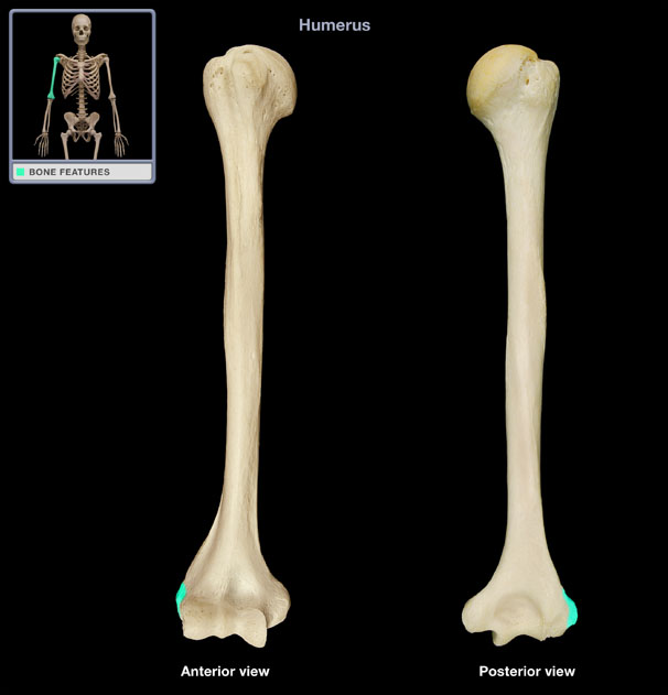
What structure is shown?
Lateral epicondyle
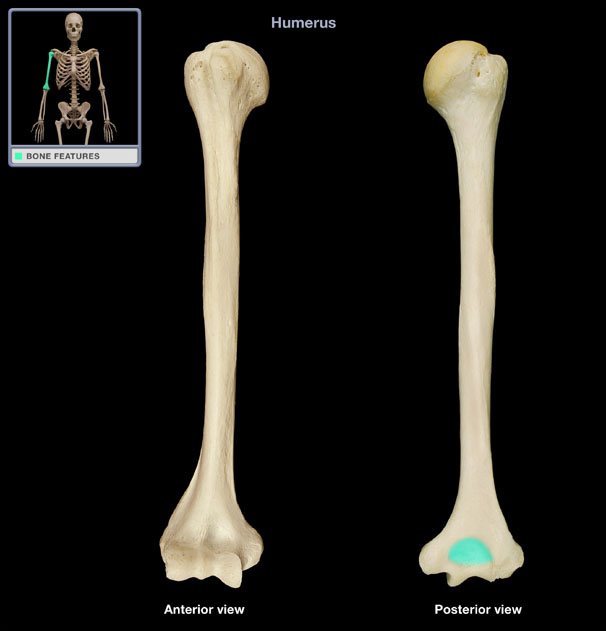
What structure is shown?
Olecranon fossa

What structure is shown?
Capitellum

What structure is shown?
Trochlea

What structure is shown?
Coronoid fossa

What structure is shown?
Ulna

What structure is shown?
Trochlear notch
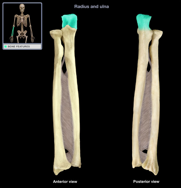
What structure is shown?
Olecranon process
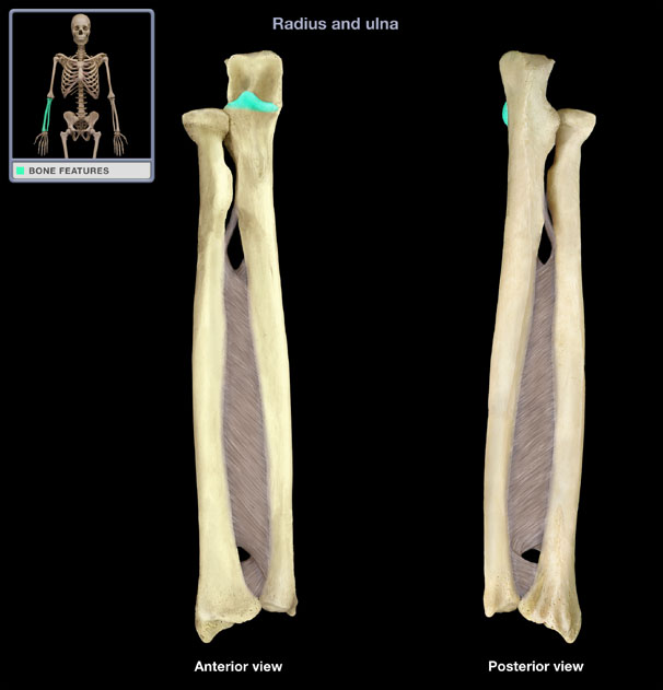
What structure is shown?
Coronoid process

What structure is shown?
Ulnar styloid process

What structure is shown?
Ulnar tuberosity

What structure is shown?
Radial notch

What structure is shown?
Radius

What structure is shown?
Head of the radius

What structure is shown?
Radial tuberosity

What structure is shown?
Radial styloid process

What structure is shown?
Carpals

What structure is shown?
Scaphoid

What structure is shown?
Lunate

What structure is shown?
Triquetrum

What structure is shown?
Pisiform

What structure is shown?
Trapezium
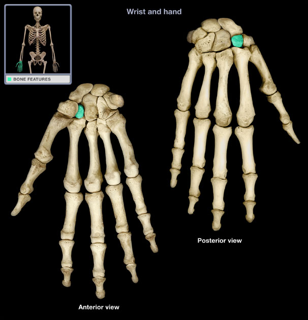
What structure is shown?
Trapezoid

What structure is shown?
Capitate

What structure is shown?
Hamate

What structure is shown?
Anatomical snuffbox

What structure is shown?
Carpal tunnel

What structure is shown?
Interosseus membrane

What structure is shown?
Triangular fibrocartilage complex (TFCC)

What structure is shown?
Extensor retinaculum
Ulna function
Primary articulation and stable base for:
Elbow flexion
Forearm pronation/supination

Interosseous membrane
Connects the radius and ulna; functionally:
Transmits force to the ulna
Protects humeroradial joint from excessive compression
Forms a fibrous “joint” that allows for functional forearm movements

Carrying angle
Natural valgus (lateral) deviation of the forearm relative to the humerus
More obvious in extension
Angulation of the articulating surfaces
Functional purpose: allows objects to be carried away from the body

Humeroulnar joint
Primary hinge joint of the elbow (trochlea → trochlear notch)
Uniaxial; allows for flexion & extension
Articulating surface & joint capsule allow for mobility
Ligamentous structures stabilize the joint (virtually no translation)

Humeroradial joint
Modified hinge joint (radial head moves about capitellum)
Technically biaxial; allows for flexion, extension, & rotation
Supports motion of the elbow
Less bony surface contact & congruency compared to humeroulnar joint
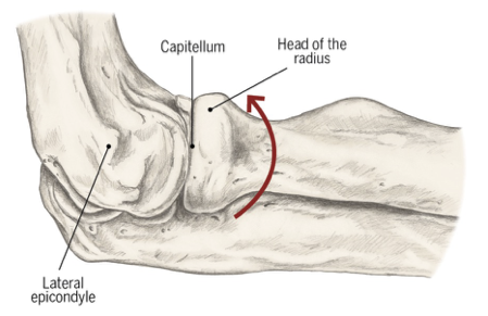
Proximal radioulnar joint
Pivot joint
Uniaxial; allows for pronation & supination
Supported by annular ligament

What three supporting ligaments stabilize the elbow?
Annular ligament
Ulnar collateral ligament (UCL)
Radial collateral ligament (RCL)
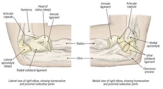
Annular ligament
Holds the radial head in place; prevents inferior displacement of the radius
Ulnar collateral ligament (UCL)
Prevents excessive valgus (lateral deviation) of the elbow
Repaired in Tommy John surgery
Radial collateral ligament (RCL)
Prevents excessive varus (medial deviation) of the elbow
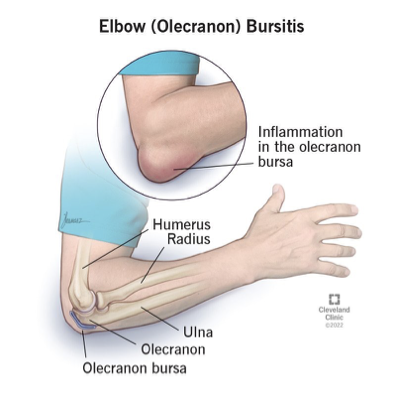
Bursae of the elbow
Sacs containing synovial fluid that help minimize friction in areas subject to stress; absorbs excessive forces that are applied to the elbow joint (e.g., higher-impact daily forces, trauma)
Seven bursa surround the elbow
Olecranon bursa is the largest
Olecranon bursitis is common from blunt force (“hitting”) the elbow
What muscles are the main flexors of the elbow?
The three B’s/three elbow benders
Biceps brachii
Brachialis
Brachioradialis
What muscles are the main extensors of the elbow?
Triceps brachii
Anconeus
What functional activity is extension of the elbow essential for?
Transfers (e.g., pushing up out of a chair)
(Scapular) depression transfers
Locked elbow joint allows scapular movements to compensate for a loss of active elbow extension to perform transfers

What are the primary rotators of the forearm?
Biceps brachii
Supinator
Pronator teres
Pronator quadratus
Biceps brachii action
Elbow flexion; forearm supination; shoulder flexion

Biceps brachii origin
Short head: coracoid process of scapula
Long head: supraglenoid tubercle of scapula
Biceps brachii insertion
Radial tuberosity & bicipital aponeurosis
Biceps brachii innervation
Musculocutaneous
Brachialis action
Elbow flexion

Brachialis origin
Distal ½ of anterior surface of humerus
Brachialis insertion
Ulnar tuberosity & coronoid process of ulna
Brachialis innervation
Musculocutaneous, small branch from radial
Brachioradialis action
Elbow flexion; assists forearm supination & pronation (when movements are resisted)

Brachioradialis origin
Proximal 2/3 of lateral supracondylar ridge of humerus
Brachioradialis insertion
Radial styloid
Brachioradialis innervation
Radial
Triceps brachii action
All heads: elbow extension
Long head: shoulder extension & adduction

Triceps brachii origin
Long head: infraglenoid tubercle of scapula
Lateral head: posterior surface of proximal ½ of humerus
Medial head: posterior surface of distal ½ of humerus
Triceps brachii insertion
Olecranon process of ulna
Triceps brachii innervation
Radial
Anconeus action
Elbow extension

Anconeus origin
Lateral epicondyle of humerus
Anconeus insertion
Olecranon process and posterior, proximal surface of ulna
Anconeus innervation
Radial
Supinator action
Forearm supination

Supinator origin
Lateral epicondyle of humerus, radial collateral ligament, annular ligament, and supinator crest of ulna
Supinator insertion
Anterior, lateral surface of proximal 1/3 of radial shaft
Supinator innervation
Radial
Pronator teres action
Forearm pronation; assists elbow flexion

Pronator teres origin
Common flexor tendon from medial epicondyle of humerus and coronoid process of ulna
Pronator teres insertion
Middle of lateral shaft of radius
Pronator teres innervation
Median
Pronator quadratus action
Forearm pronation

Pronator quadratus origin
Medial, anterior surface of distal ulna
Pronator quadratus insertion
Lateral, anterior surface of distal radius
Pronator quadratus innervation
Median
Cubital tunnel syndrome
Compression of ulnar nerve at the (medial) elbow as it passed through the cubital tunnel
Often results from overworking the wrist flexors and/or elbow extensors

Lateral epicondylitis
“Tennis elbow”; inflammatory condition related to overuse of the wrist extensors
Medial epicondylitis
“Golfer’s elbow”; inflammatory condition related to overuse of the wrist flexors
Distal radioulnar joint
Pivot joint
Uniaxial; contributes to pronation & supination
Supporting ligaments: palmar radioulnar ligament & dorsal radioulnar ligament

Radiocarpal joint
The “wrist”; an ellipsoid joint
Primary articulation between distal forearm and proximal carpals
Biaxial; allows for flexion/extension & radial/ulnar deviation

Intercarpal (midcarpal) joints
Gliding joints
Aggregated axis for wrist motion located near proximal capitate
All global wrist motions involve radiocarpal and midcarpal joints

Triangular Fibrocartilage Complex (TFCC)
Ligaments that cushion between the distal ulna & carpals
Shock absorber during weight-bearing
Stability on ulnar aspect of wrist

Where do the extrinsic wrist flexors orginate?
The medial epicondyle (minus pollicis longus)
What are the extrinsic wrist flexors?
Flexor carpi radialis
Flexor carpi ulnaris
Palmaris longus
Flexor digitorum superficialis & profundus
Flexor pollicis longus
Where do the extrinsic wrist extensors originate?
The lateral epicondyle
What are the extrinsic wrist extensors?
Extensor carpi radialis longus & brevis
Extensor carpi ulnaris
Extensor digitorum
Extensor indicis
Extensor digiti minimi
Abductor pollicis longus
Extensor pollicus longus & brevis
Flexor carpi radialis action
Wrist flexion & radial deviation; assists in elbow flexion

Flexor carpi radialis origin
Common flexor tendon from medial epicondyle of humerus
Flexor carpi radialis insertion
Bases of 2nd & 3rd metacarpals
Flexor carpi radialis innervation
Median