MCDB 165A Cell Adhesion Flashcards
 Call Kai
 Learn
 Practice Test
 Spaced Repetition
 Match
 Flashcards
 Knowt Play
1/49

There's no tags or description
Looks like no tags are added yet.
50 Terms
Cell Adhesion Complexes:
Tight Junctions, Adherens Junctions, Desmosomes, and GAP Junctions.
Junctions
Specialized molecular structures that enable cell adhesion.
Complexes often found throughout the interfaces of epithelial cells and have different functions. These junctions are polarized (apical/basal localization).
3 Categories of Skin Blisters:
Compromised junctions have many potential consequences, and one is blistering:
1) Dystrophic- tearing of the dermis (often due to defects in collagen)
2) Junctional- tearing at the dermal/epidermal surface (defects in hemidesmosomes causing a tear between the epithelial layers and the basement membrane below).
3) Simplex- tearing between epidermal cells.
Blisters- One individual had a defect in a component of the…
desmosome junction. They are prone to frequent blistering and the epidermis is ripped from the dermis.
Mouse models with the same mutated gene also have easily blistered skin (due to rubbing from birth canal).
Understanding junctions is important for understanding…
the integrity of our organs. We will discuss four cell-cell attachments.
Tight Junctions
Where two cells are tightly adhered to one another. Bilayers of two cells come close together and almost fuse (called kissing points).
Kissing points are where there are tight junction adhesive structures. In an SEM of an epithelial cell interface, you can see a maze-like network of raised structures that form kissing points.
Tight Junction Structure- Two Main Molecular Components
Occludins and Claudins. The are multi-pass transmembrane proteins that have extracellular loops for homophilic adhesion (e.x claudin on one cell interacts with claudin on another cell).
Intracellularly, occludins and claudins are attached to a complex of proteins called the ZO proteins (Tjp proteins). This connects occludins/claudins to the actin cytoskeleton.
Tight junctions prevent lateral diffusion between…
the apical and dorsolateral parts of a cell.
Epithelial cells have proteins exclusive to the apical domain and exclusive to the basolateral domain (basal and lateral sides). Tight junctions separate these proteins on their respective sides (so tight that lateral diffusion cannot occur).
The AQP4 and F-Mal domains meet at…
the tight junctions (if they were destroyed these two proteins would have the potential to intermix.)
Tight Junctions also create a…
permeability barrier (prevents molecules from the outside of the epithelium from passing through the epithelium, or a molecule from inside the epithelium passing through the outside).
An example of the permeability barrier…
Took zebrafish embryos and did multiple morpholino knockdowns of the tight junction protein ZO3. You can see with EM that the junctions are compromised.
They then put biotin outside and stained it. When the fish were injected with morpholinos, the dye has gotten through the epithelium into the animal.
Permeability fish death experiment (continuation of last one):
Put a toxin CdCl2 outside the fish that will kill it if it gets inside. Control animals do a pretty good job of surviving in a lower concentration, but tjp3/ZO2 defects have more animal death at lower concentration of the toxin.
Adherens Junctions
Sit below tight junctions. Attached to the actin cytoskeleton, which forms a belt and helps maintain the integrity of the cell.
Main molecule used for adhesion in Adherens Junctions:
Cadherin. Cadherins come from one cell and interact with a cadherin coming from a neighboring cell.
Cadherin Interactions
The cytoplasmic portion of the cadherins interact with a complex of proteins (alpha/beta catenins and ZO1), which connect cadherin to the actin cytoskeleton belt.
One function of of these cadherins is regulating…
proliferation of a cell. The Hippo pathway (or MST1/MST2) is a critical regulator of cell proliferation and misregulation can cause epithelial cells to overproliferate (tumors and cancer).
When a cell is not forming cadherin-based adherens junctions, a transcription factor called YAP can go into the nucleus and activate transcription of cell proliferation/survival.
Once an adherens junction forms, it activates kinases called MST/Hippo kinases. These kinases then activate kinases called LATS kinases, which phosphorylate YAP and prevent the transcription factor from going into the nucleus and thus prevent proliferation.
Contact Inhibition
Cells form a monolayer in culture. YAP pathway partially explains this phenomenon (cells proliferate and form adhesions, resulting in adhesion junctions that prevent YAP activity).
Potential Issues with Hippo Pathway:
In SEMs of flies with a defect in the Hippo pathway, you can see it they are full of tumors and looked like a hippo.
When the hippo pathway is blocked, the adherens junctions are still there, but the cell can no longer respond to the presence of the adherens junctions and stop growth.
Desmosomes
Can be anywhere along the basal (if a cell is underneath) or lateral membrane.
Use a different kind of cadherin-family molecule than adherens junctions (desmogleins and desmocollins).
Demosomes- Strucutre
Desmogleins and desmocollins are transmembrane molecules that interact with the molecule on the opposite cell. Intracellularly, they interact with many intermediary proteins (plakin proteins) that are very dense. These intermediary proteins connect the keratin intermediate filaments to the cadherins.
Desmosome vs. Hemidesmosome
Desmosomes are cell-cell junctions that connect two cells in the epithelium (uses cadherins). On the other hand, a hemidesmosome is a structure that connects the epithelium to the basement membrane (uses integrins).
But, both of these structures have these plaque-like density of proteins that connect molecules to the intermediate keratin filament cytoskeleton.
Desmosomes are the most important of these junctions for maintaining…
the structural integrity inside the epithelium. Defects in keratin filament proteins result in fragile cells and a simplex blister (tear through the middle of the epithelium).
GAP Junctions
Junction not thought of as being involved in adhesion, but rather cell communication.
GAP Junctions Structure
Made from genes called connexins that assemble into a hexomer (six) called a connexon. These connexins are transmembrane proteins, and they form a pore in the middle of the hexomer (allows small molecules to pass).
Hexomers in the lipid bilayer of one cell line up with a hexomer in a lipid bilayer of another cell, forming a transcellular dodecameric complex (creates a continous pore connecting the cytoplasms of both cells).
When two connexons from two different cells line up, it is called a…
GAP Junction.
There are dozens of different connexins encoded in the mammalian genome, and can assemble into…
a homomeric connexon (all 6 subunits are the same protein) or a heteromeric connexon (different subunits encoded by different genes, where the subunits are similar in structure but different in sequence, giving these connexons different properties).
These can lead to homotypic (when two cells interact, all 12 subunits are the same in the hexomers) or heterotypic (different or a mix of heteromeric subunits) interactions.
This diversity in GAP junctions gives them different properties. GAP junctions can be selective for…
size, charge, and and potentially other properties. For instance, GAP junctions can be selective through their pore size. Due to GAP junction diversity, you can have some that are specialized to allow different molecules through (ions, second messengers, metabolic molecules).
Heterotypic (the two cells have molecularly distinct connexons) GAP junctions can be…
polarized, meaning that ions that pass through the channel from neuron to neuron do so preferentially. So, these junctions are versatile, regulatable channels for direct cell communication.
Cadherin Facts
Component of adherens junctions and desmosomes. Cadherins are a family of trandmembrane glycoproteins (roughly 30) that mediate homophilic adhesion.
Different kinds of cells express different…
cadherins (E-Cadherins, N-Cadherins, etc.). Cadherins can keep adhesion between different cell types distinct.
Cadherin Strucutre
Contains 5 repeat domains on the extracellular side, aptly called cadherin repeats. The structure/activity of cadherins are regulated by calcium ions in the environment (high calcium —> rigid cadherin structure, low calcium —> rigid cadherin structure falls apart, preventing cadherins from forming an adhesion).
Integrins also require divalent cations for their rigid/active conformation. So, you can regulate cadherins and integrins at the same time by manipulating…
the concentration of calcium in the environment. In cell culture, you could disrupt adherence with each other and the substrate by lowering calcium concentration (e.x through calcium chelators).
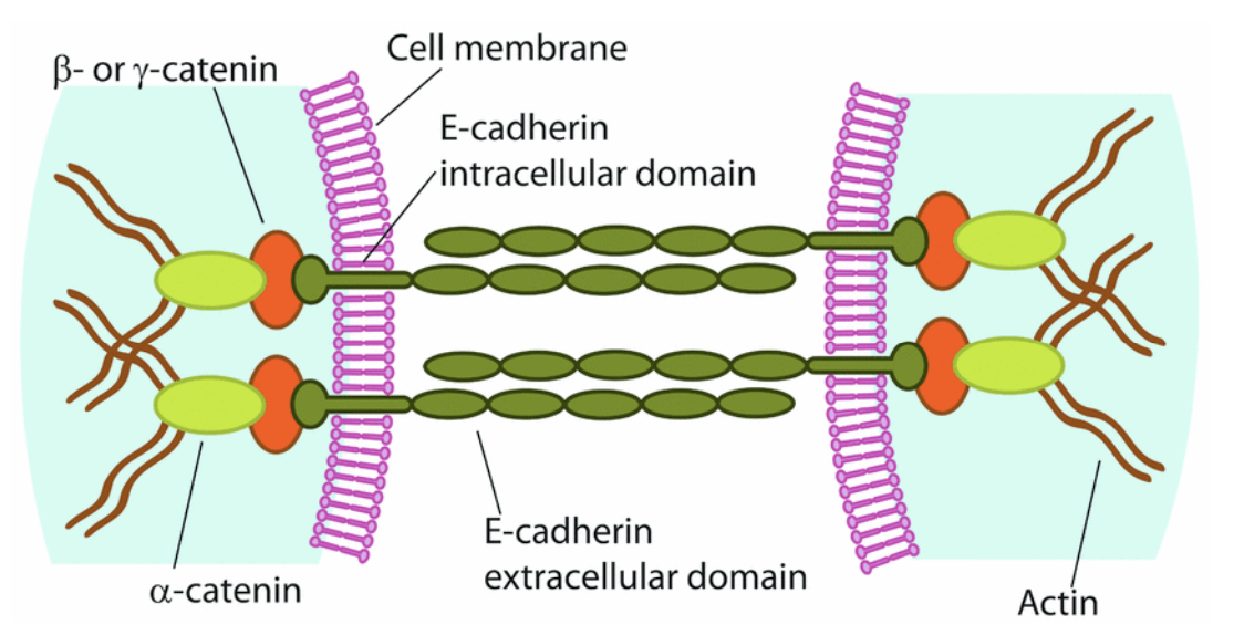
What two ways is this cartoon slightly inaccurate?
1) You only see pairs of cadherins interacting with each other. But, in addition to interacting with those on the opposite cell, cadherin molecules often interact with those on the same cell to form a cluster.
2) This cartoon implies all 5 cadherin repeats are being used for adhesion to the opposite cadherin, but, only the distal most cadherin repeat often interacts.
What technique did a group perform to characterize cadherin-based adhesions strucutre?
PALM by doing immunofluorescence against each of the cadherin-based junction components.
Initiation of Adhesion by Cadherins- Two hypotheses for how these adhesions form.
1) Two cells happen to be near each other (passive adhesion) or 2) is there an active process for cells to adhere to each other.
GFP Tagged Cadherin Experiment Results
As soon as two cell meet, a bright spot of cadherin fluorescence at the adhesion point. This adhesion point grows until you get a solid interface containing cadherin.
This process required actin polymerization. Cadherin initiates sites of contact that spread to create a tight interface; actin is required for this process.
Second Cadherin Experiment- Immunofluorescence Against Actin and Cadherins in Primary Keratinocytes
At 0 hours, you see no localization of cadherin but actin is present.
At 3 hours, cadherin begins to localize at cell-cell interfaces (spots at the interface). With more resolution, you see the cell seems to be pushing its membrane out, creating finger-like protrusions called filopodia that are filled with actin. At the tip of each of these protrusions is a spot of cadherin.
So, cells are sending filopodia out that are interacting with cadherin in a different neighboring cell that is also sending flipopodia.
At 7 hours, you still see spots, but the interface between the cells is more complete and grown (transforming into a solid band).
In the cadherin experiment above, this process reminded the authors of zippers, so they named these structures…
adhesion-zippers. In primary keratinocyte cultures, “a zipper” initiated by membrane protrusions tipped with cadherin mediate initial attachment.
Subsequent experiment testing if fillopodia are required for initial adhesion:
Treated cells with cytochalasin D drug that prevents actin polymerization. At 7 hours, the cells have not stuck to each other (no prominent spots of cadherin at the cell-cell interface). After 20 hours, the cells have proliferated so much that you get some cadherin localization at the cell-cell interfaces.
This was interpreted as a passive adhesion, compared to where the cells normally try to actively adhere to one another through fillopodia.
You need both
cadherin interactions and actin polymerization to form these initial adhesions.
Not all cadherins are the same, and that provides cells with…
the ability to sort.
Homophilic vs. Heterophilic Interactions
Homophilic- when a molecule on one cell interacts with the same kind of molecule on another cell (cadherin binding, there aredozens of cadherins, but all of them are homophilic. A particular kind of cadherin only likes to interact with another cadherin of the same type).
Heterophilic- when a molecule on one cell interacts with a different receptor on another cell (“Bride of Sevenless” (Ligand) and Sevenless (RTK)).
Classic Cell Sorting Experiment- Cadherins
Two populations of cells: one expressed one kind of cadherin (e-cadherin) + blue fluorophore and the other a different kind of cadherin (n-cadherin) + RFP.
If you allow the populations to mix and interact in culture, you’ll find the the red cells have stuck together and the blue cells will stick together, forming two distinct clumps of cells. This suggests cadherins mainly undergo homophilic binding, not heterophilic binding.
Two populations of cells both expressing e-cadherin, but different levels of e-cadherin experiment:
All the cells interact, but rhe cells with the highest levels of e-cadherin perform to interact with each other (forming the core of the clump).
More results from the cell adhesion assay (sorting); if we have e-cadherin/n-cadherin and caderhin-6b, the cells perfectly sort out into red and green clumps, driven by…
homophilic adhesion. On the other hand, e-cadherins and n-cadherins can weakly bind with each other, as the clumps join into a bigger clump.
Overall, this assay can be used to test if molecules interact homophilically and how specific this interaction is.
Challenge of sorting of neurons in the brain during development:
Fo our brain to make circuits and coherently function, it must wire neurons with exquisite specificity. Cadherins play a major role in this selective cell interactions.
Hippocampus neuron connection experiment, background and goal:
Hippocampus is organized with DG neurons that send axons to CA3 neurons to make synapses. CA1 neurons are farther along the hippocampus.
They wanted to show there was a molecular mechanism that allowed for an interaction between DG and CA3, not an anatomical interaction (maybe the DG axons grow in a certain direction and synapse with whatever cells are in their target location).
Hippocampus Neuron Connection Experiment, Methods and Results
They dissociated the structure, mixed it up, and saw if they have a selective interaction. They performed a micro-island assay (collagen islands on a plate that neurons grown/survive on).
DG, CA3, and CA1 were scattered on the plate, so their organization was not normal. They then did immunofluorescence for proteins only expressed in DG, CA3, and CA1. In DG neurons, they expressed a synaptic protein syn-GFP (so dots indicate where DG is interacting with other cells on the island).
Specificity was maintained even though anatomy was destroyed. There are few dots on the CA1 neurons (even though DG is in contact), and lots of dots on the CA3 neurons (DG is making contact).
Molecular Mechanism Hypothesis Behind the Hippocampus Neural Connection Experiment
The authors looked for cell adhesion molecules in these different populations and found cadherin 9, which is expressed in DG and CA3 neurons.
They stained the mRNA of this cadherin, so you can see that cadherin 9 is in both the DG and CA3 cells, but not CA1.
This raised the question: cadherins are involved in homophilic adhesion, and this cadherin molecule is produced in these two interacting cell types, could cadherin 9 mediate the interaction between them?
Molecular Mechanism Experiment for Hippocampus Neural Connection
The authors did an adhesion assay (that we previously discussed) to confirm cadherin 9 undergoes homophilic adhesion.
They used RNAi to knock down cadherin 9 (knocked down only in DG neurons, or only CA3 neurons, or both), you see no yellow dots compared to the control, meaning DG is not making contacts with CA3. If cadherin 9 was reinserted into these mutants, these contacts were restored (rescue).