MCB 2000 Chapter 16
 Call Kai
 Learn
 Practice Test
 Spaced Repetition
 Match
 Flashcards
 Knowt Play
1/47

There's no tags or description
Looks like no tags are added yet.
48 Terms
what is glycolysis
glucose to pyruvate, in the cytoplasm, with or without oxygen
glucose broken in two:
glycolysis takes one 6-carbon glucose and converts it into two 3-C pyruvates
it runs in the cytoplasm of every cell that has one — no organelle required
it works with oxygen present or absent, which is why it predates aerobic respiration in the evolutionary record
if oxygen is present, pyruvate can be sent to aerobic metabolism
if oxygen not present, glycolysis keeps going through fermentation

big picture of glycolysis
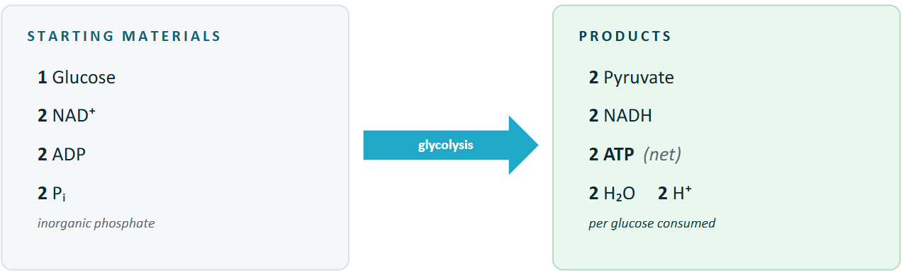
two phases
glycolysis spends ATP before it earns ATP
phase 1: energy investment
steps 1 through 5
spends 2 ATP
the cell pays up front to phosphorylate glucose and prime the molecule for splitting
the phosphorylations trap glucose inside the cell and make the six-carbon molecule easier to break
phase 2: energy payoff
steps 6 through 10
earns 4 ATP + 2 NADH
both 3-C halves are processed in parallel, each yielding 2 ATP and 1 NADH
net gain: 2 ATP per glucose
phase 1
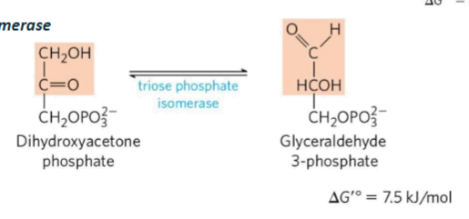
GAP continues directly into phase 2 but DHAP gets rapidly converted into GAP through triose phosphate isomerase
one glucose has become two GAP molecules, each three carbons and the cell is down 2 ATP

phase 2
starts with GAP and ends with pyruvate
oxidation of GAP makes NADH
first ATP made when high-energy phosphate is transferred to ADP, and the second in the final step when PEP donates its phosphate to ADP
both halves run in parallel. the x2 amplifier
phase 2 total: 4 ATP + 2 NADH (2 from each 3-C track)

the ledger
gross yield minus the up-front investment
net per glucose: 2 ATP +2 NADH. modest, but glycolysis only takes seconds and runs anywhere, even in cells without oxygen

first 5 steps; phase 1
ATP spent (steps 1 and 3)
3 reversible rearrangements
end with 2 GAPs ready for phase 2
step 1: irreversible regulatory step: ATP spent when phosphorylating glucose to G-6-P by hexokinase
step 2: phosphoglucose isomerase: G-6-P → F-6-P
step 3: irreversible regulatory step: ATP spent by phosphofructokinase-1 (PFK1): F6P → F-1,6-BP
step 4: aldose: splits the six-carbon molecule into DHAP and GAP
step 5: triose phosphate isomerase-catalyzed: DHAP → GAP so by the end of the pathway, 2 GAP molecules are produced

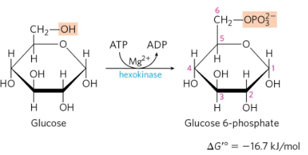
step 1
hexokinase: induced fit
glucose binds first — the enzyme closes around it and positions ATP
closure positions ATP next to glucose C6 and shuts out water, so ATP is not wasted by hydrolysis
the enzyme is trying to transfer the terminal phosphate to glucose rather than hydrolyze ATP wastefully
produces G-6-P + ADP — the phosphate gives the G-6-P a negative charge, helping to trap it in the cell and mark it for metabolism by that cell
trapped, and always primed
G6P’s negative charge prevents leaving.
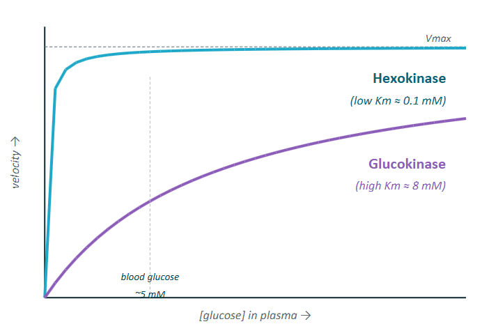
Km ≈ 0.1 mM vs intracellular glucose 1 to 5 mM means hexokinase is saturated
once glucose enters the cell, hexokinase is ready to phosphorylate it, marking it for use very quickly
induced fit = safety mechanism. no glucose, no closed site, no wasted ATP

steps 2
isomerization
phosphoglucose isomerase
G6P → F6P (aldose to ketose)
constitutional isomers, same formula, different connectivity.
no ATP cost, freely reversible
structural reason: fructose form sets up the molecule for the later cleavage into two 3-C pieces

step 3
committed step
control point
phosphofructokinase-1 (PFK-1)
F6P → F1,6BP w/ ATP
rate limiting and committed
once PFK-1 adds the second phosphate, the cell has no use for the molecule except glycolysis
G6P still has options → glycolysis, glycogen metabolism, or pentose phosphate pathway
PFK-1 is the most regulated enzyme in glycolysis

step 4
cleaves F1,6BP in half
aldose — cleaves between C3 and C4 to produce DHAP and GAP

step 5
coverts DHAP to GAP
triose phosphate isomerase
result: 2 GAP enter phase 2
Le Chatelier: as phase 2 consumes GAP, TPI pulls more DHAP across even through the mixture actually favors DHAP
effectively, both halves run
from this point on, every reaction happens twice per glucose (because 2 GAP produced)
steps 6-10: phase 2
step 6: GAPDH (glyceraldehyde 3-phosphate dehydrogenase): NAD+ reduced to NADH
step 7: phosphoglycerate kinase: makes first ATP of phase 2 by substrate level phosphorylation
step 8: phosphoglycerate mutase: moves the phosphate from C-3 onto C-2
step 9: enolase: removes water and creates PEP (phosphoenolpyruvate) which contains a very high energy phosphate donor
step 10: irreversible regulatory step: pyruvate kinase: PEP donated its phosphate to ADP to make the second ATP and pyruvate

step 6
GAPDH does two things at once: oxidizes the aldehyde and phosphorylates it in one coupled reaction
oxidizes the aldehyde group of GAP. the electrons removed from GAP are transferred to NAD+ making NADH
the chemistry:
GAP’s aldehyde is oxidized to a carboxyl-like group
Pi from solution is added at the same time, trapping the energy released by the oxidation as an acyl phosphate bond
the electrons stripped off GAP go onto NAD+, making NADH
at the same time, the dehydrogenase attaches inorganic phosphate from solution to oxidized carbon, making 1,3-bisphosphoglycerate
important feature: acyl phosphate at carbon one because acyl phosphates have a high phosphoryl-transfer potential and hydrolyzing this bond releases more free energy than it does hydrolyzing ATP
why it matters:
1,30BPG is a high-energy intermediate. its phosphate is set up to be donated to ADP in step 7, making ATP
without the energy stored here, step 7 couldn’t make ATP at all
this substrate-level phosphorylation: ATP made directly from a high-energy substrate, not from the electron transport chain
GAPDH couples oxidation to phosphorylation. it pre loads the energy that step 7 will collect as ATP

step 7
phosphoglycerate kinase
PGK transfers the high-energy phosphate from 1,3-BPG onto ADP, making ATP and the 3-phosphoglycerate carbon product
this is the first step that glycolysis produces

step 8
phosphoglycerate mutase
the phosphate moves from carbon 3 to carbon 2 to make 2-phosphoglycerate
this is a setup move: 2-PG is the right substrate for enolase, the next enzyme

step 9
enolase removes a water
the resulting phosphoenolpyruvate (PEP) holds an enol phosphate, one of the highest-energy phosphate bonds
PEP= high phosphoryl- transfer potential because after phosphate transfers, the enol form of pyruvate can tautomerize to the more stable keto form
by the end of this step, the substrate that is going to drive the final ATP producing step is created

step 10
PEP donates its phosphate. ATP out. Pyruvate locked in
why irreversible:
PEP releases ~62 kJ/mol when it loses its phosphate. only ~31 kJ/mol is needed to make ATP from ADP
pyruvate is stabilized by tautomerization
the extra energy is released as heat, locking the reaction one-way
the third regulatory step:
like hexokinase and PFK-1, pyruvate kinase is irreversible.
its regulated allosterically and hormonally, in tissue-specific ways

total irreversible steps:
hexokinase (step 1)
PFK-1 (step 3)
pyruvate kinase (step 10)
these are the regulatory checkpoints
where regulation lives
the three irreversible steps from module 2 are the regulatory checkpoints
muscle: senses energy; ATP/AMP at PFK-1. glycolysis runs when energy is low
ATP needed for contraction and often needed immediately
regulation is built around energy charge, so presence of ATP, ADP and AMP
liver: senses fuel; glucokinase +F-2, 6-BP. glycolysis runs when glucose is plentiful
managed blood glucose for the entire body — after a meal, takes it glucose and processes it but during fasting, it should preserve or release glucose instead of burning it itself
the same three enzymes regulate in both tissues. the signals they listen to are tissue-specific
muscle mainly listens to its own energy state
liver listens to glucose availability and whole-body hormonal cues

muscle strategy
muscle regulatory control centers rely on PFK-1 (glycolysis step 3), so PFK-1 is inhibited by ATP — if ATP is abundant, the cell doesn’t need to burn glucose rapidly for more ATP. PFK-1 is also activated by AMP — rises sharply when ATP is consumed faster than it is being regenerated
opposite signals, exquisite sensitivity
ATP inhibits PFK-1; AMP relives inhibition and activates glycolysis when energy is low
the trick: one enzyme, opposite signal
PFK-1 integrates ATP and AMP through allosteric regulation
high ATP favors a low-activity state; rising AMP shifts the enzyme toward high activity
why the ratio matters:
ATP is abundant at rest, AMP = low
AMP is a sensitive low-energy signal. cellular ATP greatly exceeds AMP at rest, so even a small ATP drop can produce a large relative AMP increase and rapidly disinhibit PFK-1
through adenylate kinase reaction — makes AMP an amplified signal
energy charge falls → AMP rises → PFK-1 turns on → glycolysis surges

muscle: rest vs exercise
muscle PFK-1 flips between off and on as ATP/ AMP swings
this is fast, local, no hormones involved
at rest, ATP= plentiful and AMP low
ATP inhibits PFK-1, so step 3 slows
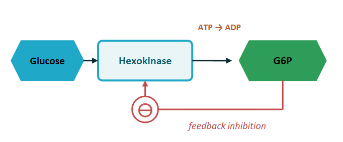
when downstream flux is slow, G6P starts to accumulate, which will feedback inhibit hexokinase (the enzyme that produces it)
during exercise, ATP is being used rapidly, so AMP rises and PFK-1 becomes more active
more F6BP produced and upstream product will also activate pyruvate kinase
that feed-forward activation helps the end of the glycolysis pathway keep pace with the beginning

hexokinase feedback
G6P inhibits its own production
if the cell doesn’t need more G6P, hexokinase backs off
depends on what happens downstream at PFK-1
at rest: PFK-1 is slow → G6P piles up → G6P inhibits HK → glucose entry stalls
during exercise: PFK-1 is fast → G6P stays low → HK is freed → glucose pours in
PFK-1 sets the pace. hexokinase follows along through the G6P feedback loop
prevents the cell from wasting ATP on glucose phosphorylation when the rest of glycolysis isn’t ready to use the product
liver strategy (VIDEO 3 AT 6:35)
the liver listens to glucose, not ATP
its job is to buffer the body’s blood sugar, not to make its own ATP
muscle (recap) : signal= energy charge
ATP inhibits PFK-1; AMP relieves it
HK has low Km, saturated at normal glucose
glycolysis runs when the cell needs ATP
goal: keep ATP up
liver: signal= glucose abundance
different step 1 enzyme: glucokinase (HK IV)
high km → activity rises with blood glucose
PFK-1 controlled by F-2,6-BP (hormone-linked)
citrate inhibits PFK-1 here too (TCA fuel signal)
goal: keep ATP up
liver jobs:
manage blood glucose for the entire body
after a meal: blood glucose and insulin levels begin to rise so liver should take up glucose, burn some and store some as glycogen and covert excess carbon into fat
during fasting, glucagon levels rise and liver should stop burning glucose, break down glycogen, make glucose by gluconeogenesis and release glucose into the blood stream for the rest of the body to use
should be able to respond to glucose abundance and hormones more so than local ATP demand — done by glucokinase (step 1) and fructose 2,6 -bisphosphate (regulates PFK-1)
same pathway, different mandate. muscle works for itself; liver works for the body
liver step 1
uses glucokinase (instead of hexokinase like muscles) (also sometimes called hexokinase IV)
glucokinase has a different Km (around 8 mM)
high Km means glucokinase activity rises with blood glucose across the normal range
not saturated at normal blood glucose levels so at around 5 mM glucose, glucokinase is active, but still has room to increase
what this means:
at normal blood glucose (~5mM):
hexokinase is already saturated. muscle uses glucose freely
glucokinase is only at about 40% of Vmax. liver pulls in glucose gradually
after a meal (~10mM)
glucokinase rises close to Vmax
liver glucose uptake accelerates exactly when blood glucose is high. that’s the buffering role
higher Km means liver glucose uptake tracks blood glucose, instead of always running flat-out
liver PFK-1
different regulators, different signals
F-2,6-BP is the dominant activator. citrate is the brake
new in liver compared to muscle: F-2,6-BP, a hormone-controlled metabolite
citrate, the downstream fuel signal, brakes PFK-1 in both tissues
liver PFK-1 is especially shaped by F-2,6-BP, on top of ATP/ AMP and citrate

three hormones, three states
Insulin (fed state)
glucagon (fasted state)
epinephrine (stress/ emergency)
the body choses on hormonal state at a time. each one routes through PFK-2 and pyruvate kinase in the liver
Insulin
fed state — after a meal
“burn the incoming glucose. store the surplus”
liver glycolysis: ON
liner glycogen synthase: ON
muscle glucose uptake: ON (GLUT4)
glucagon
fasted state — blood glucose dropping
“make and release glucose. Don’t burn it”
liver glycolysis: OFF
liver gluconeogenesis: ON
muscle: no response (no receptor)
epinephrine
stress/ emergency — fight, flight, sprint
“mobilize glucose to muscle, fast”
liver glycolysis: OFF (save glucose)
liver glycogenolysis: ON
muscle glycogenolysis: ON
the F-2,6-BP switch
one enzyme, two activities. a phosphate flips it
PFK-2 builds F-2,6-BP from F6P
FBPase-2 tears it down —F -2,6 -BP → F6P
same protein, different states
phosphorylation status of one enzyme determines whether F-2,6-BP is being built or destroyed
when liver enzyme is dephosphorylated, PFK-2 activity dominates (F-2,6-BP levels rise and PFK-1 is activated)
when the enzyme is phosphorylated, FBPase-2 activity dominates (F-2,6-BP levels fall and PFK-1 loses its strongest activator)

insulin in the liver
dephosphorylate everything, burn the glucose
insulin signals fed state. the liver shifts towards glycolysis
result: liver glycolysis is fully turned on. two of the three regulatory enzymes (PFK-1 and PK) are activated together
insulin → dephosphorylation → PFK-1 on + PK on → liver burns the incoming glucose
turns on step 3 and 10

glucagon in the liver
phosphorylate everything, save the glucose
glucagon signals fasted state. the liver shuts glycolysis down to release glucose to the body
result: liver glycolysis is shut down. the liver now releases glucose (gluconeogenesis and glycogenolysis run instead)
glucagon → phosphorylation → PFK-1 off + PK off → liver makes and releases glucose, doesn’t burn it
cAMP levels rising by the GPCR receptor signaling pathways
shuts down steps 3 and 10

muscle’s different game: same hormone, different receivers
muscle has no glucagon receptor. insulin and epinephrine do different things
muscle glycolysis is mostly allosteric. hormones in muscle act on transporters and glycogen, not on PFK-1 directly
in liver, PKA can inhibit pyruvate kinase by phosphorylation
skeletal muscle expresses a different pyruvate kinase isozyme that is not regulated in the same way so epinephrine mobilizes fuel in muscle without shutting off the glycolytic endpoint
big idea
muscle hormones control access to fuel
local ATP and AMP largely control the glycolytic enzyme activity
insulin response
GLUT4 to the surface
insulin doesn’t directly hit PFK-1 or pyruvate kinase in muscle
instead, it triggers translocation of GLUT4 glucose transporters to the muscle cell membrane
more glucose enters, allosteric regulation handles the rest
glucagon response
no response
muscle cells lack a functional glucagon receptor used for glucose metabolism
glucagon circulates past the muscle without binding
glucagon is liver-specific in its effects on glucose metabolism
epinephrine response
glycogenolysis
epinephrine binds muscle β-adrenergic receptors and activates PKA, which mobilizes muscle glycogen
glucose-1-P enters glycolysis after conversion to G6P
glycolysis floods with fuel; ATP gets made fast
muscle summary
sense self
role: make ATP for contraction
primary signal: energy charge (ATP/AMP) at PFK-1
hexokinase: low Km, saturated at normal glucose
insulin moves GLUT4 to membrane
epinephrine: drives glycogen breakdown
glucagon: no receptor, no response
liver summary
sense the body
role: buffer blood glucose
primary signal: blood glucose via glucokinase (high Km)
F-2,2-BP hub: PFK-2 phosphorylation routes hormones in
insulin: dephosphorylates, glycolysis ON
glucagon/ epinephrine: phosphorylates, glycolysis OFF
net result: liver releases glucose when fasted
pyruvate has two roads
same starting pyruvate, two destinations. oxygen makes a 15-fold difference in ATP yield

why fermentation exists
the cell runs out of NAD+ first
GAPDH needs NAD+ at step 6 of glycolysis. without it, the pathway stalls
it oxidizes GAP and reduces NAD+ to NADH
the problem: the NAD+ pool is small, two GAPDH turns per glucose drain it fast. without reoxidation, GAPDH stalls and glycolysis stops
aerobic solution: shuttle NADH electrons into mitochondria. the ETC oxidizes them and regenerates NAD+
fermentation solution: uses pyruvates itself as the electron acceptor. reducing it consumes NADH and regenerates NAD+

lactate fermentation
one step, one enzyme
lactate dehydrogenase reduces pyruvate to lactate, regenerating NAD+
per glucose, 2 pyruvates are reduced to 2 lactates and 2 NADH are converted back to 2 NAD+ so GADPH keeps running
sprinting muscle
temporary anaerobia
during intense effort, ATP demand outruns oxygen supply
muscle fibers shift to lactate fermentation, producing ATP fast while lactate builds up
once oxygen returns, the lactate is shuttled to the live via the bloodstream (Cor cycle)
red blood cells
permanent anaerobia
RBCs lack mitochondria entirely (no room and they shouldn’t consume the oxygen they are delivering)
glycolysis + lactate fermentation is their only ATP source, 24/7
the lactate is released to the blood for the liver to recycle
one enzyme. one step. the pyruvate becomes the dump site for the NADH electrons

ethanol fermentation
two steps, yeast’s solution
decarboxylate first, then reduce. same NAD+ regeneration, different product
the chemistry:
step one strips off CO2 as a gas. this is irreversible, uses no NAD+ and is not a redox step
uses thiamine pyrophosphate as a cofactor
step two is the actual reduction: NADH delivers two electrons to acetaldehyde, producing ethanol and regenerating NAD+
where it happens:
yeast and some bacteria — brewing, baking and wine
the CO2 is what makes bread rise and beer foam
the ethanol is the byproduct humans founds use for
animals do not have pyruvate decarboxylase; we cannot make ethanol from glucose so we reduce pyruvate to lactate instead /
different products, same NAD+ logic. the reduction step in both fermentation is the point

the price of no oxygen
fermentation is a 15-fold pay cut
same glucose, same glycolysis. the difference is what happens after
the ETC produces the vast majority of ATP. without oxygen, the ETC can’t run and you’re left with just substrate-level
fermentation gives up the TCA cycle and oxidative phosphorylation: about 2 vs ~30 ATP per glucose
fermentation trades efficiency for speed and independence from oxygen — useful for sprinting muscle and essential for red blood cells

where the lactate goes
the Cori cycle: muscle and liver split the work
muscle converts glucose to lactate and exports it to the blood stream
liver takes up lactate and oxidizes it back to pyruvate and then liver uses gluconeogenesis to rebuild glucose which can be released back into the bloodstream
lactate isn’t waste. it’s a shuttle.
net cost: muscle gets fast 2 ATP without waiting for oxygen. liver pays the metabolic bill with its own 6 ATP later (to remake one glucose from 2 lactate)
lactate is a metabolic loan. muscle borrows speed; liver repays in oxygen and ATP later

fructose in liver
the bypass route
hepatic fructose enters glycolysis below hexokinase and PFK-1
clinical implication:
bypassing PFK-1 means no allosteric brake on hepatic fructose metabolism
fructose flux runs largely unregulated. excess fructose floods the liver with triose phosphates, which feed acetyl-CoA and de novo lipogenesis
high chronic intake is a major driver of non-alcoholic fatty liver disease (NAFLD)
liver fructose enters glycolysis past the gatekeepers. excess fructose becomes fat
bad when high fructose concentrations

fructose in muscle and adipose
outside the liver, fructose is phosphorylated by hexokinase to F6P and enters glycolysis normally
why the difference from the liver:
muscle and adipose do not express fructokinase
their only entry point for fructose is hexokinase, which makes F6P. that F6P then must pass through PFK-1, which is fully regulated by ATP, AMP, and citrate
hexokinase has low affinity for fructose, so the rate is limited. combines with the PFK-1 gate, extrahepatic fructose metabolism is slow and well-controlled
outside the liver, fructose plays by glucose’s rules. PFK-1 still decides how fast it gets used

galactose: the Leloir pathway
three steps, four enzymes, one disease
dietary galactose (from milk lactose) is converted to G6P and enters glycolysis at step 2
clinical: galactosemia
most common form: classic galactosemia (type I), a GALT deficiency
without GALT, Gal-1-P accumulates and it toxic to liver, kidney and brain
infants fed milk develop jaundice, hepatomegaly and intellectual disability if untreated
treatment: life-long galactose-restricted diet
newborn screening catches it before symptoms appear
galactose joins glycolysis as G6P. GALT deficiency makes it medically dangerous
