Neuroscience- lab 5 structures
 Call Kai
 Learn
Learn Practice Test
 Spaced Repetition
 Match
Match Flashcards
 Knowt Play
1/74

There's no tags or description
Looks like no tags are added yet.
Name | Mastery | Learn | Test | Matching | Spaced | Call with Kai | Chat |
|---|
No analytics yet
Send a link to your students to track their progress
75 Terms
midbrain at the level of the superior colliculus
What level of the brainstem is this
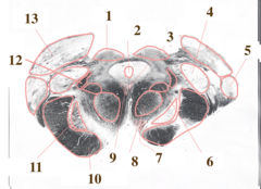
lateral geniculate nucleus
What structure is 5

oculomotor nucleus
What structure is 9

Edinger-westphal nucleus
What structure is 3
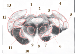
oculomotor nerve
What structure is 7

eyeball
What part of the visual system is A

optic nerve
What part of the visual system is B

optic chiasm
What part of the visual system is C

optic tract
What part of the visual system is D

superior colliculus
What part of the visual system is E

lateral geniculate nucleus
What part of the visual system is F

primary visual cortex (V1)
What part of the visual system is G

optical radiations
What part of the visual system is H

rod cell (photoreceptor)
What is A

cone cell (photoreceptor)
What is B

horizontal cell
What is C

bipolar cell
What is D

amacrine cell
What is E
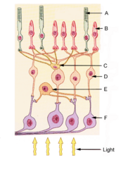
ganglion cell
What is F

light
What are the yellow arrows

optic nerve
What do the ganglion axons form

optic radiations
What structure is 1

eyeball
what structure is 2

eye socket
What structure is 3
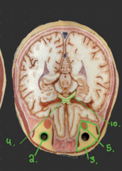
optic chiasm
What structure is 4

superior rectus muscle
What structure is 5

optic nerve (CN II)
What structure is 6

lateral rectus muscle
What structure is 7

oculomotor nerve (CN III)
What structure is 8

medial rectus muscle
What structure is 9

optic tract
What structure is 10

trigeminal nerve (CN V)
What structure is 1

horizontal brain section
What type of brain section is this

optic radiations
What structure is 1

optic radiations
What structure is 2

optic radiations
What structure is 3
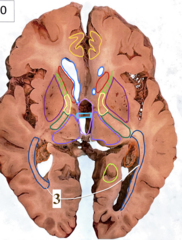
optic radiations
What structure is 4

lateral geniculate nucleus
What structure is highlighted blue

lateral geniculate nucleus
What structure is highlighted blue

optic tract
What structure is 6

optic tract
What structure is 5

optic tract
What structure is highlighted in blue
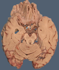
coronal brain section
What type of brain section is this

optic chiasm
What structure is highlighted in blue
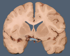
optic tract
What structure is highlighted blue
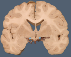
optic tract
What structure is highlighted blue

lateral geniculate nucleus of thalamus
What structure is highlighted blue

lateral geniculate nucleus of thalamus
What structure is highlighted in blue
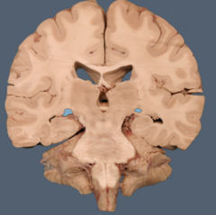
superior colliculus
What structure is highlighted in blue

brachium of the superior colliculus
What structure is the star on

brachium of the superior colliculus
What structure is 1
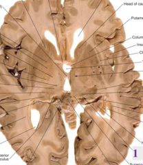
occipital lobe
What lobe is this

calcarine fissure
What structure is 6
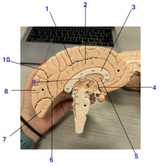
cuneus gyrus
What structure is 8

lingual gyrus
What structure is 7

primary visual cortex (V1)
What is structure A

optic nerve
What structure is 3

optic tract
What structure is 8

optic chiasm
What structure is 17

temporal retina
Label 1

nasal retina
Label 2

temporal retina
Label 3

optic nerve
Label 4

optic chiasm
Label 5

optic tract
Label 6

lateral geniculate nucleus (LGN)
Label 7

optic radiations
Label 8
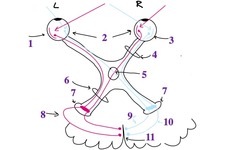
medial superior branch
Label 9

Meyers loop
Label 10

primary visual cortex V1
Label 11

central vision would remain intact whereas peripheral field of vision would be lost
What deficit would you expect to get if you lesioned the optic chiasm at midline?
loss of vision on the right side
What deficit would you get if you lesioned the left optic tract?
loss of vision on the left side
What deficit would you get if you lesioned the right optic radiation?
used to test function of individual muscles
H test

performed to determine losses in quadrants of the vision
visual field (quadrant) testing
