L5- Axial Skeleton of Vertebral Column and Thoracic Cage
 Call Kai
 Learn
Learn Practice Test
 Spaced Repetition
 Match
Match Flashcards
 Knowt Play
1/34

There's no tags or description
Looks like no tags are added yet.
Name | Mastery | Learn | Test | Matching | Spaced | Call with Kai | Chat |
|---|
No analytics yet
Send a link to your students to track their progress
35 Terms
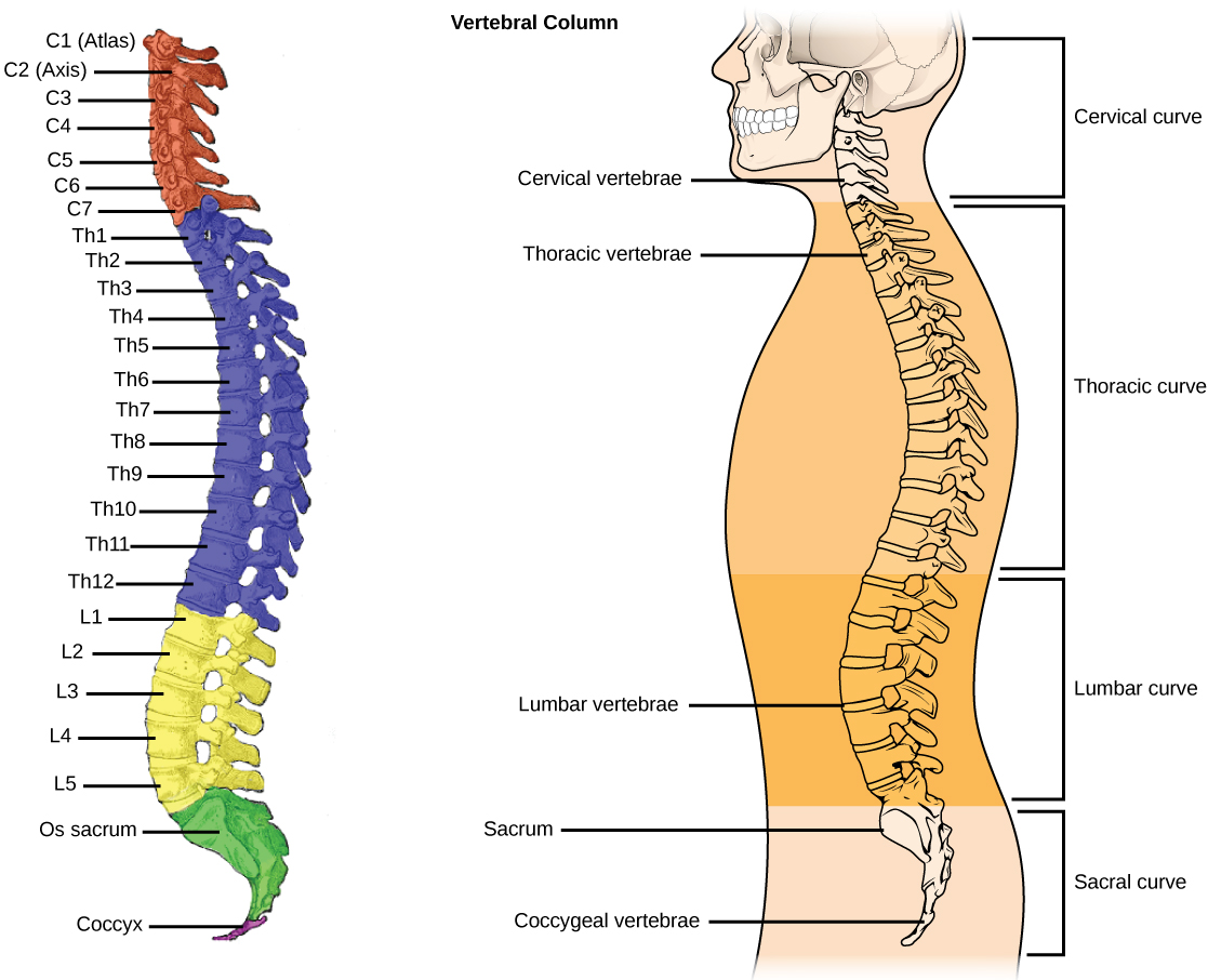
Number of Adult Vertebrate Column Bones
26 bones
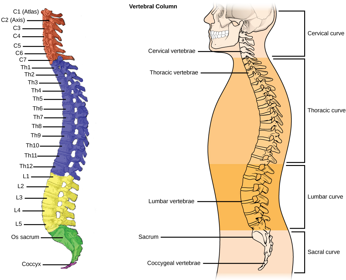
Breakdown of 24 vertebrate (from superior to inferior)
1. 7 cervical vert → anterior curve
2. 12 thoracic vert → posterior curve
3. 5 lumbar vert → anterior curve -
1 sacrum with 5 fused vert → posterior curve
1 coccyx with 3-5 fused vert → posterior curve
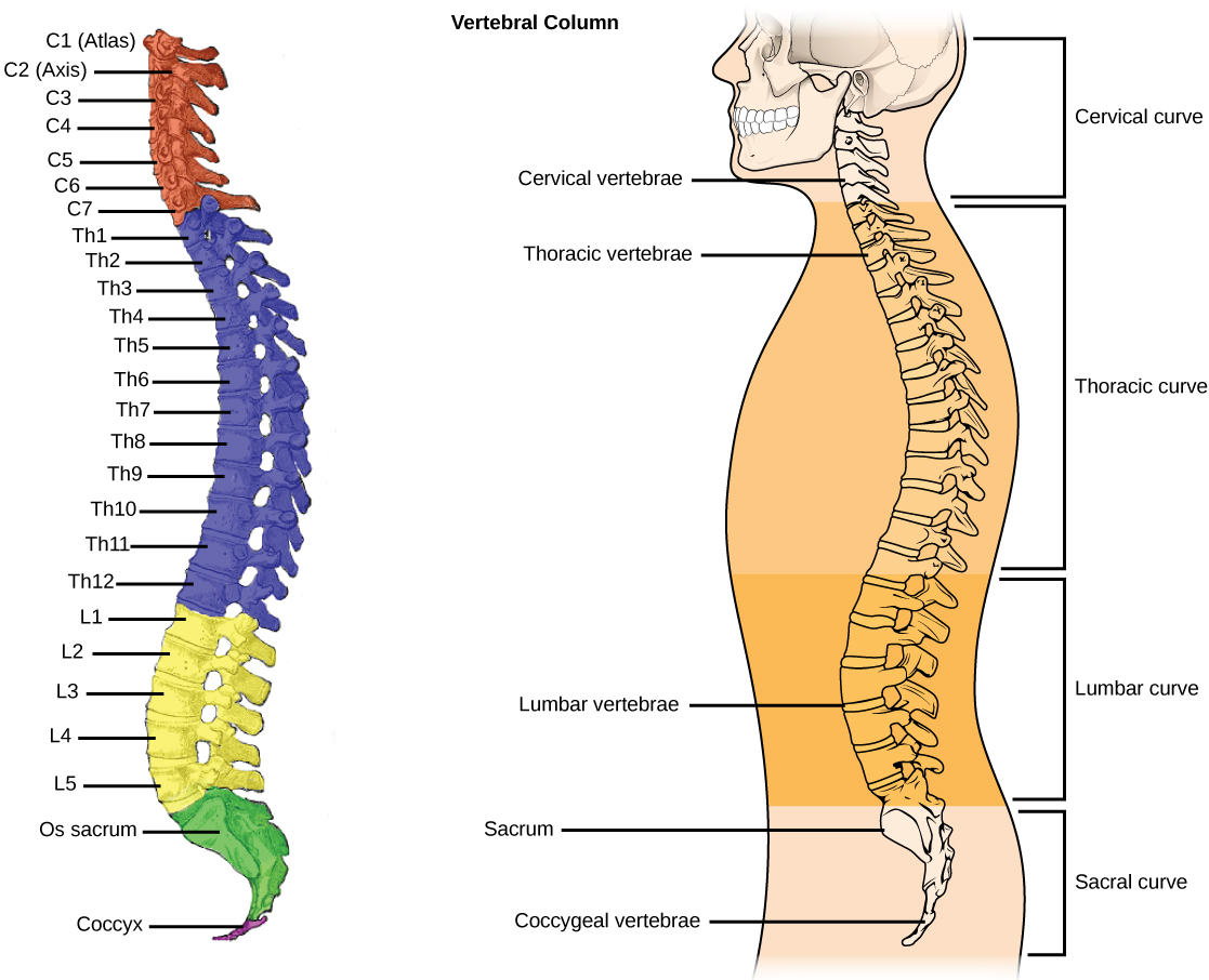
5 vertebral regions
1. Cervical region = c1-c7
2. Thoracic regions = T1-T1
3. Lumbar region = L1-L5
4. Sacrum
5. Coccyx
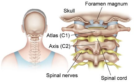
Cervical region The atlas (c1): holds up head
1. Most superior
2. Superior articular facets articulates with the occipital condyles of skull
3. Does not have a body
4. Has anterior and posterior vertebral arches and tubercles
5. Has superior and inferior articular facets
6. Has largest vertebral foramen of all vertebrae
7. Allows head to nod in yes motion
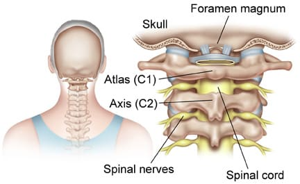
The axis (c2)
1. Has a dens projection with an anterior facet where it articulates at the axis
2. Transverse ligament binds dens to atlas allowing the no head manner
C2-C7:
Have bifid spinous processes (double headed tip off back of vert)
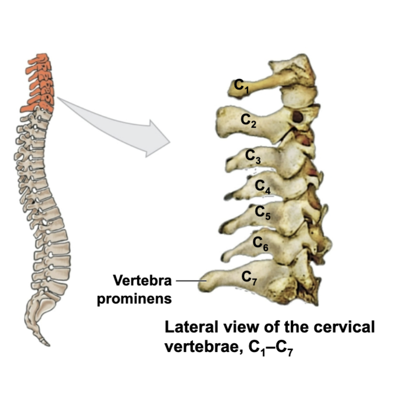
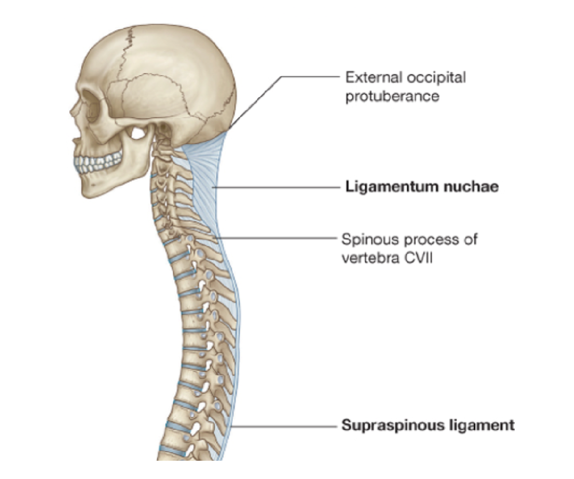
C3-C7 have most movement- Vertebral prominens (c7): long prominent spinous process)
1. Ligamentum nuchae: large elastic ligament that begins at c7 and extends to external occipital crest of skull
C7 has a long prominent spinous process
- Midsaggital (down middle of body)
- Site of attachment for cervical muscles
All vert have transverse processes fused to costal processes (rib projections
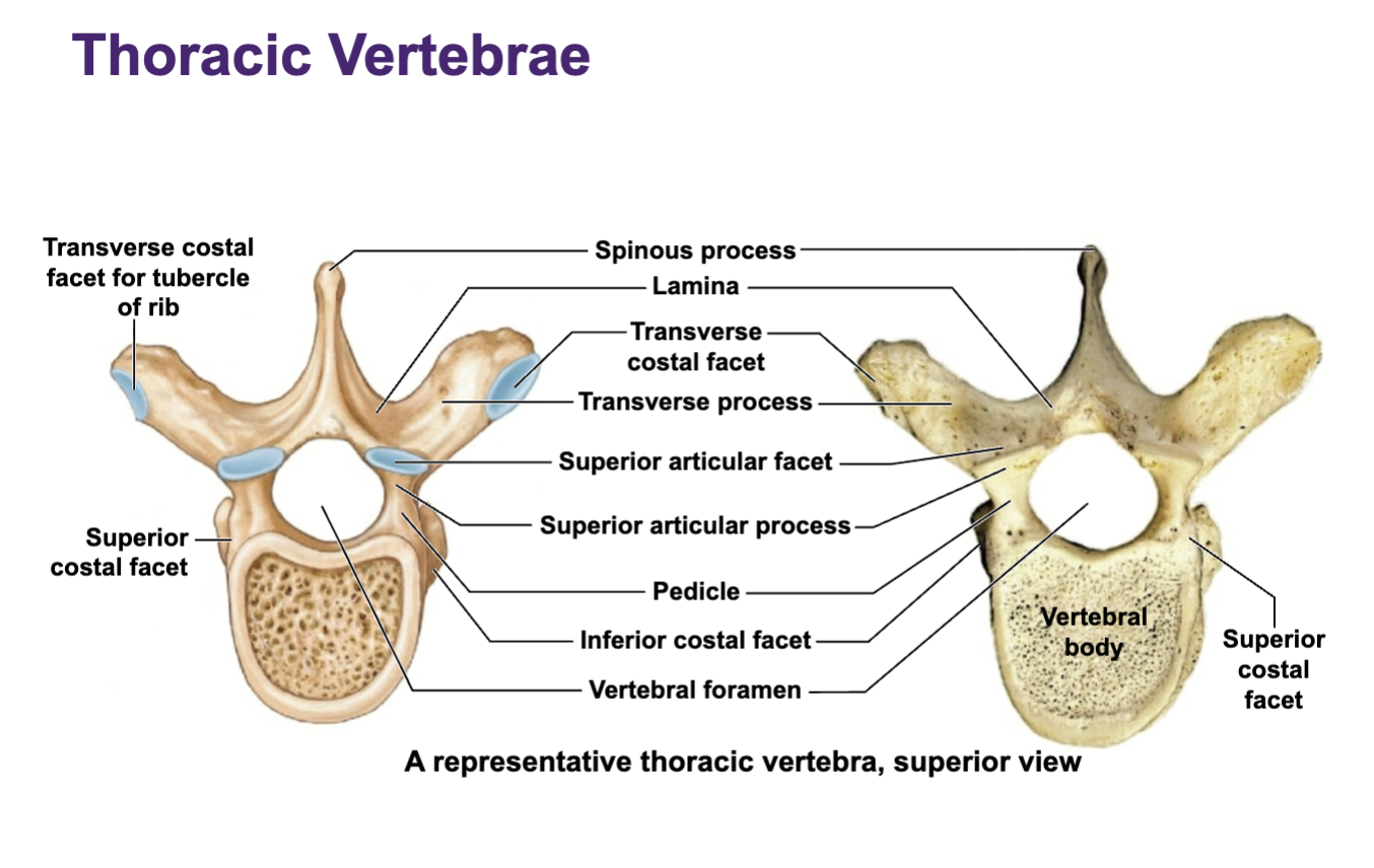
Thoracic region
1. All have rib articulation points ONLY NEED TO KNOW THIS
2. T1-T8 have superior and inferior costal facets
3. T9-T12 have 1 facet
4. T1-T10 have transverse costal facets - Articulate with tubercle of rib (posterior junction between neck and body)
5. These vert support the ribs
6. Most of the spinous processes point inferiorly (down)
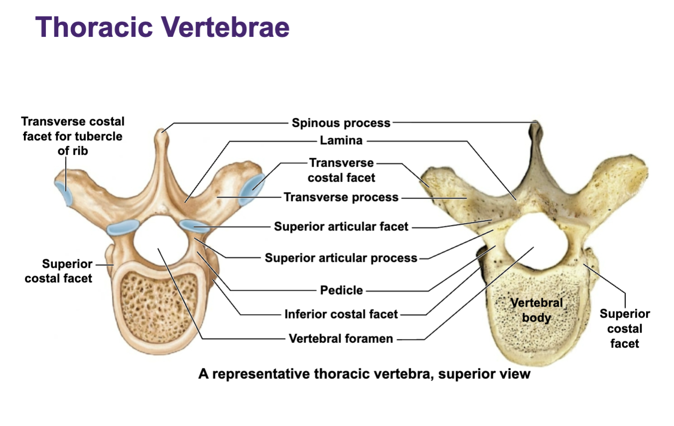
T1-T8
have superior and inferior costal facets
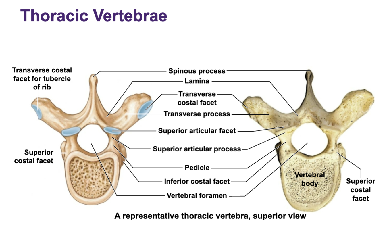
T9-T12 have
1 facet
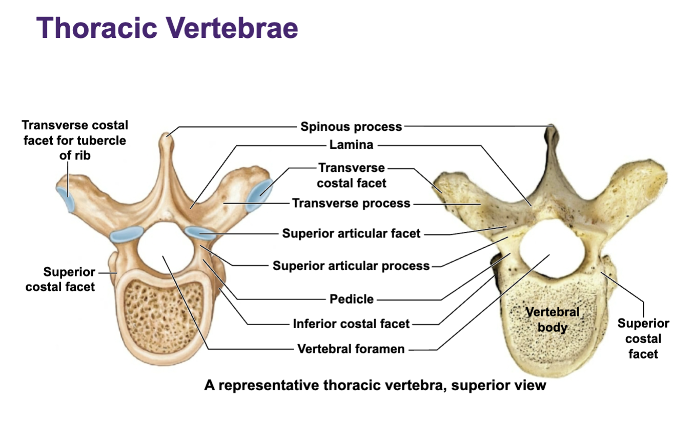
T1-T10 have
transverse costal facets
Articulate with tubercle of rib (posterior junction between neck and body)
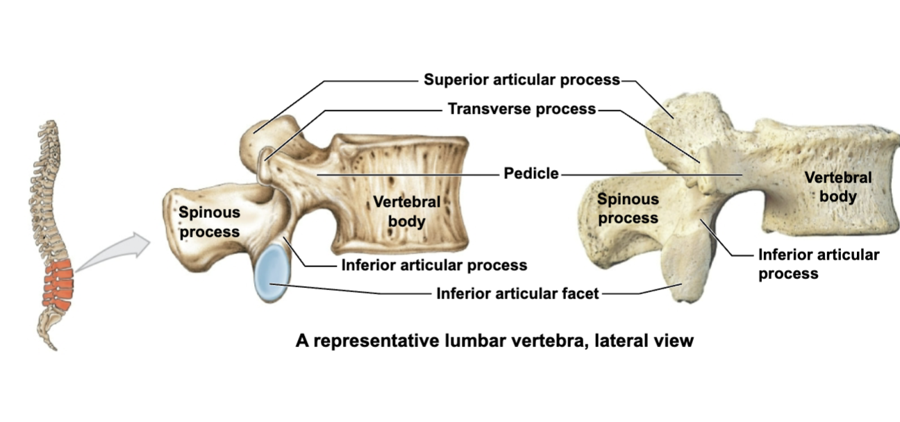
Lumbar region
There are 5
• Support the weight of the torso
• Vertebral bodies are quite large
• Spinous process points posteriorly
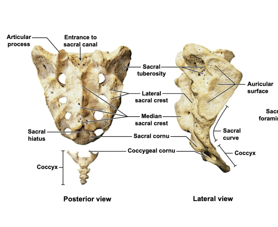
Sacrum
1. 5 fused vertebrae
2. Top of sacrum is the entrance to the sacral canal
3. Median crest = posterior mid sagittal crest protrusion
4. Sacral cornu and coccygeal cornu fit together and articulate with coccyx
5. Lots of joints: -
lumbo sacral joint = L5 articulates with sacrum (synovial joint)
Sacrum illieac joint = on each side of sacrum where sacrum articulates with pelvis (syndeosmotic = limited movement) -
Sacrococcygeal joint = where sacrum articulates with coccyx (syndesmoitc = limited movement)
6. Anterior view is concave for pelvic organs
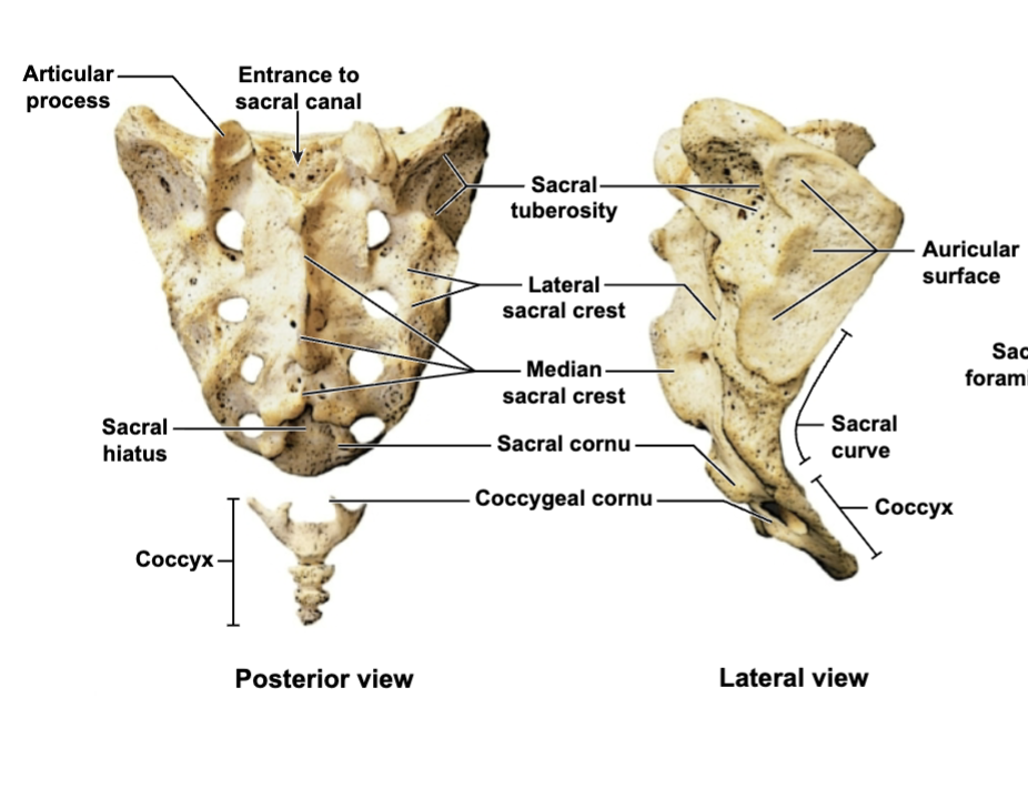
3 sacrum joints
Lumbosacral joint
Sacroiliac joint
Sacrococcygeal joint
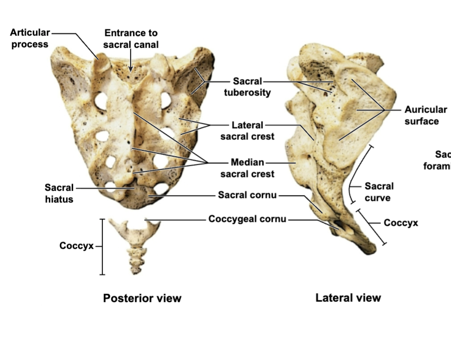
Lumbosacral joint
L5
articulates with sacrum (synovial joint)
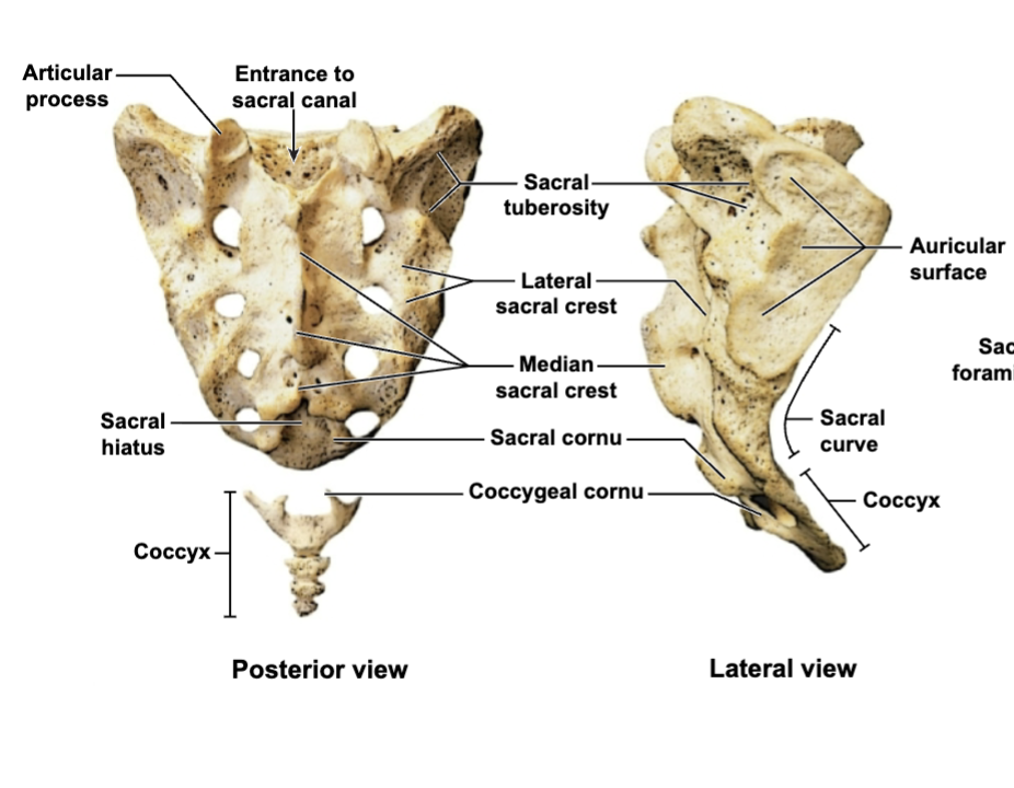
Sacroiliac joint
on each side of sacrum where sacrum articulates with pelvis
(syndeosmotic = limited movement)
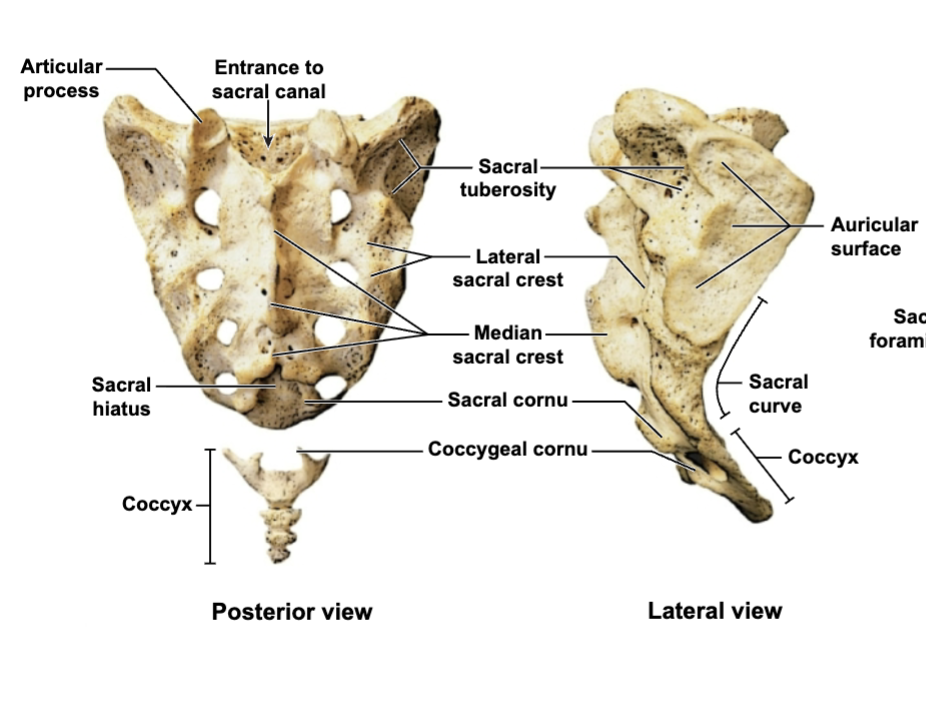
Sacrococcygeal joint
where sacrum articulates with coccyx (syndesmoitc = limited movement)
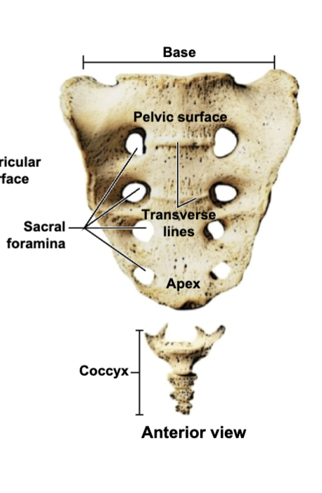
Coccyx
1. 3-5 fused vert
2. Adult male = points anteriorly
3. Adult female = points inferiorly (allows bigger opening for birth)
4. Coccyx consists of coccygeal cornu (articulation point)
Function of anatomy of vert column:
1. Closes and protects spinal cord
2. Supports skull
3. Supports weight of head, neck, and trunk
4. Transfers weight to lower limbs
5. Helps maintain upright position of body
Motions of vert column
1. Flexion and extension
2. Lateral flexion and extension
3. Left and right rotation
4 major spinal curves
1. Cervical - Develops as an infant learns to balance head on neck (sit up)
2. Thoracic - Large curve to make room for organs
3. Lumbar - Balances the segments above it over the lower limbs
4. Sacral - Accommodates for organs in pelvic regio
Structure of vertebrae
1. Between each vertebrae is a joint and intervertebral discs
Anterior side
2. Posterior side has bony projections
3. Vertebrae on top of vertebrae form the vertebral canal which is where the spinal cord can be found (basically length of spine
Intervetebral discs
1. Made of fibrous cartilage
2. Absorbs shock
3. Has 2 rings: - Inside ring = nucleus purpous (gelatinous, contains more water) - Outer ring = anulus fibrosis (contains less water)
4. Largest cartilage structure in body without vascular supply (no blood)
5. Each disc has a superior and inferior end plate to hold it in place between vert
6. Herniated disc puts pressure on spinal nerve/cord
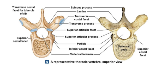
Vertebrae anatomy
Vertebral Arch
Articular processes
Vertebral articulation
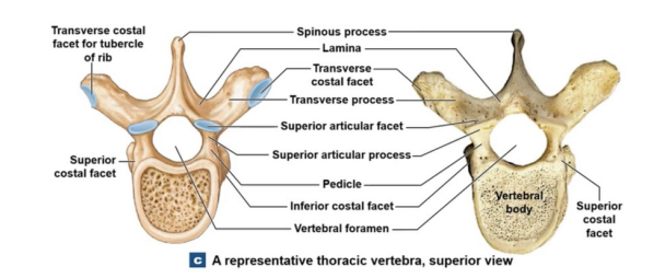
Vertebral arch -
Forms vertebral foramen: opening spinal cord goes through
Lamina and pedicle structures make this opening
Spinous process projects posteriorly: nub at top of back - Transverse process projects laterally off each side
Articulates with ribs at the superior, interior, and transverse costal facets (costal means rib)
Vertebral body has an intervertebral disc between them when stacking
Stacking Vertebrae on top of vertebrae form the vertebral canal which is where the spinal cord can be found (basically length of spine)
Intervertebral foramina = side openings where nerves protrude of spinal cord inside the stack → these can become blocked or occluded
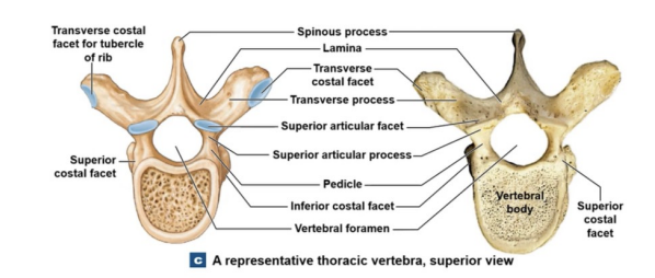
Articular processes =
projections where facets are attached -
Superior articular process
Inferior articular process
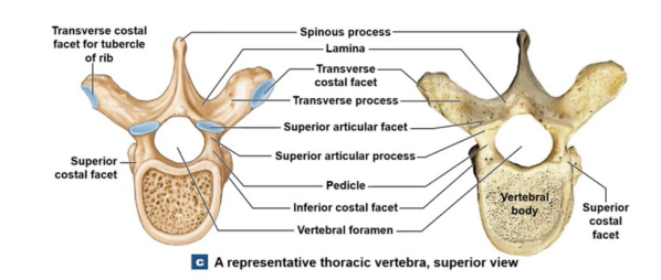
Vertebral Articulation
Tip: process means projection and facet means articulation point
Diagram = superior view → another vert can be stacked on top
Disorders
1. Spina bifida = malformation of structures that make the arch so the arch fails to close completely
2. Kyphosis = very dramatic rounding of thoracic spine
3. Lordosis = dramatic rounding in lumbar region - Dont want this - Can happen when lifting something heavy
Spina bifida =
malformation of structures that make the arch so the arch fails to close completely
Kyphosis =
very dramatic rounding of thoracic spine
Lordosis =
dramatic rounding in lumbar region - Don’t want this - Can happen when lifting something heavy
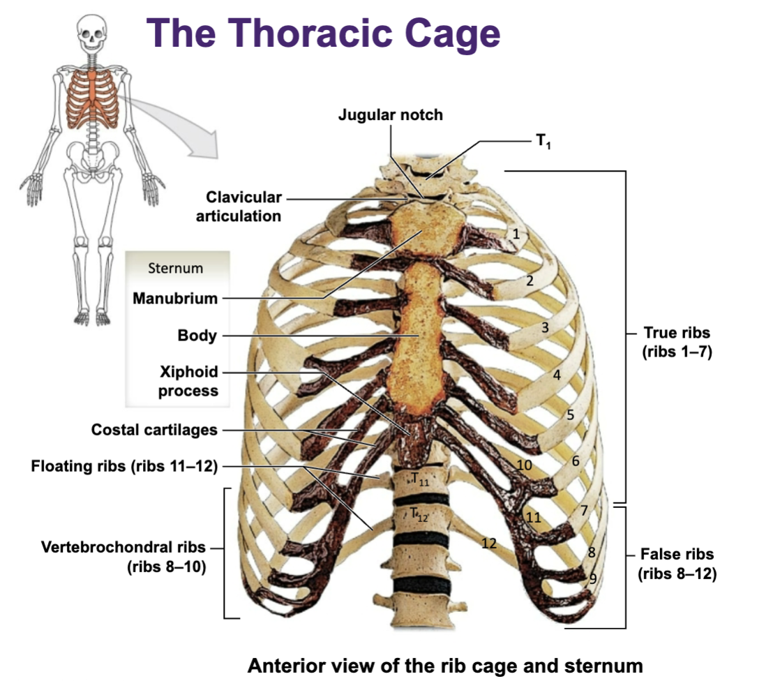
Thoracic cage functions
Thoracic cavity = heart, lungs… soft structures)
Function:
1. Protection
2. Attachment sight for muscles -
Muscles for respiration
Muscles for position
Muscles that move pectoral and shoulder girdle
Muscles that stabilize pelvis and hip
2 types of ribs:
1. True ribs/vertebrosternal ribs
ribs 1-7
Attached to the sternum via costochondral cartilage
2. False ribs/vertebrochondral ribs
ribs 8-10
Floating ribs = ribs 11-12
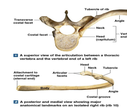
Body of rib:
Head of rib = where it articulates with costal facet of vert (vertebral end)
Neck = between head and tubercle
Tubercle = small projection that articulates with some vert at teh transverse costal facet
Angle = most curved part of rib
Costal groove = inferior side of rib where nerves and vessels run through
Body = main long portion of rib in the front
Sternal end = -superior and inferior articular facets/ interarticular crest
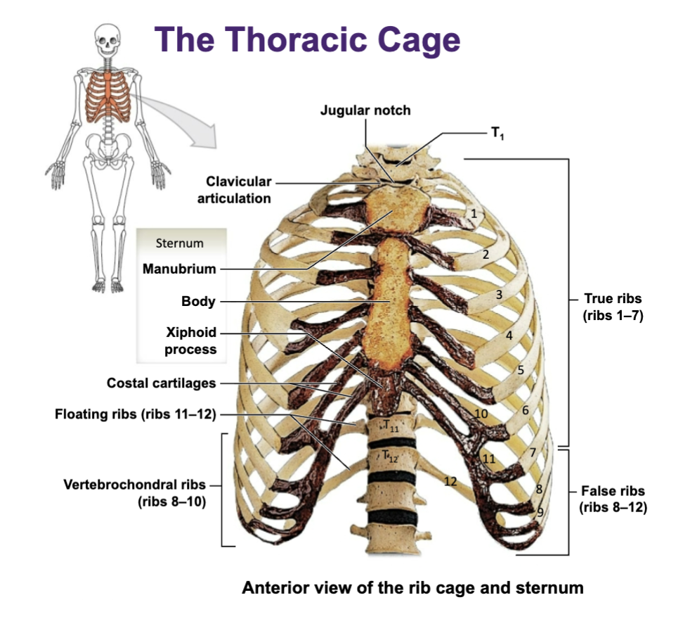
Sternum - Consists of:
1. Manubrium (very top)
2. Body
4. Xiphoid process (tip at bottom)
Want to avoid pressure here so it doesnt snap off and puncture an organ
5. Jugular notch
a large, visible, and palpable depression located on the superior border of the manubrium of the sternum, situated between the right and left clavicular notches at the base of the neck.