DI EXAM (abdomen)
 Call Kai
 Learn
 Practice Test
 Spaced Repetition
 Match
 Flashcards
 Knowt Play
1/140

There's no tags or description
Looks like no tags are added yet.
141 Terms

What can be seen in this image
normal serosal detail

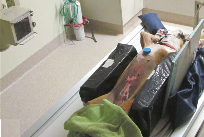
For how much gas is horizontal beam radiography sensitive in pneumoperitoneum and where is beam entered
2 - 5ml of gas, entered on diaphragm

What is shown in this image and how was it likely taken
pneumoperitoneum, horizontal beam radiography
After minor and major abdominal operation how long can we expect peritoneum
minor 24 - 48hrs, major 10d
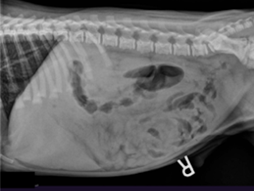
What can be seen in this image
decreased serosal detail
pneumonic for decreased serosal detail
my father says thank you
Ddx for decreased serosal detail
mass effect, fluid, skinny, technician, young
What is the next diagnostic step when seeing decreased serosal detail on radiograph?
AFAST
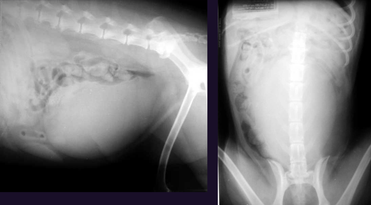
What can be seen in this image and what is the most likely Ddx
mass effect as all organs are displaced by bladder

What can be seen in this image and why?
Pneumoperitoneum, air in falciform ligament
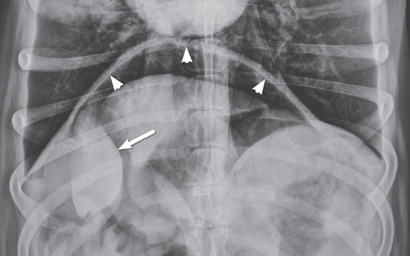
What is indicated by the arrowheads in this image and what does it indicate
diaphragm visualised from both sides (abdominal side), pneumoperitoneum
If in doubt about pneumoperitoneum what are the next diagnostic steps
horizontal beam radiography
Causes for pneumoperitoneum
ex lap, bite wounds, hit by car, gun shot, intestinal rupture
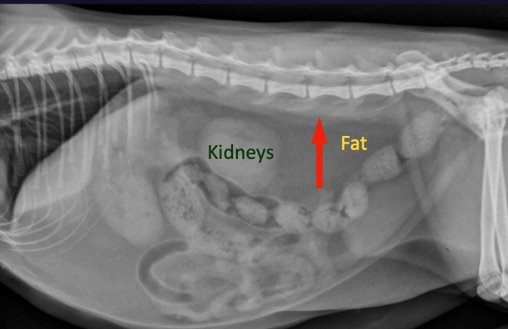
Is the the abdomen of a cat or a dog
cat

Is the the abdomen of a cat or a dog
dog

What can be seen in this image
retroperitoneal fluid

What can be seen in this image and what is likely aetiology
decreased retroperitoneal detail, haemorrhage from gun shot
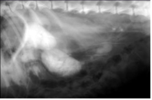
What can be seen in this image and what is the most likely cause based on the fact we have done an excretory urogram
decreased retroperitoneal detail, likely haemorrhage

What can be seen in this image and what is the most likely cause based on the fact we have done an excretory urogram
decreased retroperitoneal detail, likely ureter rupture
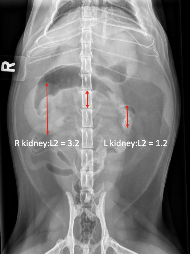
What syndrome is shown in this image of cat abdomen and what are the Ddx
Big kidney, little kidney, kidney failure, chronic pyelonephritis, hydronephrosis due to ureter obstruction or neoplasm usually of bladder neck ,

What syndrome is shown in this image of cat abdomen and what are next diagnostic steps
Big kidney, little kidney, Next steps ultrasound to confirm hydronephrosis, culture urine, CBC to assess renal function

A puppy presents with urinary incontinence. What abnormality is seen? Usually not seen on radiograph what is best modality?
Ectopic ureters, refer for CT urogram
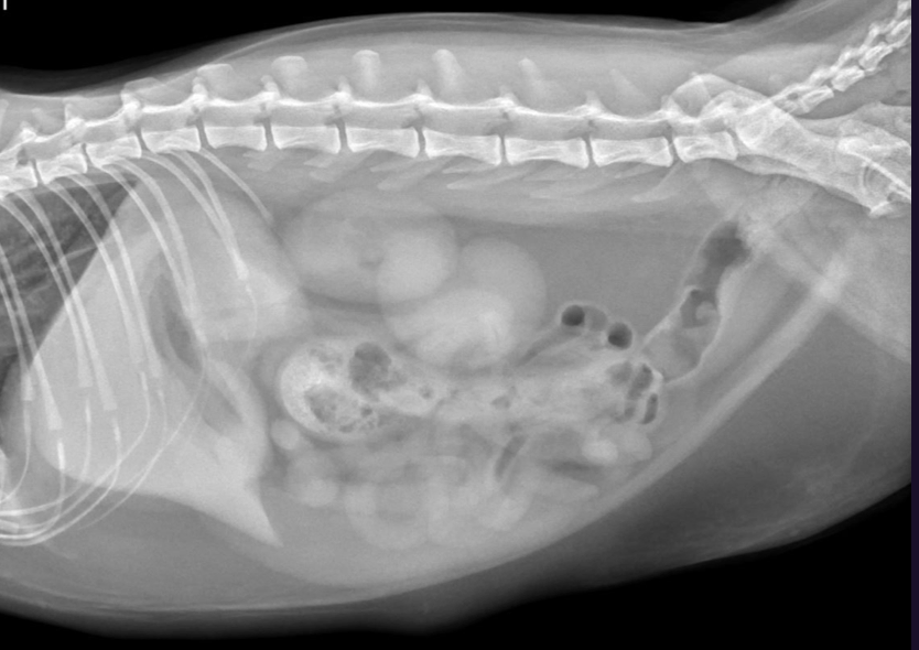
Cat abdominal X-ray, what abnormality is seen and how does feature compare to dogs.
no abnormality, cat kidneys are more parallel and mobile than dog kidneys

What is seen on this image of a cat abdomen and what does it likely indicate
small kidneys, chronic renal insufficiency, almost all old cats have it

What abnormalities are seen in this excretory urogram
none

Role of ultrasounds for patients presenting with chronic renal failure
especially pyelonephritis and ureter obstruction, rarely nodules and masses

What do we call appearance of this kind of kidney on ultrasound and what is likely Ddx
end stage kidney, chronic renal insufficiency
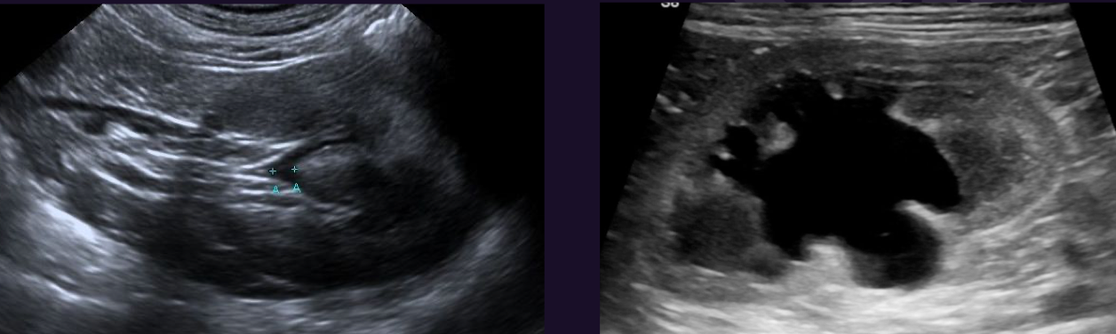
What changes of kidney are seen in this ultrasound
pyelectasis, hydronephrosis

What can be seen in this ultrasound of a cat and what is likely diagnosis, include other Ddx
pyelectasis, pyelonephritis, may be normal if on IV fluids, or renal insufficiency and all causes of PU/PD

What is seen in this ultrasound image of kidney and what does it indicate
very hyper echoic, acute renal failure

What can be seen in this US image of kidney and what are Ddx
Mass, always bad. either neoplasia primary (e.g. carcinoma), metastases (e.g. haemangiosarcoma?) or multi centric (e.g. lymphoma), or fungal granuloma (e.g. Aspergillus)

You find a kidney mass what are next diagnostic steps
FNA

What can be seen in this radiograph of a ragdoll cat and what is likely Dx
irregular margins of kidneys, polycystic kidney disease

Kidney ultrasound of a puppy, what is likely Ddx and what is it similar to
renal dysplasia, always bilateral, looks like endstage kidney which is unilateral

What is seen in this ultrasound image of cat and what is likely diagnosis and what is the only relevant Ddx
large size and dark hypoechoic rim, likely lymphoma, feline infectious peritonitis

What abnormalities are seen in this excretory urogram
ruptured ureter
Differentials for a mass in abdomen and what is a strong indication for neoplasia
CHANG: cyst, haematoma, abscess, neoplasia (irregular margins), granuloma
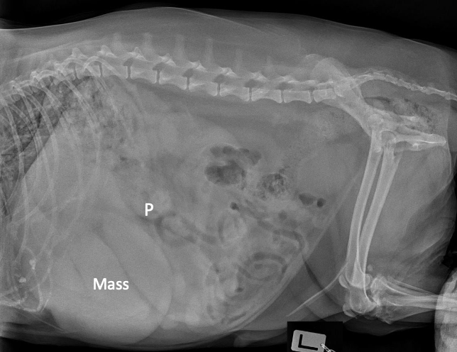
a mass in cranial abdomen is most commonly ….
liver mass

Two likely diagnosis for mass in cranial abdomen, and what masses are usually not seen on radiographs
Dilated stomach and liver mass, unlikely stomach wall, gall bladder or pancreatic mass,
How to differentiate whether cranial abdominal mass is in liver or stomach
liver mass displaces stomach, stomach mass displaces SI

Most likely Ddx of mid abdominal mass and 3 radiographic signs
Splenic mass 1. mass visible, 2. SI distended cranially, dorsally and caudally 3. haemabdomen
Can a mid abdominal mass displace liver cranially
no, cant be compressed
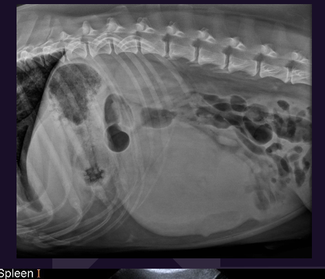
What are the differentials for splenic masses and what is prevalence
benign haematoma (1/3) malignant haemangiosarcoma (2/3)
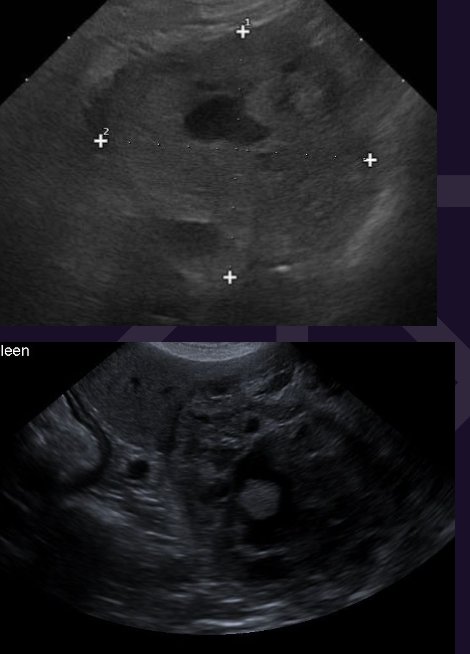
Diagnosis and treatment for large splenic mass
FNA if mass NOT capitated to rule out lymphoma, ultrasound, always treat with surgery i.e. splenectomy

How can we differentiate haematoma and haemangiosarcome
NOT ultrasound, need CT

Mass is located in mid abdomen on all views what is likely diagnosis
mesenteric mass

Most common retroperitoneal mass that is usually seen on ultrasound only
adrenal mass
Radiographic signs for retroperitoneal masses
always stay dorsal even if large, displace colon ventrally and are usually associated with kidney or adrenals
What could also displace the colon ventrally other than mass
retroperitoneal fat especially in cat


What abnormalities are seen and what is likely diagnosis
colon displaced dorsally, mass effect in abdomen, tubular structures in caudal abdomen, likely pyometra

When can pregnancy be definitively be diagnosed with radiograph in dog and cat
dog >45d, cat >35 - 45dd
What is the next step after decreased retroperitoneal detail is identified but no Hx of trauma and what is most likely cause
ultrasound (or CT) to identify adrenal mass

What is indicated by the arrow
normal liver margin dog

What is indicated by the arrow
normal liver margin cat

What is shown in this image and what is used for. What does the red arrow indicate
gastric axis, determines liver size. large amount of falciform fat in cat

Does this image show hepatomegaly
no, just a large stomach

In conjunction with a caudally displaced gastric axis on lateral , what does this VD image indicate and what is your Ddx
u shaped stomach, hepatomegaly

What is seen in this image and what are the Ddx
cranial displacement of gastric axis, indicates microhepatica, likely from cirrhosis or portosystemic shunt

What is shown in this image and what does it likely indicate?
mineralisation of wall of biliary tract, no clinical significance

What is shown by the arrow
hepatic abscess containing gas
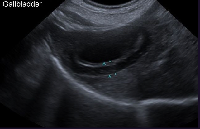
What is shown in this ultrasound and what could it indicate
increased gallbladder wall thickness: hypoproteinaemia, cholecystitis, portal hypertension e.g. RSHF, anaphylaxis

Ultrasound of dog gallbladder (left) and cat gallbladder (right), what does sludge likely indicate
dog not significant, cat cholangiohepatitis

In this abdominal radiograph of the cat what abnormality is seen, what could it indicate and what is your next step
spleen, splenomegaly, ultrasound
Causes of diffuse splenomegaly
may be normal (German shepherd), GA, IMHA, neoplasia (e.g. lymphoma), torsion (rare)
Steps to determine in which organ a mass is located
mass vs mass effect? is mass seen on left side and/or are organs displaced to the right
organ seen? if organ cant be seen mass may be located in it, if it can be seen need additional diagnostics
Steps in describing a mass
size, shape, margins, opacity, number, location
on lateral radiograph if mass is cranial to stomach where is it likely located
liver
on lateral radiograph if a mass is in mid abdomen what organ is usually involved and what are the Ddx
spleen, pedunculate liver, mesenteric mass
on lateral radiograph if a mass appears to be in dorsal abdomen
kidney or adrenal mass; or fluid from bleeding, adrenal tumor, trauma; or blood; or urine

Where is the mass located and what is most likely Ddx?
spleen, haematoma or haemangiosarcoma

Where is the mass located and what is most likely Ddx?
left abdomen, left renal mass
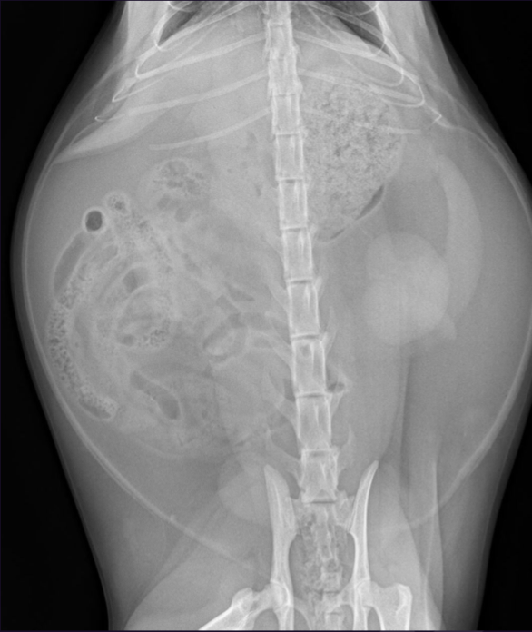
Where is the mass located and what is most likely Ddx?
no mass, normal retroperitoneal fat
on lateral radiograph if a mass appears to be in caudal abdomen
distended bladder, SL, lymph nodes, prostate, pyometra
Best imaging modality for cystitis
US and US guided cystocentesis for culture
Best imaging modality for neoplasia in urogenital tract
US and US guided catheter biopsy
Best imaging modality for calculi (urolithiasis)
US but almost all will be detected with Xray
Best imaging modality for obstruction in urogenital tract
excretory urogram

What is show in this image and what does the red arrow indicate
thickened wall most severe cranioventrally, cystitis

Why is shown in this image and what does it likely indicate
sediment, cystitis
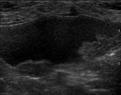
What is shown in this image and what does it likely indicate based on its location
irregular thickened wall in trigone area, bladder neoplasm,
Best imaging modality for bladder rupture
positive contrast cystogram for location and US guided abdominocentesis to determine if its urine

What is show in this image and what would be your next diagnostic step
decreased serosal detail, AFAST

What can be seen in this image and what is your diagnosis
positive contrast cystogram with contrast leaking into peritoneum and intact urethra, bladder rupture

What can be seen in this radiographic image and what are other diagnostic techniques
calculi in kidney, ureters, bladder and urethra, ultrasound or double contrast cystogram

Ultrasound image of right and left kidney of a cat what do you notice and what is the most likely Ddx
marked dilation of renal pelvis in right kidney , obstruction
What are your differentials for a mildly fluid distended renal pelvis?
could be normal for patient receiving IV fluids, or diuretic drugs, may also indicate pyelonephritis or chronic renal disease
What is the cut off for severely distended vs mildly distended renal pelvis
>2mm of fluid is severe
What features do healthy kidneys have on ultrasound
smooth margin, good cortex and medulla differentiation, no renal pelvis dilation

What abnormality is seen in this radiograph of cat and what are next steps
tail of spleen visible = splenomegaly, ultrasound to rule out lymphoma
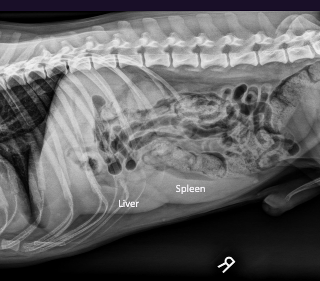
What are the differentials for diffusely enlarged spleen
normal in some breeds (German shepherd), GA/sedation, IMHA, torsion (rare)

What abnormality is seen in this radiograph of a dog
none

What 3 organs are bridged by triad
fundus, spleen, kidney

Ultrasound image of kidney in a cat, what can you see and what is likely diagnosis
fluid filled cysts that are not normal medulla, due to acoustic enhancement below it, also decreased corticomedullary differentiation, likely polycystic kidney disease

Ultrasound image of kidney of a dog, what can you see and what is likely diagnosis
increased echogenicity of renal cortex, enhanced corticomedullary differentiation, HYPOECHOIC BAND AROUND CAPSULE, very likely lymphoma

What is seen in the image of a positive contrast urethrogram and what is then diagnosis
calculi in bladder and urethra, causing rupture of urethra

What specific radiographic view is shown, why is it used and what is your diagnosis
bum view, calculi in urethra, avoid superimposition of legs on urethra, calculi causing urethral obstruction

What is shown in this image
gastric foreign body (kong)

What is shown in this image
gastric foreign body (ball)

What is shown by the arrows and what does it likely indicate
fluid distension of stomach, foreign body
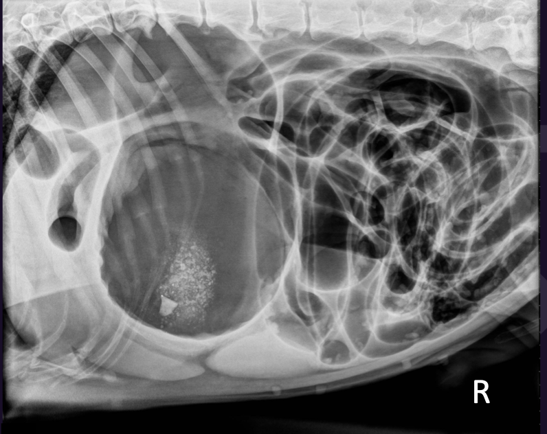
What can be seen in this image and what is your Ddx
pylorus looks like a smurf hat, GDV