3.1.2 Uses a slit lamp to examine the external eye and related structures.
 Call Kai
 Learn
Learn Practice Test
 Spaced Repetition
 Match
Match Flashcards
 Knowt Play
1/11

There's no tags or description
Looks like no tags are added yet.
Name | Mastery | Learn | Test | Matching | Spaced | Call with Kai | Chat |
|---|
No analytics yet
Send a link to your students to track their progress
12 Terms
Low magnification
x6-x10
• Lids
• Bulbar conjunctiva
• Cornea / limbus
• Tears / tear film
• Anterior chamber / iris
• Crystalline lens
Medium magnification
x20-x25
• Epithelium of cornea
• Stroma of cornea
• Endothelium of cornea
• Lens fits / surface of contact lenses
and movement on blink
High magnification
x30-x40
• Epithelium: vacuoles, microcysts, dystrophies
• Stroma: striae and folds
• Endothelium: polymegethism, blebs, dystrophies
Diffuse illumination
Allows overall examination of the eye and adnexa
Direct focal illumination
Optic section
Gives cross section of the cornea (illumination has to be perpendicular to cornea)
Parallel Piped
Width of 1mm. Used for looking at corneal nerve fibres and punctate keratitis
Conical Beam
Must be carried out in a very dark room o see if there is any light scatter back at us which shows the inflammatory cell, if any, in the anterior chamber
Observed at 45-60 degrees allowing us to observe all areas
In-direct illumination
Used for epithelial vesicles and erosions. There are two types of systems:
Coupled - when illumination and observation system swing around the same pivot point
De-coupled - when the illumination system is moved to the side therefore it no longer meets the optimal focus of the observation system
Retro-illumination
Light bounces off the iris or fundus to back illuminate the cornea (Requires de-coupled system)
Used for observing microcysts, vacuoles, cataracts, contact lens deposits
Specular Reflection
Angle of incidence of illumination is equal to angle of observation
Purkinjie image 1 - front surface of cornea
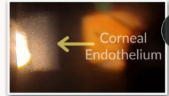
Purkinjie image 2 - endothelium, where there is a change of R.index from cornea to aqueous
Purkinjie image 3 - front surface of the lens
Purkinjie image 4 - back surface of crystalline lens
Medium to high magnification
Focus on area of interest and swing illumination beam until specular reflection achieved
Only visible through one eyepiece at a time
Tangential
Illumination at right angles to structure of interest
Low magnification
Wide angle beam but narrow
Used for iris freckles, general integrity of cornea and iris
Sclerotic scatter
Illumination slit located on limbus
You see halo of light in darkened room
Observation system de-coupled
Low magnification
Used for corneal opacities, corneal swelling, foreign bodies
Fluorescein examination
Assess staining of cornea & conjunctiva
Assess tear break up time
Assess tear meniscus height
Use Wratten filter to enhance image
Van Herick’s Technique
Low magnification Grd 4 - 1:1 open
Optic section with higher of 3-4mm Grd 3 - 1:2
60 degrees angle to observation Grd 2 - 1:4
Comparing width of cornea to anterior chamber Grd 1 - 1:<4
Grd 0 - closed
Conduct assessment also on the nasal side, if patient’s nose gets in the way lock the slit lamp at 60 degrees and move both systems approximately 5-10 degrees away from the nose, but ENSURE patient looks directly at the microscope before assessing the angle.