Anatomy 2300 Unit 4: Meninges and Dural Venous Sinuses of the Brain
 Call Kai
 Learn
 Practice Test
 Spaced Repetition
 Match
 Flashcards
 Knowt Play
1/23

There's no tags or description
Looks like no tags are added yet.
24 Terms
Meninges (Layers superficial to deep?)
The three connective tissue membranes that wrap the brain and spinal cord. From superficial to deep they are the dura mater, arachnoid mater, and pia mater.
Their job is to anchor and protect the CNS. They sit between the skull and the brain, and the spaces between them (meningeal spaces) hold or can collect fluid.

Dura mater (Layers?)
The outermost, thickest, and toughest meninges, lying just deep to the skull. In the skull, it has two layers:
Periosteal Layer - superficial layer - absent in vertebral column
Meningeal Layer - deeper layer - continuous in vertebral column as dural
sheath of spinal cord

Periosteal layer
The superficial (outer) layer, which is the periosteum (bone covering) on the inner surface of the skull. It is absent in the vertebral column, so it is only a cranial feature.

Meningeal layer
The deeper (inner) layer. It forms double-layered folds that insert into the brain's fissures (the dural septa).
Where it separates from the periosteal layer, it forms the dural venous sinuses. In the vertebral column it continues as the dural sheath of the spinal cord.

Arachnoid mater
The middle meninx, deep to the dura. It has a web-like appearance. The subarachnoid space beneath it contains CSF.
Arachnoid granulations project from it into the superior sagittal sinus and return CSF to the venous blood.
(2)

Pia mater
The deepest, thinnest meninx. It clings directly to the brain surface and follows every gyrus and sulcus, including the deep grooves.
Cerebral vessels run on top of it.
(3)

Meningeal Spaces of the Brain (Spaces?)
The spaces located around the meninges, between the skull and the brain. There are three: the extra(epi)dural space, the subdural space, and the subarachnoid space.
The epidural space (between the skull and dura) and the subdural space (between the dura and arachnoid) are only potential spaces. They don't normally hold anything and only open up if blood or fluid forces them apart, as in a hemorrhage.
The subarachnoid space (between the arachnoid and pia) is the only actual space. It contains CSF and blood vessels around the brain.

Epidural (extradural) space (Clinical significance?)
The space between the skull and the dura mater. It is a potential space, so it only exists if something (e.g., blood) forces it open. "Epi" means above.
Clinical: Extradural (epidural) hemorrhage is arterial in origin, usually from the middle meningeal artery, often after a skull fracture. Blood collects between the skull and dura as an extradural hematoma.

Subdural space (Clinical significance?)
The space between the dura and arachnoid mater. It is a potential space, so it only exists if something (e.g., blood) forces it open. "Sub" means below the dura.
Clinical: Subdural hemorrhage is venous in origin, from tearing of cerebral veins (bridging veins) as they cross toward the superior sagittal sinus.

Subarachnoid space (Clinical significance? Contains?)
The real, open space between the arachnoid and pia mater. It is an "actual space," so it contains cerebrospinal fluid (CSF) and the blood vessels around the brain. "Sub" means below the arachnoid.
Clinical: Subarachnoid hemorrhage is arterial in origin (e.g., a ruptured aneurysm of a cerebral artery). Blood mixes into the CSF.

Order of the Meninges
Order to memorize (superficial → deep): Skull → epidural space → dura → subdural space → arachnoid → subarachnoid space → pia → brain.

Dural Septa (consists of?)
Double-layered folds of the meningeal layer of the dura mater that dip down into the brain's fissures. They divide the cranial cavity into compartments and limit brain movement and support the brain inside the skull.
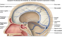
The three are the falx cerebri (between the cerebral hemispheres), the falx cerebelli (between the cerebellar hemispheres), and the tentorium cerebelli (between the cerebellum and the cerebrum above it).

Falx cerebri
A sickle-shaped fold in the longitudinal fissure, between the two cerebral hemispheres.
It runs from front (frontal) to back (occipital) along the midline, and the superior sagittal sinus runs along its top edge. The inferior sagittal sinus runs along its free bottom edge.

Falx cerebelli
A small fold between the two cerebellar hemispheres, located in the posterior cranial fossa, near the occipital bone.

Tentorium cerebelli
A horizontal fold between the cerebellum (below) and the occipital and temporal lobes of the cerebrum (above), in the transverse fissure.
It forms a tent-like roof over the cerebellum. The transverse sinuses run along its attached edge.

Dural Venous sinuses (Path?)
Channels formed where the two layers of the dura mater separate. They collect venous blood from the veins of the brain and CSF returned from the subarachnoid space, and they drain it all toward the internal jugular veins.
The path is: superior sagittal, inferior sagittal (via the straight sinus), and occipital sinuses → confluence of sinuses → transverse sinuses → sigmoid sinuses → jugular foramen → internal jugular veins.
Superior Sagittal sinus (drains into?)
Single sinus along the top midline, in the superior (attached) edge of the falx cerebri. It runs from front to back and receives cerebral veins and CSF (via arachnoid granulations).
It drains posteriorly into the confluence of sinuses.

Inferior Sagittal sinus (drains into?)
Single, smaller sinus along the free inferior edge of the falx cerebri.
It runs front to back and joins the straight sinus.

Straight Sinus (drains into?)
Single sinus where the falx cerebri meets the tentorium cerebelli.
It receives the inferior sagittal sinus and drains posteriorly into the confluence of sinuses.

Occipital sinus (drains into?)
A single (unpaired) sinus in the posterior cranial fossa, running along the attached edge of the falx cerebelli near the occipital bone.
It drains into the confluence of sinuses from below. It is one of three sinuses that feed the confluence (with the superior sagittal and straight sinuses)

Confluence of sinuses (drains into?)
The midline meeting point at the back of the skull (near the occipital bone).
It collects blood from the superior sagittal, straight, and occipital sinuses. It then sends blood left and right into the two transverse sinuses.
(5)
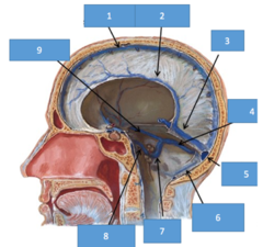
Transverse sinus (drains into?)
(2)
Runs along the edge of the tentorium cerebelli, from the confluence toward each side of the skull, along the occipital and temporal bones.
Each one drains into the sigmoid sinus on its own side.

Sigmoid sinus (drains into?)
(2)
S-shaped continuation of each transverse sinus.
It curves down along the temporal bone and exits the skull through the jugular foramen.

Internal jugular vein (drains into?)
The sigmoid sinus becomes the internal jugular vein as it passes through the jugular foramen.
The IJV carries blood from the brain down the neck, the final step of venous drainage from the head.
