Lecture 12: Acquired Valvular and Endocardial Disease
 Call Kai
 Learn
 Practice Test
 Spaced Repetition
 Match
 Flashcards
 Knowt Play
1/54

There's no tags or description
Looks like no tags are added yet.
55 Terms
acquired valvular and endocardial heart disease
myxomatous degeneration
infectious endocarditis
neoplastic
causes of mitral valve regurgitation
myxomatous mitral valve disease (MMVD)
DCM/HCM/RCM
mitral valve endocarditis
congenital mitral dysplasia
diagnostic approach to acquired valvular and endocardial heart disease
auscultation of a murmur
remainder of cardiovascular exam
ECG
thoracic radiographs
echocardiography
myxomatous mitral valve disease (MMVD)
also called endocardiosis, degeneration MV disease, chronic valvular disease
thickening and elongation of the valve leaflets, leads to valvular insufficiency and volume overload of the left atrium and ventricle
75-80% of all canine heart disease
smaller dogs <45 lbs and older dogs
males 1.5x more likely than females
disease progresses slowly
cause unclear
early MMVD
asymptomatic; incidental left apical systolic murmur
progressive MMVD
cough, exercise intolerance, tachypnea, restlessness
CHF with MMVD
dyspnea, syncope, collapse, ± ascites
MMVD PE
left apical systolic murmur
± tachycardia, arrhythmia, dyspnea, crackles, weak pulses
MMVD ECG
± sinus tachycardia, arrhythmias, large LA/LV patterns
MMVD thoracic radiographs
cardiomegaly over time (LA), with L bronchus compression
LAA bulge at 3:00 on DV
lateral projection → tracheal elevation and left atrial bulge caudodorsal aspect of heart
pulmonary venous congestion
interstitial to alveolar pattern if CHF
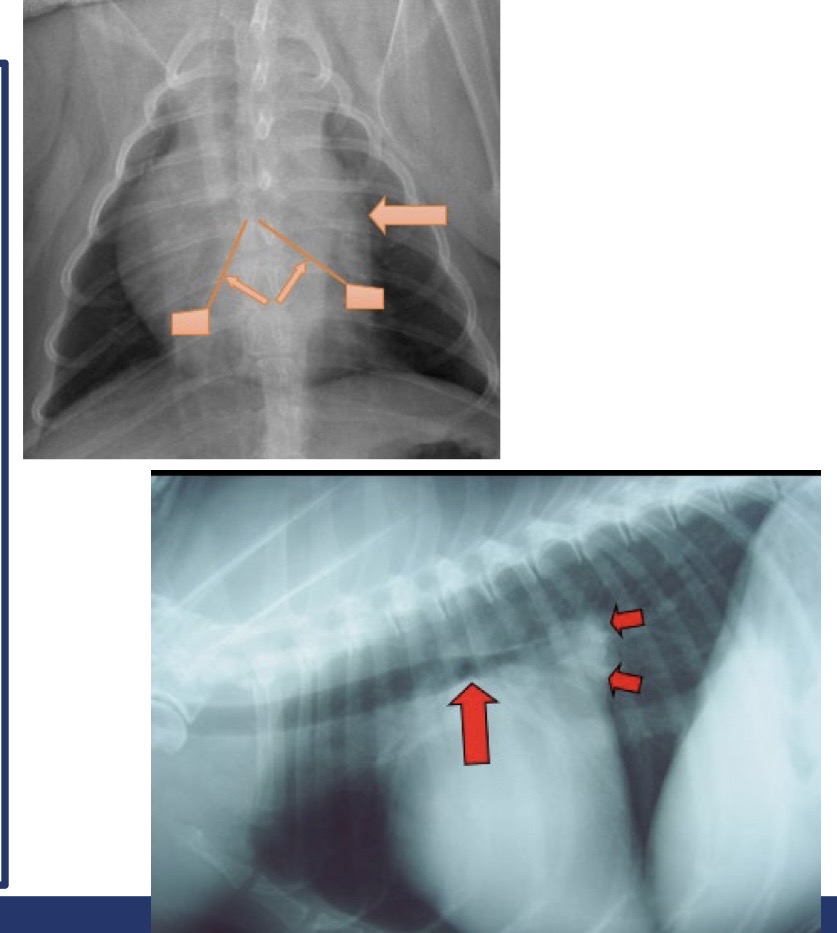
what is this?
MMVD
MMVD echocardiography
thickened mitral leaflets
mitral valve prolapse
mitral regurgitation ± tricuspid regurgitation
LV volume overload → LV, LA dilation
La:Ao ratio > 1.6 is significant

what is this showing?
MMVD → large LA

what is the yellow arrow showing?
ruptured chordae tendinae
typical presentation of ruptured chordae tendinae
acute LCHF

what are these showing?
ruptured chordae tendinae → acute LCHF
MMVD clinical syndromes
no clinical signs
cough → left main bronchus compression, early CHF
left sided congestive heart failure
ruptured chordae tendinae (acute LCHF)
syncope
endocardial split/LA tear → cardiac tamponade
MMVD lab testing
if starting ACE inhibitor → kidney values, electrolytes
if starting diuretics → kidney values, electrolytes, PCV/total solids
some dogs are also hypertensive
NT-proBNP → stage disease severity, predict imminent CHF, diagnose CHF
what are the big 4 for MMVD therapy?
furosemide
spironolactone
pimobendan
ACE inhibitor
new combo therapy for MMVD
cardalis
spironolactone/benazepril combination
for management of congestive heart failure in dogs
chewable tablets, 3 strengths
takeaways from ACVIM consensus statement
stage all MMVD patients
pimobendan in stage B2
spironolactone in CHF
ACVIM stage 1
dogs at higher than average risk for developing heart failure but without any apparent structural abnormality (no murmur etc)
diagnosis of stage A
small breed dogs should undergo regular evaluations
owners of breeding dogs or those at high risk (Cavaliers) may choose to participate in yearly screening events and conducted by board-certified cardiologists
stage A treatment
no drug treatment recommended
no dietary treatment recommended
potential breeding animals should no longer be bred if a murmur or echocardiographic evidence of mitral regurgitation is identified early
ACVIM stage B
structural abnormality (ie MMVD) but no clinical signs of heart failure
ACVIM stage B1
asymptomatic dogs with MV regurgitation from MMVD not severe enough to meet criteria used to trigger treatment
ACVIM stage B2
asymptomatic dogs with MV regurgitation from MMVD severe enough to result in cardiac remodeling (LA/LV enlargement) sufficient to recommend treatment
diagnosis for stage B2 criteria
murmur intensity >3/6
echocardiographic LA:Ao ratio > 1.6
left ventricular internal diastole diameter > 1.7
breed-adj radiographic vertebral heart score > 10.5
stage B1 treatment
no drug or dietary treatment is recommended
reeval by echocardiography or radiography in 6-12 months
stage B2 treatment
pimobendan at a dosage of 0.25-0.3mg/kg
diet → mild sodium restriction, adequate protein/calories, highly palatable
angiotensin converting enzyme inhibitors (ACEI)
beta blockade is not recommended routinely
spironolactone is not recommended for routine use to delay the onset of heart failure in dogs
cough suppressants (maybe)
surgical intervention (maybe)
ACVIM stage C
MMVD severe enough to cause current or past clinical signs of heart failure
hospitalized (acute) vs home (chronic)
diagnosis of stage C
ECG, thoracic radiographs, echocardiogram, NT-proBNP
normal/near normal [NT-proBNP] in dog with cough, dyspnea, or exercise intolerance = not CHF
treatment for stage C hospital/acute
furosemide, oxygen, pimobendan, sedation PRN, centesis PRN, nursing care, dobutamine/sodium nitroprusside/ACEi/nitroglycerin all PRN
treatment for stage C home/chronic
furosemide, pimobendan, ACEi, spironolactone
cough suppressants/bronchodilators
continue diet per stage B, promote appetite PRN, adjust K/Mg PRN
monitor BUN/Cr/lytes
increase in resting RR above normal has best predictive value for impending clinical decompensation
ACVIM stage D
patients have clinical signs of failure refractory to standard treatment for stage C heart failure from MMVD
treatment for stage D hospital/acute
furosemide higher dose or torsemide
centesis PRN, more potent afterload reduction
sildenafil PRN is pulmonary hypertension
treatment for stage D home/chronic
furosemide/torsemide, hydrochlorothiazide, pimobendan, ACEi, spironolactone
continue diet per stage B, promote appetite PRN
monitor BUN/Cr/lytes
increase in resting respiratory rate above normal baseline has the best predictive value for impending clinical decompensation
prognosis and survival for stage B1
years, may never progress
prognosis and survival for stage B2
3.5 years to CHF with pimobendan
prognosis and survival for stage C
6-12 months
prognosis and survival for stage D
weeks to months
mitral valve repair surgery (open-heart surgery)
requires cardiopulmonary bypass
available only at specialized centers
can be curative with survival often exceeding 3-5 years
high cost and requires intensive post-op care
transcatheter edge-to-edge repair (TEER/mitral clip)
minimally invasive catheter technique inspired by human mitraclip
clips anterior and posterior mitral valve leaflets to reduce regurgitation
still experimental in vet med
promising for dogs unfit for open surgery
transcatheter mitral valve replacement (TMVR)
experimental use of catheter-based prosthetic valve replacement
challenges include anchoring the valve and anatomical variability
currently limited to research environments
stem cell and gene therapy (research phase)
mesenchymal stem cells reduce inflammation and fibrosis
gene therapy targets collagen remodeling and TGF-beta signaling
bacterial endocarditis
infective endocarditis, vegetative endocarditis
bacterial infection of the endocardium → valvular endocarditis is most common
low incidence
infection leads to thickening/fibrous valvular changes
risk factors of bacterial endocarditis
pre-existing valvular disease
immunosuppressive drugs
middle aged, intact male large breed dogs
recent surgery
recent IV catheter placement
antimicrobial use
allergic factor
cardiac effects of bacterial endocarditis
mitral valve → mitral regurgitation, mitral stenosis, left sided CHF
aortic valve → aortic regurgitation, aortic stenosis, left sided CHF
pulmonic and tricuspid valve → tricuspid and/or pulmonic stenosis or insufficiency, right sided CHF, pulmonary thromboembolism
systemic effects from bacterial endocarditis
pyelonephritis
UTI
meningitis
septic arthropathy
myocarditis
immune mediated GN or polyarthritis
thrombosis can lead to infection or infarction (bland thrombus)
clinical scenarios for bacterial endocarditis
congestive heart failure
fever of unknown origin
multisystemic signs suggestive of infection or immune-mediated disease
organ embolism
common clinical findings of bacterial endocarditis
fever
leukocytosis
cardiac murmur
lameness
lethargy, anorexia, depression
diagnostic testing for bacterial endocarditis
bartonella testing
arthrocentesis → cytology and culture
blood and urine cultures
echocardiographic findings for bacterial endocarditis
thick, asymmetric, shaggy, hyperechoic vegetative valvular lesions
oscillating, vibratory, or mobile valvular lesions
cardiac chamber enlargement or hypertrophy
valvular regurgitation or stenosis
transesophageal echocardiography
treatment for bacterial endocarditis
antibiotic based on culture and sensitivity or serology
IV antibiotics for the first 5-14 days
broad spectrum antibiotics → ampicillin and enrofloxacin most common
suspicion of bartonella spp. → doxy and gentamicin or enrofloxain in dogs, doxy and pradofloxacin in cats
additional anaerobic coverage → clindamycin or metro
oral antibiotics for 4 to 8 weeks
prognosis and prevention for bacterial endocarditis
median survival 2 months
aortic valve 3 days vs mitral vale 476 days
worse prognosis if → CHF present, aortic valve affected, gram negative sepsis, renal failure, thrombocytopenia, thromboembolic complications, azotemia, treated with corticosteroids
prevention → prophylactic antibiotics in susceptible patients, human guidelines