BIO 110 Exam 2 Clark 2026 University of Rochester
 Call Kai
 Learn
 Practice Test
 Spaced Repetition
 Match
 Flashcards
 Knowt Play
1/65

Earn XP
66 Terms
Fluid mosaic model
Phospholipid bilayer is fluid (flexible)
Also has proteins embedded or attached to membrane for transport, signaling, structure
Attached carbohydrates can be recognized by complementary protein receptors on other cells
Endomembrane system
network of membrane-bound orgenelles interacting through bidirectional vesicle trafficking
ER sends new proteins/lipids to Golgi [which modifies products; eg) glycosylation = attaching carb to protein/lipid]
Golgi sends recycled ER proteins/membranes back to Golgi
Golgi sends vesicles into plasma membrane, lysosome, or to be secreted
Triacylglycerol structure and function
1 glycerol connected to 3 fatty acid tails; hydrophobic
long-term energy storage, thermal insulation, mechanical cushioning for organs
excess caloric intake of carbs/fats can be converted to this [for storage in adipocytes]
Membrane remodeling
membrane structure can change for organization and supporting endomembrane system
eg) phagocytosis
Phagosome & Phagocytosis
Phagosome = a vesicle with material ingested by phagocytosis
Phagocytosis = process where cell membrane engulfs large particles/microbes into phagosome
Primary lysosome vs. Secondary lysosome
Primary: contains digestive enzymes but hasn’t ingested material yet
Secondary: when primary lysosome fuses with phagosome or endosome to digest material
How can membrane lipids be structurally diverse? What is the point of this diversity?
any polar molecule can bind to the glycerol [or be in place of it] to make it amphipathic — any molecule can bind after that requirement
eg) phosphate group
eg in place of glycerol = shingosine
the diversity allows for specialization of membrane-bound organelles and various cell types
eg) membrane oligosaccharide antigens determine blood type
What molecule (besides cholesterol) can restrict membrane lipid movement? What are examples that keep specific lipids in the correct position?
embedded proteins
Flippase, Floppase, Scramblase
What molecules break down lipids?
enzymes
Cholesterol & function in plasma membrane
amphipathic sterol lipid with small polar -OH head (sticks slighty out of membrane)
Reduces flexibility in warm temperatures
Increases flexibility in cold temperatures (against phospholipids’ paracrystalline state)
Membrane protein types/functions
Transport proteins = selectively allow molecules to pass through membrane
Receptors = cell-to-cell communication
Enzymes = catalyze reactions at membrane surface
Cell recognition proteins = immune / cell identity recognition; often glycoproteins
Cell adhesion proteins = attach cells to each other or extracellular matrix
Structural/anchoring proteins = maintain membrane shape/stability + link membrane to cytoskeleton
generate/stabilize membrane curvature (for vesicle traffic)
Ways membrane proteins associate with membrane
Integral/transmembrane proteins = embedded in bilayer and span the membrane 1+ times; amphipathic; hard to remove without detergents
Peripheral membrane proteins = loosely attached to membrane surface or integral proteins; interact with ionic bonds or H-bonds (polar); easier to remove
Lipid-anchored proteins = covalently attached to lipids
Passive transport vs. Active transport
Passive = diffusion of molecule from high to low concentration (concentration gradient) through bilayer
simple diffusion = small, nonpolar (lipophilic) molecule goes straight through lipid bilayer
facilitated diffusion = channel/carrier proteins help large or polar molecules cross
Active = requires energy to go against concentration gradient
Glucose transporter (GLUT)
Carrier protein that performs passive transport
glucose in blood plasma binds to protein site —> conformational change that lets it into the cell —> when glucose is released into cytoplasm, it returns to original shape
Sodium-potassium (Na+K+ ATPase) pump
Membrane bound enzyme that performs active transport
3 Na+ moved out for every 2 K+ moved in = maintains negative resting potential inside cell
Valinomycin
peptide ionophore (carries specific ion across lipid membrane) that encloses K+ in a hydrophobic coat
disrupts membrane potential (voltage difference inside/outside cell)
naturally made by Streptomyces bacteria to limit growth of surrounding microbes
RNA world-hypothesis theory
RNA used to be main material of heredity, acting as both DNA and proteins
supporting evidence = RNA ability to fold and act as enzyme; ribosome composition of proteins + rRNA
Drug & target examples
small molecule designed to bind to specific target (usually protein) to change its structure [and therefore function]
target eg) enzymes, receptors, ion channels, membrane transporters, macromolecules, etc.
many drugs mimic endogenous (naturally inside organism) molecules [esp with substrates to enzymes or signaling molecules to receptors]
competitive inhibitors are common drug designs
Agonist vs. Antagonist
Agonist = initiates physiological response
Antagonist = decreases function of target [protein]
majority since it’s easier to break than “fix/activate” the enzyme
Why can some drugs be taken orally while others need to be injected?
depends on if stomach acid will break them down
Antibiotic function & types
drug that kill bacterial infections
selectively toxic (kill the microbe, not the patient)
works by binding to enzymes to inhibit their use in cell wall / nucleic acid / protein synthesis/maintenance
protein interference can be through ribosome inhibition
Bacteriostatic = inhibits growth enough for the immune system to kill
Bactericidal = kills directly
Opioids
drugs that are/mimic morphine [from the natural opium poppy plant]
eg) heroin
morphine is a mimic of endogenous opioid peptides like endorphins
released during physical exercise —> relieve pain/stress
targets opioid receptors like µ, δ, κ that activate inhibitory G proteins that decreases [pain-signaling] neurotransmitter release
produces analgesic (pain relieving) effect + ventilatory depression (NOT mental; it’s dangerously slow/little breathing that can stop heartbeat), constipation, immune suppression
Naloxone/Narcane
opioid mimic that’s an opioid antagonist
has higher affinity for opioid receptors than opioids, displacing them without activating the same pathways
can lead to symptoms of opioid withdrawal
block opioid effects for 30-90 minutes before readministration needed
administered by syringe or nasal spray
Types of diabetes, causes, and what can treat the main two
Type 1 = inability of pancreas to make/release insulin (a hormone)
cause = genetic + environmental trigger
treatment = injecting fast- or long-acting insulin (depending on needs)
Type 2 = inability of receptors to properly respond to insulin
cause = lifestyle (weight, diet, activity)
treatment = Glucagon-Like Petide-1 (GLP-1 = a hormone) mimics like semaglutide (Ozempic), which lowers blood sugar levels and appetite (by suppressing glucagon release & delaying gastric emptying)
semaglutide has 3 amino acid changes to make its effects last longer and allow for accurate binding at GLP-1 receptors
Gestational = temporary form that can occur during pregnancy
Selective serotonin reuptake inhibitors (SSRIs)
blocks the serotonin reuptake transport, increasing serotonin concentrations in the brain (since it won’t be broken down as much)
work against low levels of serotonin in depression
serotonin receptors control mood/emotions when serotonin stimulates (which is made from tryptophan)
drug eg) Prozac
Zonisamide
drug that binds to sodium channels and voltage-sensitive calcium channels to suppress neural hypersynchronization (excessive firing of electrical impulses at once)
treats seizures
Cannabinoids
chemicals that act on endocannabinoid system in body to influence neuron communication related to appetite, anxiety, learning/memory, pain
eg) THC, CBD
What did Gregor Mendel suggest?
that there’s a material of heredity in organisms
Gene
functional unit of heredity (often a contiguous {continuously touching} segment of DNA) that is transcribed into RNA [which may or may not be translated into a protein]
all include a regulatory region (regulates gene expression) and coding region (contains information to produce RNA)
all regulatory regions include a promoter & transcription factor binding sites
RNA polymerase must bind to promoter to transcribe gene
Allele
form (among many) of gene at a location on a chromosome
Autosome
non-sex chromosome
humans have 22 pairs
Sex chromosomes
X or Y chromosomes
Thomas Hunt Morgan’s hypothesis and conclusions
alleles for red/white eyes in fruit flies were carried on the X chromosome (aka X-linked)
built off Nettie Stevens’ demonstration that sex was determined by X and Y chromosome inheritance
concluded that the white-eyed phenotype was X-linked, showing that genes were carried on chromosomes
but both proteins and DNA make up chromosomes, and people believe at first that proteins’ 20 amino acids match genetic complexity better than DNA’s 4 nucleotides
Mouse experiment that showed whether DNA or proteins were the material of heredity
strains of strep bacteria:
rough/R = non-virulent (non-deadly)
can be transformed into S type by interacting with heat-killed S strain’s hereditary information
smooth/S = virulent (deadly)
can be heat-killed to be non-deadly
since destroying the S strain’s DNA was the only one to stop death, it likely contained the hereditary information for virulence
note, DNA also survives higher heats than most macromolecules

Hershey-Chase Experiment 1952
bacteriophage = virus that infects bacteria by injecting material of heredity that makes bacteria create more virus
treatment 1 radio labeled phage protein with 35S (sulfur makes up proteins)
treatment 2 radio labeled phage DNA with 32P (phosphorus makes up DNA)
after bacteriophages injected E. coli and phage ghosts were removed, radioactivity was only seen in the DNA-marked E. coli —> DNA must be the material of heredity

Chargaff’s Rules
base composition/proportion in DNA varies from species to species
different tissues in same individual have same DNA base composition
base composition of DNA doesn’t change with organism age, nutrition, or environment
in all DNA, adenosine residues = thymidine residues & guanosine residues = cytidine residues (A=T, G=C) —> knowing 1 strand’s sequence means you know the complement
Purine vs. Pyrimidine
Purine = 2 rings
A, G
Pyrimidine = 1 ring
T, U, C
X-ray crystallography
technique to find 3D protein structure
by purifying molecule —> turning it into a crystal (changing its properties to bind to itself with a regular pattern) —> shooting X-rays through it —> reverse engineering the electron diffraction paths
5’AGCT3’ sketch of DNA strand & DNA structure
DNA is an antiparallel double-stranded helix
phosphate group in sugar-phosphate backbone makes negative
major groove is where most proteins bind
free-floating nucleotides have 3 phosphates, but 2 detach when nucleotide attaches to DNA strand

How was the double helix hypothesis of DNA structure tested? What were its results?
Dickerson Dodecamer analyzes structure of chemically synthesized DNA [with known sequence] using X-ray crystallography
showed that DNA had varying bend angles (not all straight) and many slight variations in 3D structure depending on base pair sequence compared Watson & Crick’s ideal model
explained how specific proteins could find and bind to specific sequences based on structure — every DNA sequence is a different ligand (binding target)
Central Dogma
genetic information flows from DNA —> RNA —> protein
never originates from protein
DNA or RNA [in some cases] can also replicate itself
RNA can also be reverse transcribed into DNA
original dogma was provocative so people would try [and fail] to disprove
one wrong part = possible arrow from DNA straight to protein
Polymerase
enzyme that synthesizes a complement dependent on template (DNA or RNA) with very high accuracy
“{template}-dependent {product} polymerase”
works 5’ → 3’, starting from primer
requires triphosphate forms of nitrogenous bases (ATP, TTP, GTP, CTP)
breaking off of two phosphate groups provides energy needed for phosphodiester bond, attaching to daughter strand
many have exonuclease region that proofreads and removes incorrect bases during synthesis

What did Arthur Kornberg discover?
DNA requires a template for synthesis
also isolated DNA polymerase I in E. coli (he thought it was a replicating polymerase but it just proofreads)
Polymerase chain reaction (PCR)
process that amplifies small sequence of DNA into millions/billions of copies
requires DNA template (aka parent strand), nucleotides, primers, and polymerases
can be used for genetic testing
Denaturation = heats to 95ºC to fully separate DNA strands
Annealing = primer can bind with hydrogen bonds at ~55ºC and free-floating nitrogenous bases of daughter strand start making hydrogen bonds with complements
Extension = at 72ºC, heat-resistant DNA polymerase is added to synthesize 5’ → 3’ strand with phosphodiester bonds and hydrogen bonds forming between nucleotides
process repeats many cycles
DNA synthesis + proteins and parts involved
From replication fork (Y shape region where the DNA strands split):
Gyrase = type of topoisomerase that alleviates torsional stress (relaxes supercoiling)
Helicase = breaks H-bonds so DNA strands split into two
Single-strand binding (SSB) proteins = temporarily bind to single strands of DNA to prevent H-bonds from reforming
DNA primase = specialized DNA-dependent RNA polymerase that starts by adding RNA primer (since DNA polymerase can’t begin synthesis)
[DNA-dependent] DNA polymerase III = synthesizes DNA nucleotides starting from RNA primer
can continuously synthesize leading strand by following helicase
makes Okazaki fragments at multiple primers of lagging strand
DNA polymerase I = replaces RNA primers with DNA
Ligase = bonds replaced^ fragments with previous Okazaki fragment
Sliding clamp = protein that attaches to DNA polymerases to prevent them from coming off the template while adding nucleotides
Polymerases on each side (leading/lagging) are bound by a dimerization domain to prevent the leading strand from getting too far ahead too fast
Telomerase = RNA-dependent DNA polymerase that uses an internal RNA template to add complementary DNA to the parent strand (called a telomere and is unnecessary for desired product) so that the lagging daughter strand can fully replicate the essential part of the DNA template

Prokaryotic vs. Eukaryotic DNA replication
Prokaryotes have circular chromosomes —> 1 origin of replication (where unwinding & synthesis is) that terminates once the helicases at each side of replication fork meet
Eukaryotes have linear chromosomes —> multiple origins of replication that end synthesis at a termination sequence
lagging strand can’t fully be replicated because RNA primer takes up space at the end and DNA can’t be added after
DNA polymerase I can’t replace that primer unless DNA follows it
telomerase adds telomere to remedy (part which doesn’t need to be fully replicated)
Transcription parts involved and steps
Initiation = DNA-dependent RNA polymerase binds to promoter [likely with help of transcription factors] and unwinds DNA strand
—no need for helicase or primer because transcription bubble made is temporary and small)
—RNA polymerase finds where to bind from recognized signal such as TATA box (conserved DNA sequence)
Elongation = RNA polymerase moves along DNA’s 3’ → 5’ while synthesizing RNA 5’ → 3’
Termination = RNA polymerase reaches termination sequence, signaling for its release
*note, 5’ cap and poly(A) tail aren’t in prokaryotes

Which side of DNA is the template strand for transcribing a gene?
either depending on gene
Transcription factors vs. General transcription factors
Transcription factors = proteins that regulate gene expression
negative transcription factors = downregulate expression (commonly by binding to promoter to block RNA polymerase)
positive transcription factors = upregulate expression (commonly by binding to promoter to help RNA polymerase find and bind to it)
ligand often binds to repressor or activator to change its structure and stop downregulation or upregulation
General transcription factors = used to initiate transcription for almost all genes
Most and least abundant type of RNA
most = rRNA
least = mRNA
What percent of human genome is transcribed into RNA? What percent is translated into proteins? Why?
~25% into RNA
~1% into proteins
vast majority is non-coding or regulatory region
What main type of RNA does each RNA polymerase correspond to?
RNA polymerase I = rRNA
RNA polymerase II = mRNA
RNA polymerase III = tRNA
this prob isn’t important
Prokaryotic vs. Eukaryotic transcription
Prokaryotes’ mRNA can go straight from transcription to translation since there’s no nucleus in between
Eukaryotes need to modify by adding a 5’ cap and poly(A) tail [to 3’] to protect mRNA from degradation on the way (leaving nucleus) to translation and help it find/interact with the ribosome
Intron splicing is also much more common = removing noncoding regions
Intron splicing vs. Alternative splicing
Intron splicing = introns are all removed by proteins, leaving exons to connect
Alternative splicing = some introns aren’t removed or some exons are removed
contributes greatly to protein diversity since the same gene could code for many different proteins
explains why DNA can seem less complex than proteins at first but really isn’t
How was the genetic code experimentally found?
E. coli was grinded to just include ribosomes, tRNAs, and [radio-labeled] amino acids [and existing was mRNA removed]
synthesized mRNA sequence was added with known nitrogenous bases
eg) UUUUUU…
eg) U and As in any order UUAAUUUAUAUAAAAUUUUAU…
reverse engineer codons (sequence of 3 bases) with the amino acids in the peptide they coded for to match to each possible combination
then start radio-labeling only certain amino acids to isolate single codon from possibilities
*this worked despite the lack of a start codon due to structural changes that came with excess Mg+ in the test tubes
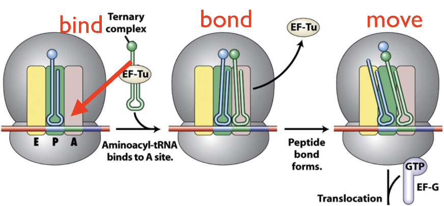
Translation steps and parts involved
enzymes attach each amino acid to corresponding tRNA
Initiation = ribosome’s small subunit binds to mRNA and initiation factors (helper proteins) —> fMet tRNA anticodon binds to start codon [at P site] —> large subunit binds and locks in place + initiation factors leave
Elongation = anticodon of next tRNA matches/binds at A site —> amino acids in P and A sites bond —> tRNA at A site moves to P site; tRNA at P site moves to E site to exit
Termination = stop codon doesn’t code for an amino acid, so an RFI (release factor) binds at its place to signal for peptide release and ribosome disassembly
A = decoding site; P = peptidyl site; E = exit site
hydrolysis of GTP provides energy for process
mRNA is translated 5’ → 3’ (ribosome would go right in the picture = towards 3’)
Glycosylation & Phosphorylation are examples of…
post-translational modifications to a peptide
Glycosylation = adding sugar to peptide
Phosphorylation = adding phosphate group to peptide
What makes each of your cells unique?
the regulation of their expressed genes
(NOT the genes they have — most all cells in one organism have the same DNA)
Benefit of regulating genes early (at transcription) compared to late (post-translation)
early avoids wasting energy or letting minimal expression slip through later in the process
late helps if you still need a gene’s products quick and may just be temporarily disabling them
eg) blood clotting proteins
Prokaryotic vs. Eukaryotic gene expression
Prokaryote genes tend to be “turned on” by default
negative regulation is more common
Eukaryote genes tend to be “turned off” by default
positive regulation is more common
*BOTH still use BOTH positive and negative regulation though
Key requirements for regulating gene expression
being able to sense changing environmental condition
being able to activate/repress genes in response to changes
Operon
string of genes on chromosome with a single promoter that regulates their transcription
often since each coded protein works together and can be regulated together
most common in prokaryotes
Arabinose operon [is an example of what?]
example with AraC protein that can be both a repressor (when arabinose doesn’t bind) and an activator (when arabinose binds)
Tryptophan operon [is an example of what?]
example of negative feedback, where the final product made by its enzymes (tryptophan), signals for the gene’s downregulation
Nucleosomes & role in gene expression
Eukaryotic nuclear/linear DNA strands [which are negative] wrapped tightly around histone proteins [which are positive] (like string around a spool)
make up chromatin = mixture of DNA and proteins that makes up chromosomes
Heterochromatin = tightly bound histones —> genes are less likely to be transcribed (transcriptionally silent)
Euchromatin = less condensed —> genes are more likely to be transcribed
Some methods of regulating gene expression
transcription factors
nucleosomes condensing DNA
intron splicing or alternative splicing (depending on proteins that splice)
microRNAs helping selectively destroy mRNA that comes from a certain gene
environmental conditions