The Cardiovascular System
 Call Kai
 Learn
Learn Practice Test
 Spaced Repetition
 Match
Match Flashcards
 Knowt Play
1/77

There's no tags or description
Looks like no tags are added yet.
Name | Mastery | Learn | Test | Matching | Spaced | Call with Kai | Chat |
|---|
No analytics yet
Send a link to your students to track their progress
78 Terms
What is the primary function of the cardiac skeleton regarding electrical signals of the heart?
It acts as an electrical insulator, preventing signals from passing directly from the atria to the ventricles
The right side of the heart is responsible for this circuit.
Pulmonary circuit
The left side of the heart is responsible for this circuit
Systemic circuit
Two arteries at the base of the aorta are responsible for this circuit
coronary circuit
Heart strings (chordae tendinea) function to _____
prevent the AV valves from opening into the atria
What is important in preventing backflow of blood?
chordae tendineae, papillary muscles, AV valves
Which valve separates the left atrium from the left ventricle?
bicuspid/mitral
List the valves in the correct order starting with deoxygenated from the body arriving at the atrium
tricuspid, pulmonary semilunar, bicuspid, aortic semilunar
the myocardium would be thickest in the _____
left ventricle
Which structures in the fetus heart have connections to bypass the lungs?
right and left atria; aorta and pulmonary trunk
The heart contains two different cardiocyte types. What for?
Conducting cardiocytes — setting the pace
Contracting cardiocytes — pumping blood
What is the main difference between the action potentials of conducting and contracting cardiocytes?
Conducting cardiocytes have no resting potential
Contracting cardiocytes have a plateau phase
What are the three calcium channels and their main function?
Voltage-gated calcium channels — depolarization of conducting cardiocytes
Slow calcium channels — plateau phase of contracting cardiocytes
Calcium channels in sarcoplasmic reticulum — contraction of cardiocytes
What ion is responsible for the slow depolarization that NEVER allows a resting potential in conducting (pacemaker cells)?
sodium
Which portion of the ECG corresponds to repolarization of the ventricles?
T wave
At what point are the atria repolarizing on an action potential?
At the peak
Which component of the heart conduction system would have the fastest rate of firing?
sinoatrial node
The cardiac cycle consists of a distinct relaxation and contraction phase. Which term is typically used to refer ventricular contraction while no blood is being ejecvted?
isovolumetric contraction
Most blood enters the ventricles during _____
atrial and ventricular diastole
The first heart sound of represents which portion of the cardiac cycle?
closing of the atrioventricular valves
Ventricular relaxation immediately follows _____
ventricular repolarization
The heart pumps blood, which is crucial for delivering oxygen and nutrients to the body's tissues. The volume of blood the heart pumps per minute is called cardiac output. Which two factors can change the heart’s output to respond to stress, low blood pressure, etc.?
heart rate and stroke volume
The Autonomic Nervous System influences the heart’s cardiac output. It ______-
affects both the heart rate and stroke volume
The force the heart must overcome to pump blood is known as _____
afterload
The cardiovascular centers are located in which area of the brain?
medulla oblongata
What happens when venous return to the heart is high (e.g. due to exercise)?
the preload increases, which leads to increased contraction force
What happens to preload when there is venous constriction in the veins?
it increases
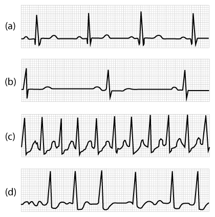
This question refers to ECG traces in the figure below. If the trace in panel (a) depicts normal activity, which trace would be expected in a patient with a severed (non-functioning) vagus nerve?
C
Which of the following statements about arteries and veins is not correct?
A. Arteries contain a lot of elastin, veins contain a lot of collagen
B. Arteries constrict and dilate to regulate blood flow, veins act as blood storage
C. Arteries experience higher pressure than veins
D. Arteries contain more of the blood volume than veins
D. Arteries contain more of the blood volume than veins
True or False: Arteries contain a lot of elastin, veins contain a lot of collagen
true
True or False: Arteries constrict and dilate to regulate blood flow, veins act as blood storage
true
True or False: arteries experience higher pressure than veins
true
True or False: Arteries contain more of the blood volume than veins
false
What is the main function of the main artery (e.g. aorta)?
elastic, conducting blood away from heart
What is the main function of the medium sized artery?
Muscular, distributing to specific organs
What is the main function of the Arteriole/Metarteriole?
Connect to capillaries, control perfusion
What is the main function of the Capillary?
gas and nutrient exchange
What is the main function of the vein?
capacitance/storage vessel
Which type of arteries are responsible for keeping the blood pressure high, even when ventricles are in diastole (relaxation)?
elastic arteries
Which of the following is most common capillary type in the human body (the “normal” type used for perfusion of most tissues)?
continuous
True or false: All capillary beds receive the same amount of blood at any given time
false
What would be an accurate description of the tunica media?
the layer responsible for changing the diameter of the blood vessel
Match each blood vessel type to its description.
Artery — distribute blood
Vein — carry blood toward the heart
Capillary —carry blood within tissues to allow for exchange
Flow is directly proportional to pressure, and indirectly inversely proportional to resistance. Since blood flow is essential for perfusion, your cardiovascular system works hard to control the flow. What can be said about the blood vessels and heart pressure?
the blood vessels influence the resistance by vasodilation/vasoconstriction
the heart can influence the pressure by increasing the stroke volume
The flow of blood through a vessel is _____
decreased if the resistance is increased
Which of the following describes the relationship between atherosclerosis (buildup/plaque on arterial walls) and resistance to flow?
Atherosclerosis increases resistance to flow by narrowing blood vessels and generating turbulent
What is true about resistance to blood flow?
resistance to flow increases with increased blood viscosity
What is responsible for monitoring for fluctuations in arterial blood pressure?
baroreceptors
Match each portion of the cardiovascular center in the Medulla Oblongata to a description of its activity
Vasomotor center — stimulation leads to vasoconstriction/vasodilation on throughout the body
Cardioacceleratory center — stimulation leads to increased heart rate and contractility
Cardioinhibitory center — stimulation leads to decreased heart and contractility
What would be the effect of decreased stroke volume on blood pressure?
blood pressure would drop
What is the function of venous valves?
ensure unidirectional flow of blood through low-pressure veins
You are studying an organ that exchanges large amounts of matieral with its blood supply; however, this organ does not allow cells to move back and forth between its tissues and the blood supply. Which type of capillary would you expect to find serving this organ?
fenestrated
Autoregulation means metabolites in a tissue can act as vasodilators, ensuring more blood flow to the area. What would result in vasodilation of arterioles serving a systemic capillary bed?
increased carbon dioxide levels in local tissues
The interstitial fluid that is not recovered at the venous end of the capillary bed is _______
returned to the venous system by lymphatic vessels
Low blood pressure is sensed by baroreceptors, located in the _____, leads to a/an _____ in sympathetic stimulation
aorta; increase
What is the driving force for movement of fluid out of the capillary into the intracellular space?
arterial blood pressure
True or False (is it a function of blood)? Carrying oxygen and CO2 to and from cells
true
True or False (is it a function of blood)? regulating body temperature
true
True or False (is it a function of blood)? Making red blood cells
false
True or False (is it a function of blood)? Initiating the blood-clotting process.
true

After centrifuging a blood sample from a patient, you will see three distinct layers. What are the contents?
Dark layer on bottom (~45%) — Erythrocytes (red blood cells)
Small middle layer (<1%) — Leucocytes (white blood cells)
Yellow layer on top (~55%) — plasma with proteins
Match the three proteins with their functions.
Albumin — most abundant protein; transports things
Globulin — transports things; fights infections (antibodies)
Fibrinogen — least abundant protein; help with blood clotting
The hematocrit is the ratio of _____ to whole blood volume, and a normal value is ______.
erythrocytes (RBC); 45%
What are the advantages of the erythrocytes (RBC) disc shape?
Allows bending and twisting to get through tiny capillaries
Losts of surface area to quickly attach/detach O2
Allows forming stacks for better flow through tiny capillaries
Predict what would happen if a person was unable to consume iron (FE) in thier diet?
the oxygen-carrying capacity of whole blood would be decreased
Iron released from (recycled) hemoglobin is transported through the blood by _____ to the ______.
transferrin; bone marrow
What part of hemoglobin ultimately gives feces its brown color and urine its yellow color during the recycling process?
Heme
What is the primary function of erythropoietin (EPO) in the human body?
stimulates red blood cell production in the bone marrow
The heart contains 3 main circuits. What are they?
systemic/body, pulmonary/lungs, coronary/heart
Intercalated discs between cardiocytes contain desmosomes and gap junctions. What are their functions?
anchoring cells together/ion movement from cell to cell
Order the pacemaker structures (Master of signaling goes on top)
Sinoatrial node
Atrioventricular node
AV bundle
Bundle branches
How do the funny channels keep you alive?
the funny channels prevent resting in pacemaker cells
True or False: an electrocardiogram (ECG) measures electrical activity of ALL cardiocytes (conducting and contracting)
true
What happens when both atria and ventricles are in diastole?
passive filling of the heart
Ventricular systole eventually creates enough pressure to open the ______
semilunar valves
True or False: the end-diastolic volume is higher than the end-systolic volume
True
The first heart sound is due to ______, while the second heart sound is due to ______.
closing of the AV valve, closing of the semilunar valve
What could calcium-induced calcium release mean?
Ca2+ responsible for action potential trickers Ca2+ release at SR