Respiratory system - all
 Call Kai
 Learn
 Practice Test
 Spaced Repetition
 Match
 Flashcards
 Knowt Play
1/53

There's no tags or description
Looks like no tags are added yet.
54 Terms
what is the word to help remember what causes the Bohr shift?
MCCOAT
what does MCCOAT stand for?
M - muscle activity increases
C - curve moves to the right
C - carbon dioxide increases
O - oxygen decreases
A - more acidity (increase in lactic acid - low pH)
T - increase temperature
why does an increase in muscle activity cause the oxygen dissociation curve to shift to the right?
So more dissociation happens so the muscles get more oxygen.
why is a warm up important in terms of partial pressure?
A warm up is important as partial pressure of oxygen will decrease so more oxygen will disassociate, when temperature rises 37 to 39 degrees C.
what happens in relation to the Bohr shift during recovery and why?
During recovery the oxyhaemoglobin curve shifts back to the left.
This returns haemoglobin saturation with oxygen to it's original relationship.
This allows a greater uptake or association of oxygen to haemoglobin at the alveoli - oxygenating the blood stream to flush waste.
how many molecules of oxygen can haemoglobin carry?
4
what depends on the partial pressure of oxygen (pO2)? (in terms of haemoglobin)
the amount that associates
What happen when the partial pressure is high? (in terms of haemoglobin)?
oxygen readily associates
therefore blood leading to the alveoli is 98% saturated with oxygen
this relationship is shown in the oxyhaemoglobin dissociation curve
what is the oxyhaemoglobin dissociation curve?
a curve which shows how haemoglobin saturation varies as partial pressure of oxygen changes.
E.g. during exercise pO2 in the muscles drops to 5 and the saturation of haemoglobin drops to 25%. 75% of oxygen is released to the muscle cells in order to supply the increased demand of oxygen required for respiration.
where is the RCC located?
In the medulla oblongata
what 2 centres is the RCC made up of?
the inspiration centre
the expiration centre
what happens in the inspiration centre?
Has intercostal nerve and phrenic nerve which work together to control diaphragm and intercostals.
Works at both rest and exercise.
what happens in the expiration control centre?
Only kicks in during exercise.
what does the RCC do during exercise?
uses the receptors to detect change:
Chemoreceptors: detect ↑CO₂, ↑H⁺, ↓O₂
Thermoreceptors: detect ↑blood temperature
Proprioceptors: detect ↑muscle and joint movement
These receptors stimulate the inspiratory centre (IC)
IC increases force of diaphragm & external intercostals
IC recruits sternocleidomastoid & pectoralis minor → ↑depth of inspiration
Baroreceptors: monitor lung inflation
Excessive stretch stimulates expiratory centre (EC)
EC recruits internal intercostals & abdominals → forced expiration
Combined IC & EC activity → ↑breathing rate and depth
what are the 4 receptors which help regulate the respiratory system during exercise and what do they measure?
baroreceptors: located in lung tissue and bronchioles and inform the state of lung inflation.
proprioceptors: inform of motor activity in the muscles and joints.
thermoreceptors: inform of an increased blood temperature.
chemoreceptors: located in aorta and carotid arteries and pick up an increase in blood acidity, increases in CO2 conc and decrease in O2 conc.
what does the RCC do during rest?
IC is responsible for the rhythmic cycle of breathing.
Nerve impulses are generated and stimulate the inspiratory muscles causing them to contract via the: intercostal nerve to the external intercostals and the phrenic nerve to the diaphragm.
This causes thoracic activity volume to be increased.
Approx. 500ml of air will be inspired.
After approx. 2 seconds, stimulation stops and the inspiratory muscles relax.
Lung tissues recoil, causing passive expiration.
This will be repeated approx. 12-15 times per minute.
The expiratory centre is inactive during rest due to the natural relaxation of the diaphragm and external intercostals.
when is the phrenic nerve stimulated and what does it cause?
Stimulated by the inspiratory centre (IC) within the respiratory control centre when chemoreceptors detect ↑CO₂, ↑H⁺ and/or ↓O₂ (at exercise)
Sends motor impulses to the diaphragm
Diaphragm contracts and flattens
Thoracic cavity volume increases
Lung pressure decreases
Air is drawn into the lungs, initiating and increasing inspiration
when is the intercostal nerve stimulated and what does it cause?
Stimulated by the inspiratory centre (IC) within the respiratory control centre when chemoreceptors detect ↑CO₂, ↑H⁺ and/or ↓O₂ (at exercise)
Sends motor impulses via the intercostal nerve to the external intercostal muscles
External intercostals contract
Rib cage moves up and out
Thoracic cavity volume increases
Lung pressure decreases
Air is drawn into the lungs, increasing inspiration
define association (in terms of haemoglobin)
the combining of oxygen with haemoglobin to form oxyhaemoglobin.
define dissociation (in terms of haemoglobin)
the release of oxygen from haemoglobin.
define oxyhaemoglobin (in terms of haemoglobin)
a bright red substance formed by the combination of haemoglobin with oxygen, present in oxygenated blood.
define haemoglobin
a red protein responsible for transporting oxygen in the blood of vertebrates. Its molecule comprises four subunits, each containing an iron atom bound to a haem group.
define saturation (in terms of haemoglobin)
the extent to which haemoglobin in the blood is bound with oxygen
define external respiration
the process of exchanging gases between the alveoli in the lungs and the blood
define internal respiration
the exchange of gases, between the body's tissue cells and the blood
define partial pressure
the pressure exerted by an individual gas held in a mixture of gas.
define diffusion
the movement of gases across a membrane down a gradient from an area of high pressure (or conc) to an area of low pressure (or conc)
define diffusion gradient
the difference in areas of pressure (or conc) from one side of a membrane to the other.
define pulmonary ventilation
the inspiration and expiration of air from the atmosphere behind us.
define gaseous exchange
the extraction of oxygen from the air into the blood stream and then into the muscle tissues
define breathing frequency
the number of breaths taken per minute.
what percent of blood is plasma and what percent is blood cells?
blood consists of:
55% plasma
45% blood cells
what percent of blood combines with haemoglobin to produce oxyhaemoglobin and what happens to the rest?
97% combines with haemoglobin to produce oxyhaemoglobin (HbO2)
the other 3% is dissolved within the blood plasma
how is carbon dioxide transported in the blood?
70% dissolved in water and carried as carbonic acid
23% combines with haemoglobin to create carbaminohaemoglobin
7% dissolved in blood plasma
define tidal volume
Tidal volume is the volume of air inhaled or exhaled in one normal breath.
define minute ventilation
Minute ventilation is the total volume of air inhaled or exhaled per minute.
resting and maximal value for breathing frequency
resting = 12 - 16
maximal = 40 +
what are the resting and maximal for tidal volume
resting = 0.5 litres
maximal = 3-5 litres
resting and maximal for minute ventilation
resting = 6-8 litres
maximal = 200+ litres
how does external respiration work at rest?
Occurs in the alveoli of the lungs
O₂ diffuses from alveoli → capillary blood
High O₂ in alveoli, low O₂ in blood
CO₂ diffuses from capillary blood → alveoli
High CO₂ in blood, low CO₂ in alveoli
Diffusion gradients are moderate
Meets low oxygen demand at rest
how does external respiration work during exercise?
Breathing rate and depth increase
O₂ diffuses from alveoli → blood at a faster rate
Steeper gradient due to ↓blood O₂
CO₂ diffuses from blood → alveoli at a faster rate
Steeper gradient due to ↑blood CO₂
Ensures sufficient oxygen supply to working muscles
how does internal respiration work during rest?
Occurs at muscle capillaries
O₂ diffuses from blood → muscle cells
High O₂ in blood, low O₂ in muscles
CO₂ diffuses from muscle cells → blood
High CO₂ in muscles, low CO₂ in blood
Diffusion gradients are small
Matches low metabolic demand
how does internal respiration work during exercise?
Muscles increase aerobic respiration
O₂ diffuses rapidly from blood → muscles
Very steep gradient due to ↓muscle O₂
CO₂ diffuses rapidly from muscles → blood
Very steep gradient due to ↑muscle CO₂
Increased temperature and CO₂ maintain steep gradients
Supports increased energy production
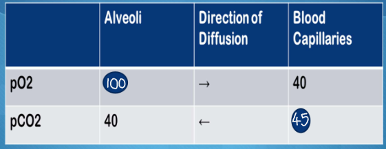
what are the partial pressures at external respiration at rest?
PO2 in alveoli = 100
PO2 in blood capillaries = 40
so diffusion of O2 goes from alveoli to blood capillaries
PCO2 in alveoli = 40
PCO2 in blood capillaries = 45
so diffusion of CO2 goes from blood capillaries to alveoli
what are the partial pressures at internal respiration at rest?
PO2 in muscle cell = 40
PO2 in blood capillaries = 100
so diffusion of O2 goes from blood capillaries to muscle cell
PCO2 in muscle cell = 45
PCO2 in blood capillaries = 40
so diffusion of CO2 goes from muscle cell to blood capillaries
what are the partial pressures at internal respiration during exercise?
PO2 in muscle cell = 5
PO2 in blood capillaries = 100
so diffusion of O2 goes from blood capillaries to muscle cell
PCO2 in muscle cell = 80
PCO2 in blood capillaries = 40
so diffusion of CO2 goes from muscle cell to blood capillaries
Describe how oxygen and carbon dioxide are exchanged between the lungs, blood, and body tissues at rest (including exact values).
At rest:
Blood arriving at the lungs has low O₂ (40) and high CO₂ (45).
In the alveoli, air has high O₂ (105) and low CO₂ (40).
Because of these differences, O₂ diffuses into the blood and CO₂ diffuses out (external respiration).
Blood leaving the lungs (pulmonary veins → left heart → arteries) now has high O₂ (100) and low CO₂ (40).
When this oxygen-rich blood reaches tissues, the tissues are low in O₂ and high in CO₂ because they are constantly using oxygen and producing carbon dioxide.
So O₂ diffuses from blood to tissues and CO₂ diffuses from tissues to blood (internal respiration).
Blood returning to the right heart (veins → vena cava) now has low O₂ (40) and high CO₂ (45–50) again, completing the cycle.
Describe how oxygen and carbon dioxide are exchanged between the lungs, blood, and body tissues during exercise. (including exact values)
At exercise:
Blood arriving at the lungs during exercise has very low O₂ (~20–30) and high CO₂ (~50–60) because muscles are using huge amounts of oxygen and producing lots of CO₂.
In the alveoli, ventilation increases, so air has high O₂ (~105) and low CO₂ (~35–40) or even lower.
Because of these differences, O₂ diffuses into the blood rapidly and CO₂ diffuses out even faster (external respiration).
Blood leaving the lungs (pulmonary veins → left heart → arteries) still has high O₂ (~100) and low CO₂ (~38–40) because the lungs keep up with demand.
When this oxygen-rich blood reaches actively contracting muscles, the tissues are extremely low in O₂ and very high in CO₂ due to intense metabolism.
So O₂ diffuses from blood to tissues even more strongly, and CO₂ diffuses from tissues to blood at a higher rate (internal respiration).
Blood returning to the right heart (veins → venae cava) now has very low O₂ (~20–30) and high CO₂ (~50–60) again, completing the exercise cycle.
when is breathing in and breathing out passive or active?
inspiration rest = active
expiration rest = passive
inspiration exercise = active
expiration exercise = active
what happens during inspiration at rest?
Diaphragm flattens and moves down
External intercostal muscles contract
Rib cage moves up and out
Volume of thoracic cavity increases
Pressure of thoracic cavity decreases
Air moves from higher pressure outside to lower pressure in the lungs
what happens during expiration at rest?
Diaphragm relaxes and returns to a dome shape
External intercostal muscles relax
Rib cage moves down and in
Volume of thoracic cavity decreases
Pressure of thoracic cavity increases
Air moves from higher pressure to lower pressure outside lungs
This process is passive.
what happens during inspiration during exercise?
The diaphragm contracts and flattens more than at rest
External intercostal muscles contract more that at rest
Additional muscles are recruited: sternocleidomastoid, scalene and pectoralis major
Rib cage moves up and out further than at rest
Volume of thoracic cavity increases more than at rest
Pressure of the thoracic cavity decreases more than at rest
More air than at rest moves from higher pressure outside to lower pressure in the lungs.
what happens during expiration during exercise?
The diaphragm relaxes
External intercostal muscles relax
Additional muscles are recruited: rectus abdominus, external obliques, internal obliques contract
Rib cage moves down and in further than at rest
Volume of the thoracic cavity decreases more than at rest
Pressure of the thoracic cavity increases more than at rest
More air moves from higher pressure outside to lower pressure in the lungs