Introduction to Movement
 Call Kai
 Learn
 Practice Test
 Spaced Repetition
 Match
 Flashcards
 Knowt Play
1/61

There's no tags or description
Looks like no tags are added yet.
62 Terms
How does movement begin?
sensory systems → continuously getting sensory feedback as you move
internal representation that gets translated into motor commands
How is movement organized?
higherarchically
cortex → brainstem → spinal cord
Parallel
core pyramidal pathway
extrapyramidal pathway
Which functions are associated with the brainstem?
respiration
chewing
facial expressions
stereotyped behaviour
Which functions are associated with the spinal cord?
protective reflexes
locomotion
What makes up the extrapyramidal motor system?
The cerebellum and the basal ganglia nuclei
How is the frontal lobe organized along its rostral-caudal gradient?
Anterior: planning and goals (executive function)
Middle: translating goals into movements
Posterior: sending commands to execute movement
Which areas make up the motor cortex?
Area 4 (M1)
Area 6 (lateral premotor cortex, SMA and pre-SMA)
What is the result of a lesion in the primary motor cortex?
Contralateral paralysis and increased muscle tone (spasticity), especially for dexterous movements
What deficits follow lesions to SMA vs. premotor cortex?
SMA: alien limb syndrome (loss of control of limbs)
Premotor: loss of ability to use sensory information to reach for objects
What did Wilder Penfield's work show about primary motor cortex?
It contains a map of the body musculature paralleling the somatotopic map in primary somatosensory cortex
What are larger regions of the map dedicated to? smaller regions?
larger regions:
hands for fine motor control
face for the complexity of facial muscles
smaller regions:
torso
legs
feet
What are the firing patterns of neurons in the primary motor cortex?
fire before and during voluntary movements of contralateral muscles
because activity in the brain precedes the movement
What is the role of pre-motor and supplemental motor areas?
organize the motor programs for complex voluntary movements
What do reciprocal parietal-premotor connections accomplish?
Sensory-motor transformations that let sensory information guide interaction with objects
dorsal stream from the visual cortex is combined with somatosensory information in the parietal lobe
axons from the parietal lobe then go to the lateral pre-motor cortex
information is translated into correct command to enable movement
What network directs arm movements toward objects (reaching)? How was this shown?
parietal lobe
dorsal premotor cortex
primary motor cortex
This was shown by recording neurons while monkeys reached out and grabbed objects
What do neurons in the parietal lobe respond to?
both tactile and visual stimuli with receptive fields that are spatially in register
neurons are involved in contraction of a peripersonal spatial map used to guide goal directed movements
How do premotor neurons fire? What does this suggest?
fire during the movement and during an imposed delay prior to the movement
this suggests that they are involved in the planning and preparation to move
What happens if the premotor neuron is given the same cue but is preceded by instruction not to move?
the neurons do not fire during the delay
shows that they are not sensory neurons → Because the same cue doesn't make them fire when no movement is required, they are not simply sensory neurons
shows that they are connecting the sensory input to the appropriate action
How do premotor and primary motor neurons differ regarding which limb is used?
Premotor:
neurons fire during preparation for a planned movement and during execution, regardless of which arm is used
Primary motor:
neurons fire only during execution and only for the contralateral arm
the network that is involved in shaping the hand to grasp and object includes what structures? What is it indicative of?
parietal lobe
ventral premotor cortex
primary motor cortex
as you reach out, hand takes shape which is indicative of two parallel pathways
What do ventral premotor neurons respond to?
they respond to preferred shapes
fires during cue and action
What are mirror neurons? What do they suggest?
Premotor neurons that fire when a monkey reaches for an object and when it watches someone else do so (but not to the object alone or non-goal-directed arm movements)
suggests that the monkey understands what the experimenter is doing
what is the SMA involved in?
internally generated movements → free will movements
learned complex movement sequences
What happens the more forward in the frontal lobe you go?
things are more directly involved in movement but things also become more abstract
What happens if the SMA and pre SMA are lesioned?
paradoxical effects on volitional movement including alien limb syndrome or less of spontaneous movement
the mechanism for coordinated movement are still intact but the ability of the mind to voluntarily control has been decoupled
What is alien limb syndrome?
rare neurological disorder where a person's hand or limb moves involuntarily and purposefully, as if it has a mind of its own, while the person lacks conscious control over those actions
What might pre-SMA be involved in?
the connection between thought and action
When is SMA active?
active during complex learned movement sequences even when they are just imagined
What were the three tasks that demonstrated the time of activity of the SMA?
Task 1: single finger flexion → tell the person to tap index finger and thumb together
motor region that corresponds to the fingers lights up
somatosensory region that corresponds to the fingers lights up
Task 2: sequential finger movement → show complex movement sequence of fingers
both previous regions light up
SMA also lights up → complex sequence involved
Task 3: mental rehearsal of sequential movement → don’t actually move hand just imagine it
SMA lights up
What are SMA neurons selectively responsive to?
select components of learned movement sequences
for example in one case the neuron is active prior to the turning motion in a sequence of movements, but only when its followed by a pull movement
What is the readiness potential?
an EEG signal that is recorded from the medial frontal lobes (SMA) starting around 1 second before voluntary movement
there is a build up of electrical activity that always precedes voluntary movement in the SMA
What did the early work by Wildren Penfield show?
the primary motor cortex contained a map of the body musculature that paralleled the somatotpic map in the primary somatosensory cortex
extensive connections between the two regions
What does the somatotopy suggest about the primary motor cortex?
it is the final common pathway for motor output from the cortex
How much motor output does M1 contribute to the brainstem and spinal cord
only 1/3 of cortical motor output
Where does the remaining cortical output come from?
premotor cortex, SMA and S1
what do projections from motor cortex to the spinal cord form?
corticospinal tract
evolutionarily advanced
What do projections from the motor cortex to the brainstem form?
Corticobulbar tract
older pathway
What is the corticospinal tract important for?
moving the fingers
what is the pathway for motor fibres?
they pass within the internal capsule → form the cerebral peduncles and the pyramids
some fibres from the corticobulbar tract end in the brainstem
cotricospinal fibres project to the spinal cord
Where do the motor fibers cross over?
where the brainstem turns into the spinal cord at the pyramidal decusation → why left side of brain controls right side of body and vice versa
Where do the fibers start?
in layer 5 of the motor cortex
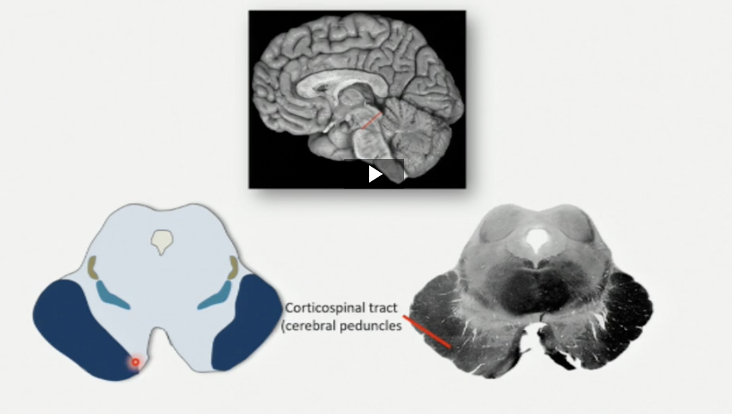
midbrain: corticospinal tract

Pons: corticospinal tract

rostral medulla: corticospinal tract (pryramids)

caudal medulla: corticospinal tract (pyramids)
what happens when the pyramids are cut?
no corticospinal tract intact
moneys were fine for the most part after recovery → they could move arms and legs around after surgery
loss control of individual fingers
What does the lesion in the pyramids indicate?
there is an alternate pathway through the brainstem → parallel pathway
what does the brainstem control?
stereotyped movements of the head
through descending connections with the spinal cord it, contributes to the control of voluntary movements of the body
what do some corticobulbar projections control?
cranial nerves involved in voluntary movements of the head
facial expression
tongue movement
jaw movement
eye movements
What do medial brainstem pathways innervate?
axial muscles that control posture and balance
what is the pathway of the medial brainstem fibres? Where do they terminate?
they travel down the ventral spinal cord white matter and terminate in the ventromedial regions of the cord gray matter
Where does the medial pathway originate from?
tectospinal tract in the midbrain
reticulospinal tract in the pons
vestibulospinal tract in the medulla
where do the medial pathway fibres get input from?
from the cortex
cerebellum
basal ganglia
What is the main lateral pathway?
rubrospinal tract
where does the rubrospinal tract originate?
red nucleus of the midbrain
how does the rubrospinal tracts descend and where does it terminate?
descend in the contralateral dorsolateral column of the spinal cord
terminates in the dorsolateral gray matter
What does the rubrospinal tract control?
voluntary limb movement
what does the complex circuity of the spinal cord gray matter enable?
rhythmic and coordinated movements of the body
Where do sescenting projections terminate in the spinal cord?
on premotor neurons or directly on ventral horn motor neurons
What are direct connections important for?
control of individual digits
what brain regions act in parallel with the central pattern generators?
basal ganglia
cerebellum