CB. 18 Meiosis and karyotypes
 Call Kai
 Learn
 Practice Test
 Spaced Repetition
 Match
 Flashcards
 Knowt Play
1/29

Earn XP
Description and Tags
• To describe karyotypes and their analysis • To describe the purpose and mechanism of cell division to produce gametes (meiosis • To describe the range of chromosomal abnormalities • To outline the mechanism for the generation of aneuploidies • To explain gene translocations and their relevance
30 Terms
What is a chromosome and how many do we have?
A chromosome is a structure made of DNA and associated proteins. It carries genes, which contain instructions used by cells to make functional products, including proteins.
Humans typically have 46 chromosomes in most nucleated somatic cells, arranged into 23 pairs: 22 pairs of autosomes, numbered 1–22. One pair of sex chromosomes, usually XX or XY.
A typical diploid human cell therefore contains two copies of each autosome, one inherited from each biological parent.

What is the basic order of how DNA is condensed/packaged into chromosomes?
Histone = protein
Nucleosome = DNA + histone octamer (H2A, H2B, H3, and H4).
Chromatin = basic repeating unit of chromatin is the nucleosome, which consists of approximately 146 base pairs of DNA wrapped around a histone. Role: package/organise DNA so it fits into the nucleus.
Chromosome = made up of chromatin. Chromosomes protect DNA from shearing.
What is a karyotype? Typical male and female karyotype? Down syndrome karyotype?
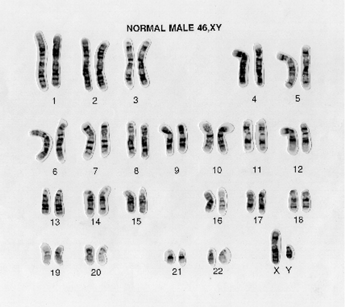
A karyotype is an organised display of an individual's chromosomes, usually arranged into homologous pairs according to their size, centromere position and banding pattern.
For example, a typical male karyotype is written as 46,XY, while a typical female karyotype is written as 46,XX.
An individual with trisomy 21 may have a karyotype written as 47,XX,+21 or 47,XY,+21, depending on the sex chromosomes.
Why are karyotypes important?
Allows scientists and clinicians to investigate chromosome number and large structural changes.
Demonstrate: Extra or missing chromosomes. Large deletions or duplications. Certain chromosome translocations. Chromosomal causes of developmental conditions or reproductive difficulties. Some acquired chromosome abnormalities in cancers.
How are karyotypes produced in the lab? E.g. using WBCs
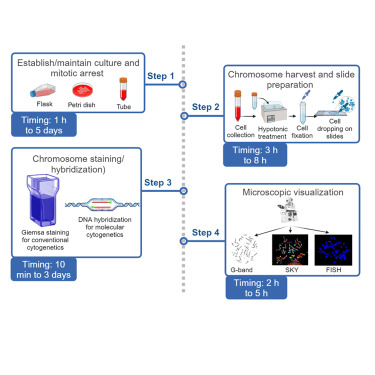
Step 1: Stimulate cell division; White blood cells, typically lymphocytes, are cultured with a mitogen, a substance that stimulates them to divide.
Step 2: Arrest cells in metaphase: A mitotic inhibitor, commonly a spindle inhibitor, prevents normal chromosome separation. Chromosomes are highly condensed and easier to see at this stage.
Step 3: Stain the chromosome: Giemsa staining produces characteristic light and dark bands. Each chromosome has a recognisable banding pattern – reflecting composition and organisation; it is not simply a direct measurement of how much DNA each region contains.
Step 4: Arrange and analyse: Chromosomes are photographed, paired and examined for abnormalities in number and structure.

When karyotyping why are the cells halted/arrested in metaphase?
During metaphase, chromosomes are condensed and individually visible. This makes them easier to distinguish and count.
Giemsa staining can be used to produce bands that help identify individual chromosomes.
What is the difference between conventional karyotyping and using FISH?
Conventional karyotyping examines the overall chromosome structure, whereas FISH can target specific sequences or chromosome regions. Neither method detects every possible DNA change.
Fluorescence in situ hybridisation (FISH) uses fluorescent DNA probes that bind to complementary DNA sequences on chromosomes.

What is the difference between “homologous pair” vs “sister chromatids”?
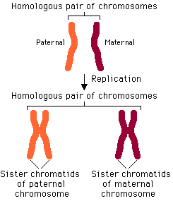
Homologous chromosomes (a homologous pair) =Two chromosomes that carry the same genes in the same position (same gene different allele). One comes from your mother and one from your father. They pair up during meiosis I.
Sister chromatids = Two copies of a single chromosome made during DNA replication. They are joined at a region called the centromere. They are usually genetically identical before crossing over. They separate during mitosis and meiosis II.
REMEMBER: Homologous chromosomes = a matching pair, one from each parent. Vs. Sister chromatids = two copies of the same chromosome joined together.
What is meiosis?
Meiosis is a specialised type of cell division that reduces the chromosome number by half and generates genetic variation.
Its main purpose in humans is to produce haploid gametes for sexual reproduction.
Diploid cells have two sets of chromosomes: 2n (46 chromosomes)
Haploid cells have one set: n (23 chromosomes)
When a haploid sperm and haploid egg fuse during fertilisation, their chromosome sets combine to produce a diploid zygote.
What are the stages of meiosis?
Meiosis consists of one round of DNA replication followed by two successive nuclear divisions, meiosis I and meiosis II.
(1) DNA Replication
(2) Meiosis I: separation of homologous chromosomes – Prophase 1, (prometaphase 1), metaphase 1, anaphase 1, telophase 1.
(3) Meiosis 2: separation of sister chromatids. Prophase 2, (prometaphase 2, ) metaphase 2, anaphase 2, telophase 2,
+ followed by cytokinesis.

Compare the different products of mitosis, meiosis 1 and meiosis 2?
Mitosis: separates sister chromatids.
Meiosis I: separates homologous pairs.
Meiosis II: separates sister chromatids again.

Explain the steps of Meiosis 1
Separation of homologous chromosomes = reductional division because it reduces the chromosome-set number from diploid to haploid.
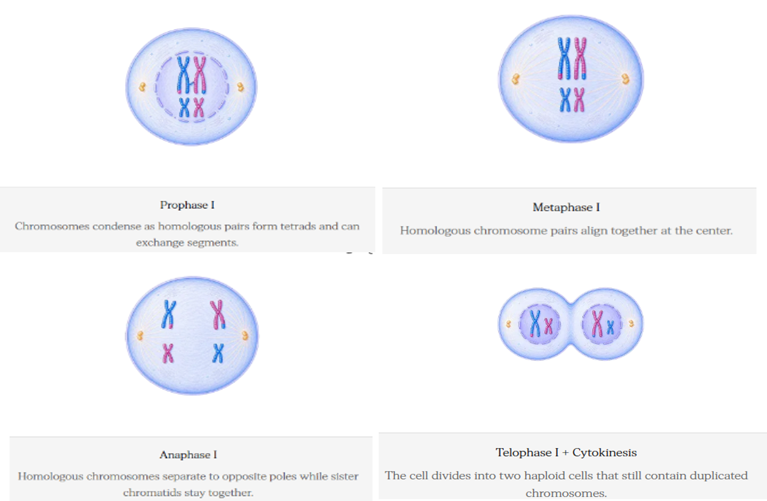
Prophase I = Homologous chromosomes pair up. THEN there is crossing over can occur between non-sister chromatids. DNA segments are exchanged between the homologues.
(+Prometaphase is the stage between prophase and metaphase in which: the nuclear envelope breaks down. Spindle microtubules attach to chromosomes at their kinetochores.)
Metaphase I = Homologous chromosome pairs line up at the cell equator. Their orientation is random, creating opportunities for different maternal and paternal chromosome combinations.
Anaphase I = Homologous chromosomes separate and move to opposite poles. Sister chromatids remain joined at their centromeres.
Telophase I and cytokinesis =The cell divides, usually producing two cells. Each cell has one chromosome from each homologous pair, but each chromosome still consists of two sister chromatids.
Explain the steps of Meiosis 2
Separation of sister chromatids aka equational division because sister chromatids separate while the haploid chromosome-set number remains the same.
Prophase II: Chromosomes condense and the spindle forms.
(+Prometaphase 2: nuclear envelope breaks down, spindle attaches at kinetochore)
Metaphase II: Chromosomes line up individually at the cell equator.
Anaphase II: Sister chromatids separate and move to opposite poles.
Telophase II and cytokinesis: The cells divide, typically producing four haploid cells.


How does meiosis generate genetic variation?
(1) Crossing over (recombination) within prophase I, homologous chromosomes pair. DNA segments are exchanged between non-sister chromatids. This produces new combinations of alleles on chromosomes.
(2) Independent assortment. During metaphase I, each homologous pair can orient in either direction, independently of the other pairs. The chromosome inherited from the mother or father of the individual undergoing meiosis can therefore end up in different combinations in different gametes. With 23 chromosome pairs, independent assortment alone gives: 2^23=8,388,608 possible combinations of whole maternal and paternal chromosomes, before considering crossing over.

What are chromosomal abnormalities and what are the two broad types?
Number/structure of chromosomes differ from usual pattern.
Numerical abnormalities: an extra chromosome, a missing chromosome or an abnormal number of complete chromosome sets. (Aneuploidy).
Structural abnormalities: a chromosome has lost, gained, rearranged or exchanged genetic material.

Numerical chromosomal abnormalities. What is Aneuploid, trisomy or monosomy?
Aneuploidy = the cell has an abnormal number of individual chromosomes, rather than a whole extra set. E.g. typical human cell has 46 chromosomes. A cell with 47 chromosomes because of an extra chromosome 21 has an aneuploidy.
There are two important types of aneuploidy (loss/gain of chromosomes) are trisomy and monosomy.
Trisomy = a cell has three copies of a particular chromosome instead of the usual two. Many autosomal trisomies are incompatible with continued embryonic development and result in pregnancy loss.
Monosomy = one copy of a particular chromosome is missing. For example, a typical human cell has two sex chromosomes. In Turner syndrome, one X chromosome is missing or structurally altered the karyotype is:45,X
What are three important trisomy’s?
Trisomy 21 = Downs syndrome: instead of two copies of chromosome 21, affected cells typically have three, giving a total of 47 chromosomes.
Trisomy 13 — Patau syndrome: associated with severe developmental abnormalities.
Trisomy 18 — Edwards syndrome: associated with severe growth and developmental abnormalities.

Why does an extra chromosome cause problems? E.g. explain the pathophysiology of Downs Syndrome and risk of dementia.
Relates to gene dosage.
Genes provide instructions for producing functional products, including proteins. Having an extra chromosome can increase the dosage of many genes simultaneously. This disrupts the normal balance between gene products and can interfere with development.
For example, chromosome 21 contains the APP gene, which encodes amyloid precursor protein. An extra copy of chromosome 21 contributes to increased APP gene dosage and is associated with increased susceptibility to Alzheimer-related pathology in people with Down syndrome.

Name an important condition called by monosomy?
In Turner syndrome, one X chromosome is missing or structurally altered the karyotype is:45,X
Turner syndrome can affect growth, ovarian function and other aspects of development. Some individuals have mosaicism, meaning that different cells have different chromosome complements.

Why is the loss of a chromosome harmful?
Many genes normally have two copies, and losing one copy can reduce the production of important gene products. This is called gene dosage imbalance or, for particular genes, haploinsufficiency.
Haploinsufficiency is a genetic condition in which one functional copy of a gene cannot produce enough gene product to maintain normal biological function, leading to a disease phenotype even though one allele remains intact.
What is polyploidy and triploidy?
Aneuploidy: an individual chromosome is gained or lost.
Polyploidy: an additional complete set of chromosomes is present. In humans, the normal diploid chromosome number is 46. Triploidy means that there are three complete chromosome sets: 3n=69. A triploid cell has 69 chromosomes rather than 46.
Triploidy can arise, for example, if an egg is fertilised by two sperm or if a gamete with an abnormal chromosome complement contributes an extra complete set. Triploidy usually causes severe developmental abnormalities and pregnancy loss. Complete extra chromosome sets are generally incompatible with survival to birth in humans.


What is nondisjunction?
Nondisjunction is the failure of chromosomes or sister chromatids to separate properly during cell division. It is a major mechanism responsible for aneuploidy.
What can nondisjunction in meiosis 1 cause? What would the 4x resulting gametes contain and what would happen if 1 of them fused with a normal gamete?
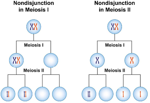
Nondisjunction during meiosis I prevents the normal process of homologous chromosomes separating during anaphase I.
The nondisjunction means BOTH homologues may move to the same pole instead of separating.
After meiosis II, the resulting gametes typically contain: An extra copy of the chromosome, giving n+1. OR No copy of that chromosome, giving n−1.
If one of these gametes fuses with a normal gamete, the resulting zygote may have trisomy or monosomy.
What can nondisjunction in meiosis 2 cause? What would the 4x resulting gametes contain?
Nondisjunction during meiosis 2 prevents the normal process of sister chromatids separating during anaphase II.
If nondisjunction occurs, the resulting gametes typically include: One with an extra chromosome, n+1. OR One missing that chromosome, n-1. OR Two with the normal chromosome number, n

Why is nondisjunction more common in female gametogenesis?
Association with maternal age.
Human oocytes begin meiosis before birth and can remain arrested in prophase I for many years. The machinery responsible for holding chromosomes together and ensuring their accurate separation can become less reliable with age.
Abnormal crossing over (recombination) patterns can also increase the likelihood of chromosome segregation errors.
Explains why the risk of certain trisomies, including trisomy 21, increases with maternal age.
What is translocation and the two different types?
Creates a structure chromosomal abnormality rather than numerical abnormality.
A translocation occurs when a chromosome segment moves to a different chromosome.
Two types of translocation abnormalities:
Balanced translocation: chromosome segments are rearranged without a substantial net gain or loss of genetic material. The carrier may have no obvious symptoms, although reproductive risks can arise.
Unbalanced translocation: genetic material is gained or lost, potentially affecting development and health.

Explain the pathophysiology of the Philadelphia chromosome and chronic myeloid leukaemia.
Philadelphia chromosome: is the translocation between chromosomes 9 and 22. It is written as: t(9;22)
Genetic material exchanged between chromosomes 9 and 22 creates a fusion gene called BCR: ABL1, commonly associated with chronic myeloid leukaemia (CML).
BCR: ABL 1 fusion gene produces an abnormal protein with persistently active tyrosine kinase activity. The abnormal signalling promotes cell proliferation and survival. Abnormal blood-forming cells accumulate, contributing to CML.

How does the pathophysiology of the Philadelphia chromosome relate to the treatment of CML?
The BCR:ABL1 fusion protein is also a therapeutic target. Tyrosine kinase inhibitors, such as imatinib, can block its abnormal signalling and are an important treatment for CML.
